

 PDF
PDF Citation
 Print
Print


INTRODUCTION
Clostridioides difficile, formerly known as Clostridium difficile, is a gram-positive, spore-forming, obligate anaerobic bacterium that is the leading cause of antibiotic-associated diarrhea worldwide [1]. There has been a substantial increase in the incidence and severity of C. difficile infections (CDIs) since the early 2000s, especially in elderly patients in the United States and other industrialized countries [2]. In Asia, the incidence of CDI is 5.3 per 10,000 patient-days, similar to the rates reported from North America and Europe [3].
A rapid and accurate diagnosis is essential to guide treatment and prevent transmission [4]. An effective diagnosis of CDI requires the presence of symptoms (typically diarrhea) and either a positive stool test for C. difficile toxins or detection of toxigenic C. difficile, or colonoscopic or histopathologic findings revealing pseudomembranous colitis (PMC) [5, 6]. Several laboratory tests are recommended for the diagnosis of CDI, but the best standard laboratory test for diagnosis has not been established [5, 7]. Laboratory diagnostic tests for CDI include the toxigenic culture of C. difficile, cell culture cytotoxicity assay, enzyme immunoassay (EIA) for glutamate dehydrogenase (GDH) and toxins A and/or B, and nucleic acid amplification test (NAAT) for 16S RNA, toxin genes, and GDH genes [5, 8-13]. The toxigenic culture of C. difficile and cell cytotoxicity assay have been considered the gold standards for diagnosis; however, these tests are not clinically practical because of their high turnaround time and labor intensiveness. Hence, these tests are infrequently used for routine clinical diagnosis [7, 12]. Currently, EIA and NAAT, which give quick results, are commonly used to detect CDI [14].
Scoring systems composed of some potential factors for correlation with disease severity or treatment outcome have recently been developed and validated [6, 15-19]. We selected three scoring systems based on a combination of simple clinical and laboratory elements [17-19]. In this study, we aimed to investigate whether laboratory diagnostic methods using fecal C. difficile toxin EIA and tcdB NAAT correlate with the disease severity scoring systems for CDI patients.
Go to : 
MATERIALS AND METHODS
We reviewed the records of inpatients who had been tested with both the C. difficile toxin A/B EIA assay and the multiplex PCR test including C. difficile tcdB gene at Incheon St. Mary’s Hospital from December 2015 through May 2018. CDI was defined as antibiotic-related diarrhea with positive stool test for toxin A/B EIA and/or tcdB NAAT, or PMC found on endoscopic examination. We collected the following routinely available patient data: blood test results obtained within 3 days of receiving stool samples (white blood cell (WBC) count, C-reactive protein (CRP), serum albumin, estimated glomerular filtration rate (eGFR), and serum creatinine), age, gender, history of antibiotics use and intensive care unit (ICU) admission, and colonoscopic evidence of colitis. This study was approved by the ethics committee of the institute (OC18RESI0078).
1. Identification of C. difficile toxin
Stool specimens were submitted to the clinical laboratory, where they were stored at 4°C and processed within 24 hours. Stool specimens were examined for toxins A and B by using a VIDAS toxin A/B enzyme-linked fluorescent assay (BioMerieux SA, France) according to the manufacturer’s instructions. The results were interpreted as positive (≥0.37), equivocal, or negative (<0.13), according to the intensity of fluorescence.
2. Identification of TcdB gene
The C. difficile tcdB gene was detected using the multiplex PCR assay (Seeplex® Diarrhea ACE Detection, Seegene, Seoul, Korea). The Seeplex Diarrhea ACE Detection kit contains reagents to identify 10 bacteria (Salmonella spp., Shigella spp., Vibrio spp., Campylobacter spp., C. difficile toxin B, Clostridium perfringens toxin, Yersinia enterocolitica, Aeromonas spp., Escherichia coli O157:H7, and verotoxin-producing E. coli). When the results of mixed infection appeared in this test, CDI was determined by referring to the results of the EIA and C. difficile culture test (if performed) and clinical diagnosis through medical record review.
3. CDI severity scoring systems
Three CDI severity scoring systems—the C. difficile associated diarrhea (CDAD) severity score of Zar et al. [17], the ATLAS score of Miller et al. [18], and the Clostridium difficile severity score (CDSS) of Na et al. [19]—were included in this study (Table 1). The CDAD severity score is a severity assessment score for stratification of disease groups into mild and severe CDAD using 6 factors (age, temperature, leukocyte count, albumin, hospitalization in the ICU, and presence of PMC) [17]. The ATLAS scoring system is composed of five clinical and laboratory variables (age, treatment with systemic antibiotics, leukocyte count, and albumin and serum creatinine as a measure of renal function) measured at the time of CDI diagnosis [18]. The CDSS score includes three binary variables: age >65 years, peak serum creatinine >2 mg/dL, and peak peripheral blood leukocyte count of >20,000 cells/μL [19]. The mild and severe disease groups were determined according to the criteria of each scoring system; however, for the ATLAS scoring system, we used patients with >6 points with an actual cure rate of less than 70% in the previous report as the severe disease group [18]. For the CDAD scoring system, patients with >2 points were considered to have severe disease. Since the optimal cut-off score was not clear in the ATLAS scoring system, we considered patients with >6 points to have severe disease based on the actual cure rate in the previous study [18]. For the CDSS score, disease severity was divided into a low severity score (0 or 1) and a high severity score (2 or 3) [19].
Table 1
Comparison of three CDI severity assessment scoring systems
| Scoring system | Parameter | Point scale | Reference |
|---|---|---|---|
| CDAD | age (≥65 years, 1 point), temperature (≥38.3°C, 1 point), leukocyte count (>15,000 cells/mm3, 1 point), albumin (<2.5 mg/dL, 1 point), hospitalized in the ICU (yes, 2 points), and presence of pseudomembranous colitis (yes, 2 points) | 0-6 | Zar et al. [17] |
| ATLAS | age (60-79 years, 1 point; ≥80 years, 2 points), treatment with systemic antibiotics (yes, 2 points), leukocyte count (16,000-25,000 cells/mm3, 1 point; >25,000, 2 points), albumin (2.6-3.5 mg/dL, 1 point; ≤ 2.5 mg/dL, 2 points), and serum creatinine (121-179 μmol/L, 1 point; ≥ 180 μmol/L, 2 points) | 0-10 | Miller et al. [18] |
| CDSS | age (≥65 years, 1 point), temperature (≥38.3°C, 1 point), leukocyte count (>15,000 cells/mm3, 1 point), and serum creatinine (≥2 mg/dL, 1 point) | 0-3 | Na et al. [19] |
![]()
4. Statistics
Statistical analyses were performed using MedCalc for Windows, version 19.4 (MedCalc Software, Ostend, Belgium). The Chi-square test was used for comparison between two groups. The Mann-Whitney U test was used to correlate the combined results of toxin EIA and tcdB NAAT with three CDI severity scores for patients with CDI. P-values <0.05 were considered statistically significant.
Go to :
RESULTS
1. Clinical and laboratory findings of CDI patients
Ninety-one patients were included as defined CDI cases in this study. The median age of the patients was 73 years (range: 29-87 years) and 56% were female. The clinical findings—including fever, admission to the ICU, history of systemic antibiotic treatment and presence of PMC—and laboratory findings—including WBC, CRP, serum albumin, eGFR, and serum creatinine—for five groups categorized according to the results of EIA and NAAT are summarized in Table 2. Among the 91 patients, 43 (48%) tested positive for both toxin EIA and tcdB NAAT, whereas 36 (40%) only tested positive for tcdB NAAT. Nine patients (10%) tested equivocal for toxin EIA and positive for tcdB NAAT. Two patients (2%) tested equivocal for toxin EIA and negative for tcdB NAAT, and one patient only tested positive for toxin EIA. Among the 88 NAAT positive patients, 17 (19.3%, 17/88) had fever and 10 (58.8%, 10/17) tested positive or equivocal for the toxin EIA assay. Furthermore, 16 patients (18.2%, 16/88) were hospitalized in ICU and 13 patients (72.2%, 13/16) tested positive or equivocal for the toxin EIA assay. The presence of PMC was observed in 4 patients, and all of them tested positive or equivocal for the toxin EIA assay. Among the 88 tcdB NAAT positive patients, 15 exhibited additional positive results for other bacterial pathogens in the multiplex PCR assay. The pathogens detected with tcdB were C. perfringens toxin (N=11), Aeromonas spp. (N=3), Salmonella spp. (N=1), and Campylobacter spp. (N=1).
Table 2
Summary of clinical and laboratory findings of 91 CDI patients according to combined results of toxin EIA and tcdB NAAT
| EIA+/NAAT+ N= 43 | EIA−/NAAT+ N= 36 | EIA equivocal/NAAT+ N= 9 | EIA equivocal&NAAT− N= 2 | EIA+/NAAT− N= 1 | |
|---|---|---|---|---|---|
| Age (yr)* | 73± 12.9 | 66.6 ± 13.4 | 76.4 ± 5.9 | 68± 14.1 | 76± 0 |
| Gender, female/male | 29/14 | 15/21 | 4/5 | 2/0 | 1/0 |
| Fever ( ≥ 38.3°C) | 8 | 7 | 2 | 0 | 0 |
| Hospitalized in the ICU | 11 | 3 | 2 | 1 | 0 |
| Treatment with systemic antibiotics | 43 | 14 | 7 | 2 | 1 |
| Presence of pseudomembranous colitis | 3 | 0 | 1 | 0 | 0 |
| White blood cell (cells/mm3)* | 12.7 ± 11.9 | 10.5 ± 7.4 | 11± 5.8 | 8.6 ± 6.7 | 9.2 ± 0 |
| CRP (mg/L)* | 66.6 ± 57 | 82.1 ± 60.8 | 70.8 ± 59.6 | 48.6 ± 63.7 | 28.7 ± 0 |
| Albumin (g/dL)* | 2.9 ± 0.6 | 2.9 ± 0.6 | 2.7 ± 0.6 | 2.8 ± 0.4 | 2.5 ± 0 |
| eGFR (mL/min/1.73 m2)* | 89.2 ± 60.6 | 102.6 ± 79.8 | 68.6 ± 40.5 | 85.1 ± 46.6 | 73.2 ± 0 |
| Creatinine (mg/dL)* | 1.1 ± 0.9 | 1.3 ± 1.5 | 1.6 ± 1.6 | 0.8 ± 0.4 | 0.7 ± 0 |
![]()
2. Correlation between results of toxin EIA and CDI severity scores
To determine the correlation between toxin EIA and CDI severity scores, we divided patients according to the toxin EIA results. Eighty-eight patients who were tcdB NAAT positive were divided into the toxin EIA negative group and the toxin EIA positive group. We performed statistical analysis firstly by including the equivocal EIA results in the negative results, and then by including them in the positive results.
The patients had a median CDAD severity score of 1.5 (range: 0-6), and 50.0% (44/88) of them were included in the mild group (scores 0 and 1). They had a median ATLAS score of 4 (range: 0-10), and 92.0% (81/88) of them were included in the mild group (scores 0-6). The patients had a median CDSS score of 1 (range: 0-3), and 83.0% (73/88) of them were included in the mild group (scores 0 and 1).
When toxin EIA equivocal results were included in the negative results, the toxin EIA and NAAT positive group showed significant correlation with the ATLAS score (P=0.005) but did not show a correlation with the CDAD severity score (P=0.107) and CDSS score (P=0.534) (Table 3 and Fig. 1). However, when toxin EIA equivocal results were included in the EIA positive results, the EIA and NAAT positive group showed significant correlation with the CDAD severity score (P=0.03) and ATLAS score (P<0.001) but did not show a correlation with the CDSS score (P=0.169). There was no statistical significance between two categorical groups of the mild and severe cases in the three CDI severity scoring systems (Table 3).
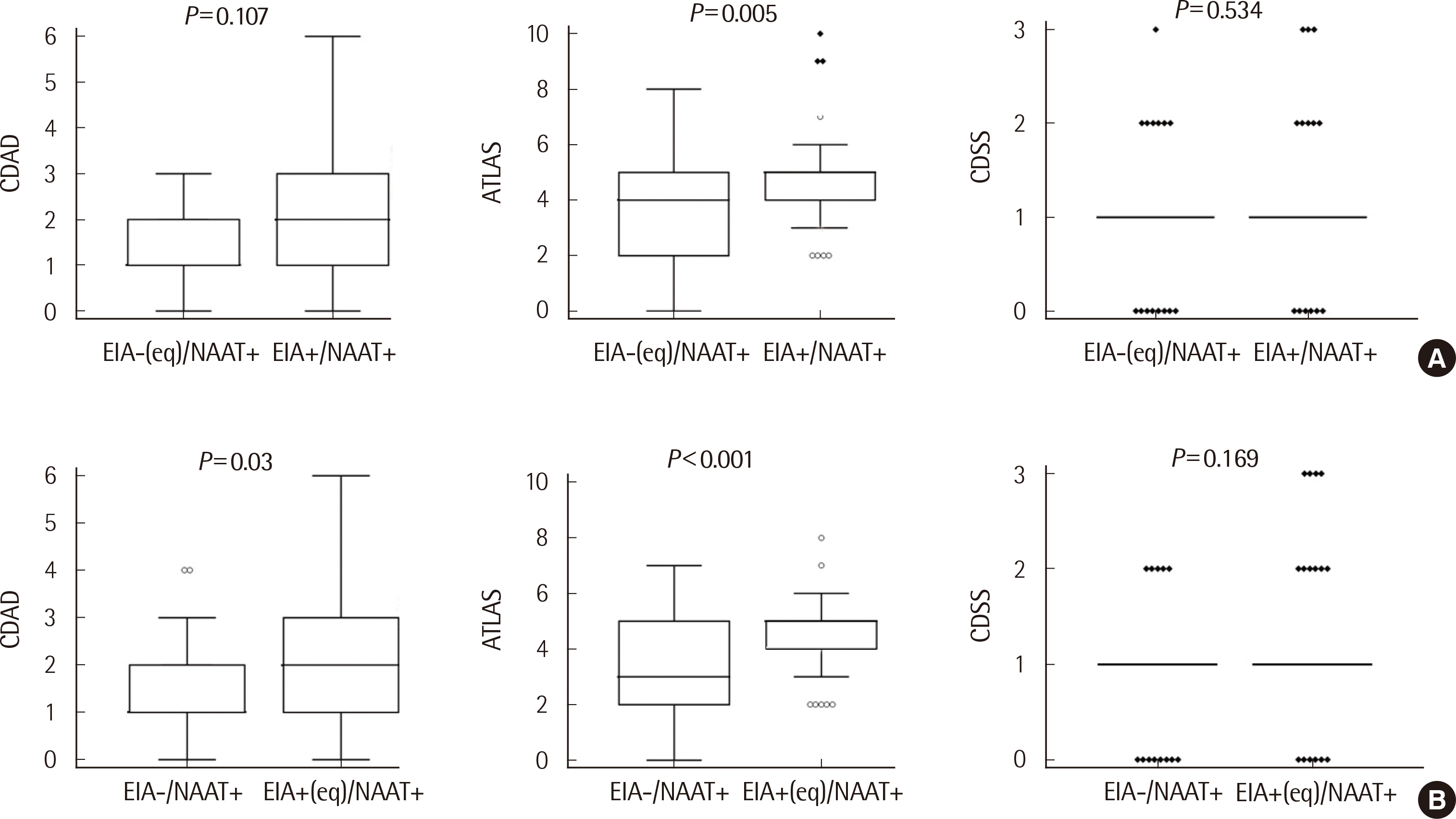 | Fig. 1Correlation between the combined results of toxin EIA and tcdB NAAT and three CDI severity scores. (A) Toxin EIA negative including equivocal results. (B) Toxin EIA positive including equivocal results. All data are median and interquartile ranges. Mann-Whitney U test, P-value < 0.05 was considered statistically significant.
Abbreviation: CDI, C. difficile infection; CDAD, C. difficile associated diarrhea; CDSS, C. difficile severity score; eq, equivocal. Acronym: ATLAS, age treatment leukocyte albumin serum creatinine.
|
Table 3
Correlation between combined results of toxin EIA and tcdB NAAT and three CDI severity scores for 88 CDI patients
| CDI severity | Score | EIA (including equivocal)−/NAAT+ | EIA+/NAAT+ | P* | EIA−/NAAT+ | EIA (including equivocal)+/NAAT+ | P* | N (%) |
|---|---|---|---|---|---|---|---|---|
|
|
|
|
|
|
|
|||
| N (%) | 45 (51.1) | 43 (48.9) | 36 (40.9) | 52 (59.1) | 88 | |||
| CDAD | ||||||||
| Mild (0, 1) | 0 | 4 | 4 | 0.138 | 4 | 4 | 0.085 | 8 (9.1) |
| 1 | 22 | 14 | 18 | 18 | 36 (40.9) | |||
| Severe (2-6) | 2 | 12 | 11 | 10 | 13 | 23 (26.1) | ||
| 3 | 3 | 6 | 2 | 7 | 9 (10.2) | |||
| 4 | 2 | 4 | 2 | 4 | 6 (6.8) | |||
| 5 | 2 | 3 | 0 | 5 | 5 (5.7) | |||
| 6 | 0 | 1 | 0 | 1 | 1 (1.1) | |||
| P† | 0.107 | 0.03 | ||||||
| ATLAS | ||||||||
| Mild (0-6) | 0 | 2 | 0 | 0.6497 | 2 | 0 | 0.491 | 2 (2.3) |
| 1 | 3 | 0 | 3 | 0 | 3 (3.4) | |||
| 2 | 10 | 4 | 9 | 5 | 14 (15.9) | |||
| 3 | 6 | 3 | 5 | 4 | 9 (10.2) | |||
| 4 | 9 | 10 | 7 | 12 | 19 (21.6) | |||
| 5 | 8 | 18 | 6 | 20 | 26 (29.5) | |||
| 6 | 4 | 4 | 2 | 6 | 8 (9.1) | |||
| Severe (7-9) | 7 | 2 | 1 | 2 | 1 | 3 (3.4) | ||
| 8 | 1 | 0 | 0 | 1 | 1 (1.1) | |||
| 9 | 0 | 2 | 0 | 2 | 2 (2.3) | |||
| 10 | 0 | 1 | 0 | 1 | 1 (1.1) | |||
| P† | 0.005 | < 0.001 | ||||||
| CDSS | ||||||||
| Mild (0, 1) | 0 | 8 | 6 | 0.7054 | 8 | 6 | 0.518 | 14 (15.9) |
| 1 | 30 | 29 | 23 | 36 | 59 (67.0) | |||
| Severe (2, 3) | 2 | 6 | 5 | 5 | 6 | 11 (12.5) | ||
| 3 | 1 | 3 | 0 | 4 | 4 (4.5) | |||
| P† | 0.534 | 0.167 | ||||||
*P-values were calculated using Chi-square test for mild and severe groups. †P-values were calculated using Mann-Whitney U test for methods and CDI severity scores. Abbreviations: CDI, C. difficile infection; EIA, enzyme immunoassay; NAAT, nucleic acid amplification test; CDAD, C. difficile associated diarrhea; CDSS, C. difficile severity score; ATLAS, age treatment leukocyte albumin serum creatinine.
![]()
Go to :
DISCUSSION
In this study, three CDI severity scoring systems were used; CDAD, ATLAS, and CDSS scores [17-19]. Among them, the toxin EIA results best correlated with the ATLAS score and the positive EIA results including equivocal results correlated with the CDAD and ATLAS severity scores. Antimicrobial therapy remains the treatment of choice for CDI, and specific antimicrobial therapy guideline recommendations should be based on the severity of the disease [7]. In a large, prospective, and randomized study by Zar et al. [17], the CDAD scoring system was developed based on clinical criteria for stratifying patients into mild and severe disease groups. Metronidazole and vancomycin were equally effective for the treatment of mild CDAD, but vancomycin was superior for treating patients with severe CDAD [17]. The ATLAS scoring system composed of five simple and commonly available clinical and laboratory variables was suggested by Miller et al. [18] to predict response to therapy. This scheme highly correlates with the treatment outcome (R2=0.95; P<0.001) and might be useful in stratifying CDI patients so that appropriate therapies can be chosen to maximize cure rates [18]. The CDSS was developed as a prediction tool for severe outcomes in CDI [19]. The CDSS comprised three binary variables that proved to be independent factors associated with severe CDI among 263 CDI patients from a Boston cohort [19]. In the entire cohort (Boston, Dublin, and Huston) of 596 individuals, those with a CDI severity score of 0, 1, 2, and 3 were associated with a 16.3%, 34.9%, and 46.9% risk of severe clinical outcomes of CDI, respectively [19].
The recently updated clinical guidelines for CDI by IDSA and SHEA recommend using a stool toxin assay as part of a multistep algorithm (i.e., GDH plus toxin; GDH plus toxin, arbitrated by NAAT; or NAAT plus toxin) rather than NAAT alone for all specimens received in the clinical laboratory when there are no pre-agreed upon institutional criteria for patient stool submission [6]. The European Society of Clinical Microbiology and Infectious Diseases strongly recommends using a two-step algorithm for diagnosis of CDI instead of a single stand-alone assay [20]. The algorithm should start with either the NAAT or GDH assay, and specimens testing positive in the first step should be tested further with the toxin A/B EIA [20].
We aimed to determine the correlation between the diagnostic methods for toxigenic C. difficile and the disease severity scoring systems for CDI mentioned above. The laboratory tests used for the detection of toxigenic C. difficile in this study were EIA for toxins A/B and NAAT for the tcdB gene. CDI was diagnosed based on NAAT on a multiplex diarrhea panel (Seeplex Diarrhea ACE detection kit) and/or a toxin test (VIDAS C. difficile toxin A/B assay). Recent reports indicate that the sensitivity of the VIDAS C. difficile toxin A/B test ranges from 48% to 90% and specificity ranges from 97% to 99%, depending on the pathogen present [21, 22]; moreover, the sensitivity of the Seegene multiplex PCR for C. difficile detection is 93.9%, which is higher than that of the BD MAX C. difficile kit, a singleplex PCR that detects the tcdB gene [23]. The sensitivity and specificity of the Seegene multiplex PCR for C. difficile detection has been reported to be 40-90% and 96-100%, respectively, both higher than that of toxigenic C. difficile culture [24-26].
In this study, the correlation between EIA toxin positivity and CDI severity differed according to the scoring system, but it was best expressed by the ATLAS score. In addition, EIA toxin positivity, including equivocal results, was more closely related to the ATLAS and CDAD severity scores. The severity score in the CDAD and ATLAS scoring systems was higher for the EIA positive (including equivocal) group than for the EIA negative group. Therefore, the equivocal results of the EIA toxin assay may provide positive meaning for the risk stratification of CDI. In a recent study by Cohen et al. [27], the fecal C. difficile toxin level correlated with CDI severity and conferred an increase in the risk of 30-day mortality. A large prospective study by Planche et al. [28] showed that toxin positivity correlates with clinical outcome, and hence that the detection of toxins is an essential step in the diagnosis of CDI. On the other hand, a retrospective study by Humphries et al. [29] indicated that the presence of stool toxin measured by EIA does not correlate with disease severity of CDI.
However, NAAT positivity seems less relevant to disease severity. In a large prospective study by Polage et al., the toxin immunoassay positive and PCR positive (Tox+/PCR+) patients had a longer duration of diarrhea than Tox−/PCR+ patients and Tox−/PCR− patients, but Tox−/PCR+ patients and Tox−/PCR− patients had a similar risk of diarrhea on most days [30]. In a study by Kumar et al. [31], a positive C. difficile toxin EIA stool sample was associated with both significantly higher WBC and CRP but NAAT positivity was not. NAAT is rapid and highly sensitive; however, this test detects the DNA of the toxin gene of C. difficile rather than the presence of the toxin in stool samples [31]. Diagnosis of CDI by PCR can also lead to misclassification of some cases of C. difficile carriers as CDI cases [32].
Among C. difficile tcdB positive cases, C. perfringens (11/88, 12.5%) was most commonly detected by the multiplex PCR assay in our study; this is consistent with the results of Kim et al. [33], in whose study C. perfringens was commonly detected with C. difficile tcdB in CDI stool specimens by using the Seeplex Diarrhea kit. These patients were included as CDI cases because they had a history of antibiotic treatment before symptoms of diarrhea and/or the C. difficile culture results were positive.
Our study has several limitations. We only evaluated the clinical and laboratory data at the time of CDI diagnosis by retrospective design and could not follow up on the clinical outcomes of the patients after treatment. Several factors can influence the severity and the clinical outcome of CDI, including the virulence of the infecting strain and the host immune responses [19]; however, these factors were not evaluated in this study. In addition, we could not perform the reference methods of the cell cytotoxicity assay or cytotoxigenic culture. Instead, we used the EIA and NAAT tests, which are readily available methods in routine laboratories.
In conclusion, the C. difficile toxin EIA assay may be clinically useful in assessing the severity of disease in CDI patients, especially in correlation with ATLAS severity scores. Further prospective studies are needed to validate the association between diagnostic tools and clinical outcomes of CDI.
Go to :
 XML Download