

 PDF
PDF Citation
 Print
Print


INTRODUCTION
Mycobacterium kansasii is a common non-tuberculous mycobacterial (NTM) species with relatively high pathogenicity [1, 2]. It can cause severe lung diseases, similar to that caused by Mycobacterium tuberculosis [3, 4]. Seven subtypes of M. kansasii have been described, based on restriction fragment length polymorphism analysis of hsp65 [5-7]. PCR-restriction enzyme analysis of rpoB and tuf has also been used to distinguish subtypes [8, 9]. Among these subtypes, M. kansasii sensu stricto is considered the most pathogenic and is isolated most frequently [4, 10-13]. Reports of human diseases caused by subtype 2 are rare, demonstrating its higher association with immunosuppression than subtype 1 [7, 14]. There is a consensus that the other subtypes (subtypes 3–7) are not human pathogens, although this opinion is controversial owing to the limited evidence caused by the paucity of isolates of these subtypes [4]. “Mycobacterium kansasii complex” has been proposed as an inclusive term comprising all subtypes of M. kansasii and M. gastri, which is a closely related species but indistinguishable from M. kansasii by 16S rRNA sequencing [4, 15].
Although the difference in the pathogenicity of M. kansasii subtypes has been recognized, these subtypes have recently been differentiated into distinct species [15, 16]. Unlike subspecies, subtypes are not a part of the standard taxonomic classification system, and reporting of subtypes in mycobacterial identification by clinical laboratories is not mandatory [17, 18]. However, the recently published M. kansasii-derived species, which were formerly classified as M. kansasii subtypes, have not been included as target species of commercial kits for NTM identification [15]. Genotyping of at least one discriminatory target, such as rpoB and hsp65, is required for accurate species identification.
The detection frequency of M. kansasii subtypes in clinically relevant populations have been reported; however, only a few studies have compared the clinical characteristics among infections caused by M. kansasii subtypes [4, 13]. Several studies on the clinical relevance of M. kansasii have not separately analyzed the subtypes, resulting in substantial variability (17%–88%) in the reported rate of pulmonary diseases caused by M. kansasii isolates and diversity in radiological findings [1, 2, 19–24].
To fill this knowledge gap, we reanalyzed the sequencing trace files of all isolates reported to involve M. kansasii, from an up-to-date database of reference sequences, and compared the clinical characteristics of infections caused by different M. kansasii subtypes.
Go to : 
MATERIALS AND METHODS
Participants and samples
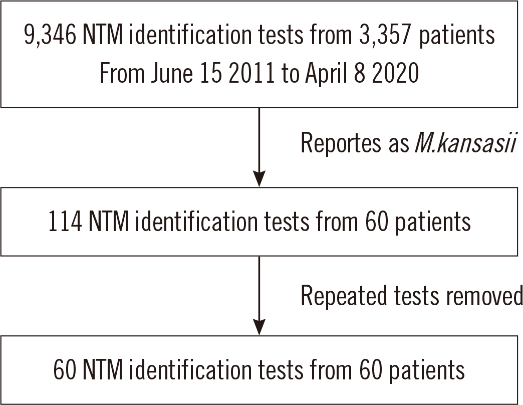
This study was approved by the Institutional Review Board of Seoul National University Hospital (SNUH), Seoul, Korea. We reviewed the medical records of 60 consecutive patients with M. kansasii infection, diagnosed based on a routine NTM identification testing performed from June 15, 2011 to April 8, 2020 at SNUH. The routine NTM identification was performed by in-house method which was based on PCR amplification of the two target regions, the 5’ end of the 16S rRNA gene (about 500 bp) and a part of rpoB gene, and subsequent Sanger sequencing. Informed consent from patients was not obtained, as this was a retrospective study performed using medical records and raw data files. The patient selection process is shown in Fig. 1. For patients with NTM identification testing being requested two or more times, we preferentially selected the tests with drug susceptibility testing results. For patients with no drug susceptibility testing results or two or more drug susceptibility testing results, earlier NTM identification testing results were chosen. All samples included in the analysis were cultured colonies from sputum (53, 88.3%), bronchial wash or bronchoalveolar lavage fluid (6, 10.0%), and joint fluid (1, 1.7%).
Identification of M. kansasii subtypes
Sequencing data generated from routine identification testing were used. For the 60 patients, percent identity score was calculated based on curated reference sequences of each subtype of M. kansasii. The reference sequences of the 16S rRNA gene and rpoB were curated from the List of Prokaryotic Names with Standing in Nomenclature (http://www.bacterio.net) and NCBI Genbank (https://www.ncbi.nlm.nih.gov/genbank/), respectively. SnackNTM software (https://github.com/Young-gonKim/SnackNTM, last accessed August 27, 2020) was used for aligning sequencing data to the curated reference sequences.
Grouping of patients
The patients were divided into two groups for comparison. Group 1 comprised patients infected with M. kansasii subtype 1, the most pathogenic subtype [4, 10–13]. Group 2 comprised patients infected with other subtypes, M. kansasii subtypes 2, 3, and 6. Baseline characteristics of patients, clinical manifestations, outcome, and drug susceptibility of the isolates were compared between the groups.
Review of medical records
Patient characteristics including age, sex, body mass index, and smoking history were retrieved from the medical records. Medical histories of tuberculosis, malignancy, diabetes mellitus, liver diseases, kidney diseases, and immunocompromising diseases were reviewed. Radiologic findings and pulmonary function test results including forced expiratory volume in 1 second (FEV1), forced vital capacity (FVC), and FEV1/FVC were retrieved. Clinical course of NTM isolation, such as co-infection with other NTM organisms, presence of NTM pulmonary diseases, and treatment initiation were reviewed. The drug susceptibility test results were also reviewed.
Statistical analysis
Statistical analysis was performed using R software (version 4.0.2, R Foundation for Statistical Computing, Vienna, Austria). For quantitative variable comparison, Shapiro test was used to evaluate the normality of data. Student’s t-test was used when the normality assumption was satisfied; otherwise, the non-parametric Wilcoxon rank-sum test was used. Fisher’s exact test was used to compare categorical variables between groups. P<0.05 were considered statistically significant.
Go to :
RESULTS
Among the 60 isolates included in the analysis, 13 were reclassified as one of the newly reported M. kansasii-derived species (21.7%), including 10 (16.7%) isolates of M. persicum (former subtype 2), 2 (3.3%) isolates of M. pseudokansasii (former subtype 3), and 1 (1.7%) isolate of M. attenuatum (former subtype 6). The remaining 47 isolates were classified as subtype 1.
The baseline characteristics of the two patients’ groups are shown in Table 1. FVC was significantly lower in Group 1 than in Group 2 (88.0% vs. 97.5% predicted, P=0.025), leading to a significantly higher FEV1/FVC ratio in Group 1 (74.0 vs. 70.5, P=0.038). Non-cavitary nodular bronchiectatic lesions were commonly observed in Group 2 (34.0% vs. 76.9%, P=0.010) and fibrocavitary lesions were observed only in Group 1 (38.3% vs. 0%, P=0.006).
Table 1
Patients’ characteristics including comorbidities, pulmonary function, and radiologic findings
![]()
The clinical course of the two groups is summarized in Table 2. Co-infection with M. avium complex was more frequent in Group 2 (19.1% vs. 53.8%, P=0.029). The proportion of patients who satisfied the NTM pulmonary disease (NTM-PD) diagnostic criteria did not differ between the groups (85.1% vs. 76.9%, P=0.675). However, the proportion of treated patients was significantly higher in Group 1 than in Group 2 (55.3% vs. 7.7%, P=0.003).
Table 2
Comparison of co-infection rate and clinical courses between Groups 1 and 2
| Group 1: M. kansasii former subtype 1 (N = 47) | Group 2: M. kansasii former subtypes 2, 3, and 6 (N = 13) | P | |
|---|---|---|---|
| Co-infection with other organisms, N (%) | 16 (34.0) | 7 (53.8) | 0.215 |
| With M. avium complex | 9 (19.1) | 7 (53.8) | 0.029 |
| With M. abscessus complex | 3 (6.4) | 0 (0) | 1 |
| With other NTM | 9 (19.1) | 2 (15.4) | 1 |
| Met diagnostic criteria of NTM-PD, N (%) | 40 (85.1) | 10 (76.9) | 0.675 |
| Observed without treatment, N (%) | 21 (44.7) | 12 (92.3) | 0.003 |
| Spontaneous conversion, N (%) | 6/14 (42.9) | 3/7 (42.9) | 1 |
| Treatment initiation within three yrs, N (%) | 26 (55.3) | 1 (7.7) | 0.003 |
| Microbiologic cure* n/N (%) | 18/20 (90.0) | 1/1 (100) | 1 |
| Surgical treatment, N (%) | 5 (10.6) | 0.0 | 0.575 |
![]()
The in vitro drug susceptibility testing results of the available isolates are presented in Table 3. Among the 60 patients, 32 (25 from Group 1 and 7 from Group 2) had drug susceptibility testing results. Among the eight drugs, whose breakpoints are published in the CLSI guidelines [25], resistance to four drugs(ciprofloxacin, ethambutol, rifampin, and trimethoprim/sulfamethoxazole) was detected. Susceptibility frequencies for all the four drugs were higher in Group 1 than in Group 2. The frequency of ciprofloxacin susceptibility was significantly higher in Group 1 than in Group 2 (80.0% vs. 28.6%, P=0.019). However, there was no significant difference in the MICs of the antimicrobial drugs whose breakpoints are not available in the CLSI guidelines (cefoxitin, doxycycline, imipenem, and tobramycin) between the groups.
Table 3
Comparison of in vitro drug susceptibilities between Groups 1 and 2
![]()
Go to :
DISCUSSION
In this study, 21.7% of isolates previously identified as M. kansasii were reclassified as new species with reportedly lower pathogenicity than M. kansasii sensu stricto. As conventional line probe-based commercial kits cannot discriminate the species, a considerable proportion of isolates identified as M. may actually belong to different species with significantly different clinical implications [15]. Even sequencing-based methods cannot detect new species unless at least one discriminatory target, such as hsp65 and rpoB, is incorporated in the test.
Most reports on M. kansasii subtype 1, sensu stricto, being the most pathogenic subtype are based on its detection frequency in clinically relevant populations [4, 10, 12, 13]. However, a recent report indicated that a specific genetic element, the espACD operon, is the main source of pathogenicity of this subtype [13]. We did not find a difference in the detection frequency of isolates that met the criteria for NTM-PD between Groups 1 and 2. However, other results suggested that subtype 1 isolates are more pathogenic than other subtypes. The significantly lower values of pulmonary function test parameters and FVC in Group 1 further support the higher pathogenicity of subtype 1, considering that lung destruction can decrease the FVC. Distinct radiographic findings were obtained for the two groups: Group 1 showed a higher frequency of fibrocavitary lesions, and Group 2 showed a higher frequency of non-cavitary nodular bronchiectatic lesions. These results support the assumption that the disease caused by subtype 1 is more aggressive.
Only a few studies have examined the drug susceptibility of M. kansasii subtypes [11, 12, 14]. The CLSI recommends susceptibility testing of clarithromycin and rifampin as first-line treatment, and isoniazid, ethambutol, streptomycin, amikacin, cotrimoxazole, moxifloxacin, linezolid, ciprofloxacin, and others as second-line treatment, because M. kansasii isolates are generally susceptible to these drugs [12]. Indeed, all isolates included in this study were susceptible to amikacin, clarithromycin, linezolid, and moxifloxacin. In contrast to the present study, a previous study found that the drug resistance frequency was consistently higher for subtype 1 isolates and attributed this result to the selection pressure due to higher exposure to drugs owing to a high frequency of treatment [14].
Our study had some limitations. First, the utilization of only two target regions, the 16S rRNA gene and rpoB, would not have provided enough information for distinguishing all M. kansasii subtypes. No single target could accurately distinguish all M. kansasii subtypes, requiring whole genome sequence-based approach for reliable subtyping [4]. It is possible that we have missed rarer subtypes of M. kansasii due to the limitation of the target regions used. Second, the number of isolates was limited, especially for non-subtype 1 M. kansasii isolates. We included only 2 subtype 3 M. kansasii isolates and 1 subtype 6 M. isolate. The adoption of laboratory methods capable of distinguishing these subtypes can provide more data.
In conclusion, approximately one-fifth of the isolates identified as M. kansasii were newly designated species derived from M. kansasii but with lower pathogenicity. Non-subtype 1 M. kansasii species should be identified by routine testing in clinical laboratories to select appropriate treatment strategies.
Go to :
 XML Download