

 PDF
PDF Citation
 Print
Print


Go to : 
INTRODUCTION
Epithelioid hemangioendothelioma (EHE) is a rare tumor composed of cords of epithelioid cells on a background of myxohyaline stroma. The 2002 World Health Organization classification described EHE as lesions with metastatic potential [1,2]. Hepatic EHE is a rare borderline vascular tumor, with an aggressiveness graded between hemangioma and hepatic hemangiosarcoma [3,4]. Because many patients with hepatic EHE are asymptomatic, these lesions are frequently detected incidentally [4-7]. Due to their rarity and protean behavior, the optimal treatment of hepatic EHE has not yet been standardized [8]. Partial hepatectomy has been recommended for patients with unilobar hepatic EHE, although aggressive recurrences have been reported after hepatectomies [9,10]. Liver transplantation (LT) is indicated for patients with advanced liver involvement, with approximately 200 such patients being reported in literature, mostly from the United States, Europe, and Canada [11-13]. To our knowledge, only three patients in Korea have undergone LT cases for hepatic EHE [14,15]. This study describes the clinicopathological features and prognosis of patients with hepatic EHE who underwent living donor liver transplantation (LDLT) at a single center over a 10-year period.
Go to :
METHODS
The study protocol was approved by of the Institutional Review Board at Asan Medical Center (IRB No. 2019-1347), which waived the requirement for informed consent due to the retrospective nature of this study. This study was performed in accordance with the ethical guidelines of the World Medical Association Declaration of Helsinki 2013.
Patients
The LT database at our institution was searched to identify patients who had been diagnosed with hepatic EHE and underwent LDLT over a 10-year period from January 2007 to December 2016. During this study period, 3,467 patients had undergone adult LT at our center. The medical records of these patients were retrospectively reviewed, with all patients followed up until July 2020.
Preoperative Evaluation, Surgical Procedures, and Postoperative Follow-up
Routine preoperative evaluation of primary liver tumors has been described elsewhere [16]. The protocol for ABO-incompatible LDLT includes desensitization with rituximab and plasmapheresis [17]. The LDLT recipients were followed up every month during the first year, every 2 months for the next 4 years, and every 3 months thereafter. Patients with recurrent liver tumors were treated as described [18].
Immunohistochemical Staining
Formalin-fixed paraffin-embedded tissue samples were immunohistochemically stained for CD34 (1:500, QBEND10; Immunotec Inc., Monrovia, CA, USA), CD31 (1:800, JC70; Cell Marque, Rocklin, CA, USA) and coagulator factor VIII-related antigen (FVIII:Ag) (1:2000; DAKO, Glostrup, Denmark) using a Benchmark autostainer (Ventana Medical System, Tucson, AZ, USA). The diagnosis of hepatic EHE was based on the histological features and immunohistochemical profiles described in the 2010 WHO classification of liver tumors [19].
Hepatic EHE-LT Score for Assessing Risk of Posttransplant Tumor Recurrence
Hepatic EHE-LT scores were calculated using the following formula: 5×(pathological macrovascular invasion)+3×(waiting time ≤120 days)+2×(pathological invasion hilar lymph node). Hepatic EHE-LT scores of 0–2, 3–5, and 6–10 were regarded as indicating low, intermediate, and high risk, respectively, for posttransplant tumor recurrence [20].
Calculation of Model for End-Stage Liver Disease Score
The model for end-stage liver disease (MELD) score is calculated using the following formula: “9.57×loge (creatinine, mg/dL)+3.78×loge (total bilirubin, mg/dL)+11.2×loge (INR)+6.43” [21].
Statistical Analysis
Numerical data are presented as mean and standard deviation. Survival rates were estimated using the Kaplan-Meier method. Statistical analyses were performed using IBM SPSS ver. 22.0 (IBM Corp., Armonk, NY, USA).
Go to :
RESULTS
Patient Demographics and Preoperative Diagnosis
During the 10-year study period, four patients (0.11% of the 3,647 patients who underwent adult LT) underwent LDLT for hepatic EHE. The clinicopathological features of these four patients are described in Table 1. The four patients included one man and three women, of mean age 41.3±11.1 years. One (25%) was positive for hepatitis B virus infection, but none were positive for hepatitis C virus infection or had alcoholic liver disease. Three patients (75%) were experiencing jaundice or abdominal pain at the time of their initial visit to the outpatient clinic, which led to further examination.
Table 1
Clinical profiles of the recipients
HBV, hepatitis B virus; MELD, model for end-stage liver disease; AFP, alpha-fetoprotein; DCP, des-γ-carboxy prothrombin; CA 19-9, carbohydrate antigen 19-9; HEHE, hepatic epithelioid hemangioendothelioma; LT, liver transplantation; R/O, rule out; EHE, epithelioid hemangioendothelioma; Mx, metastasectomy; CTx, chemotherapy; HCC, hepatocellular carcinoma.
![]()
Imaging modalities indicated suspected preoperative diagnoses of hepatic EHE or hepatocellular carcinoma (Fig. 1). Preoperative liver biopsy resulted in a pathologic diagnosis of hepatic EHE in all four patients. Analysis of mean blood concentrations of preoperative tumor markers showed a mean alpha-fetoprotein concentration of 2.2±1.4 ng/mL (reference, 7.5 ng/mL), a mean des-γ-carboxy prothrombin (DCP) concentration of 31.0±16.8 mAU/mL (reference, 40 mAU/mL), and a mean carbohydrate antigen 19-9 (CA19-9) concentration of 22.7±13.2 ng/mL (reference, 37 ng/mL). Only one patient presented with slightly elevated levels of CA19-9 and DCP (Table 1).
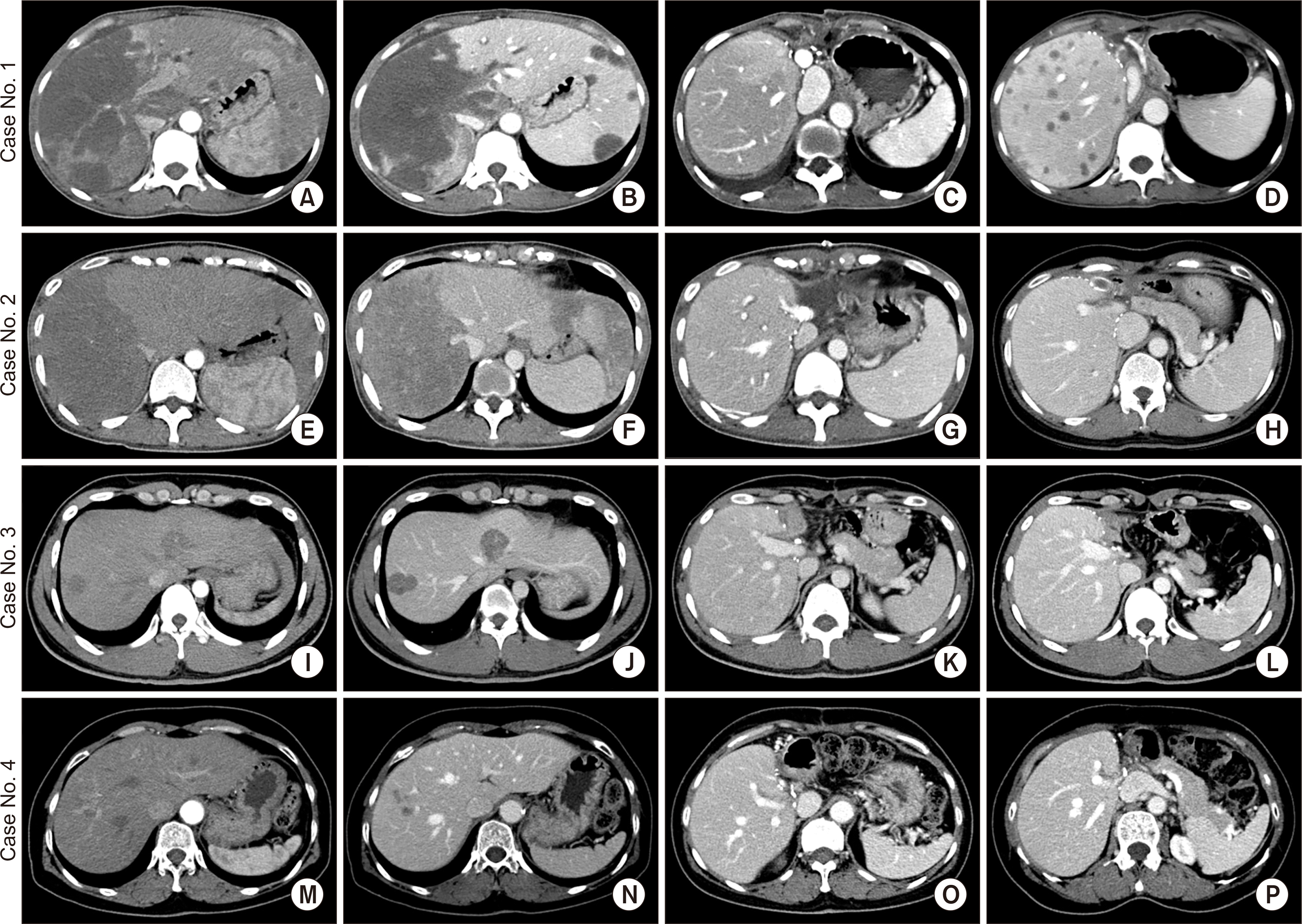 | Fig. 1Pretransplant and posttransplant computed tomography (CT) findings. Case No. 1 (A-D): pretransplant (A) arterial-phase and (B) portal-phase CT images show a large tumor and small masses, along with involvement of the right portal vein branches. (C) CT scan taken at 1 month after transplantation shows the usual posttransplant findings. (D) CT scan taken at 7 months after transplantation shows multiple liver metastases. Case No. 2 (E-H): pretransplant (E) arterial-phase and (F) portal-phase CT images show a large tumor and small masses. CT scans taken at 1 month (G) and 7 years (H) after transplantation show the usual posttransplant findings. Case No. 3 (I-L): pretransplant (I) arterial-phase and (J) portal-phase CT images show multiple small tumors. CT scans taken at 1 month (K) and 4 years (L) after transplantation show the usual posttransplant findings. Case No. 4 (M-P): pretransplant (M) arterial-phase and (N) portal-phase CT images show multiple small tumors. CT scans taken at 1 month (O) and 3 years (P) after transplantation show the usual posttransplant findings.
|
Peritransplant Clinical Courses
All four patients were indicated for LT because they had unresectable multiple tumors scattered throughout the entire liver (Fig. 1). Their mean MELD score was 10.8±5.7. Because none had any likelihood of undergoing deceased donor liver transplantation (DDLT), all four underwent LDLT using modified right liver grafts. Three donors (75%) were siblings of the recipient, and one (25%) was a daughter. The mean graft-recipient weight ratio was 1.11±0.19 (Table 2). All patients recovered uneventfully from LDLT operation without major surgical complications (Fig. 1).
Table 2
Profiles of living donors and transplantation
![]()
Explant Pathology
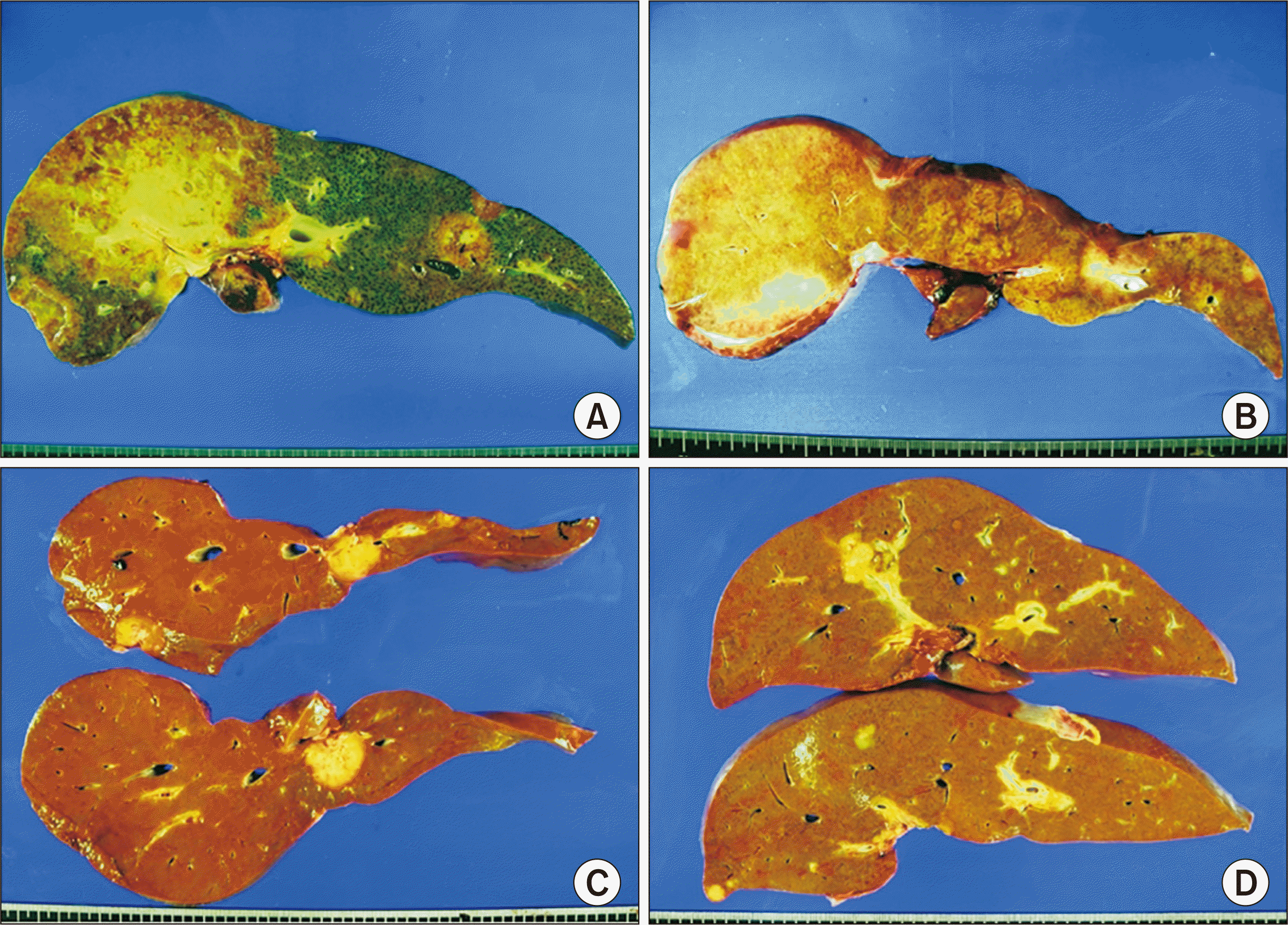
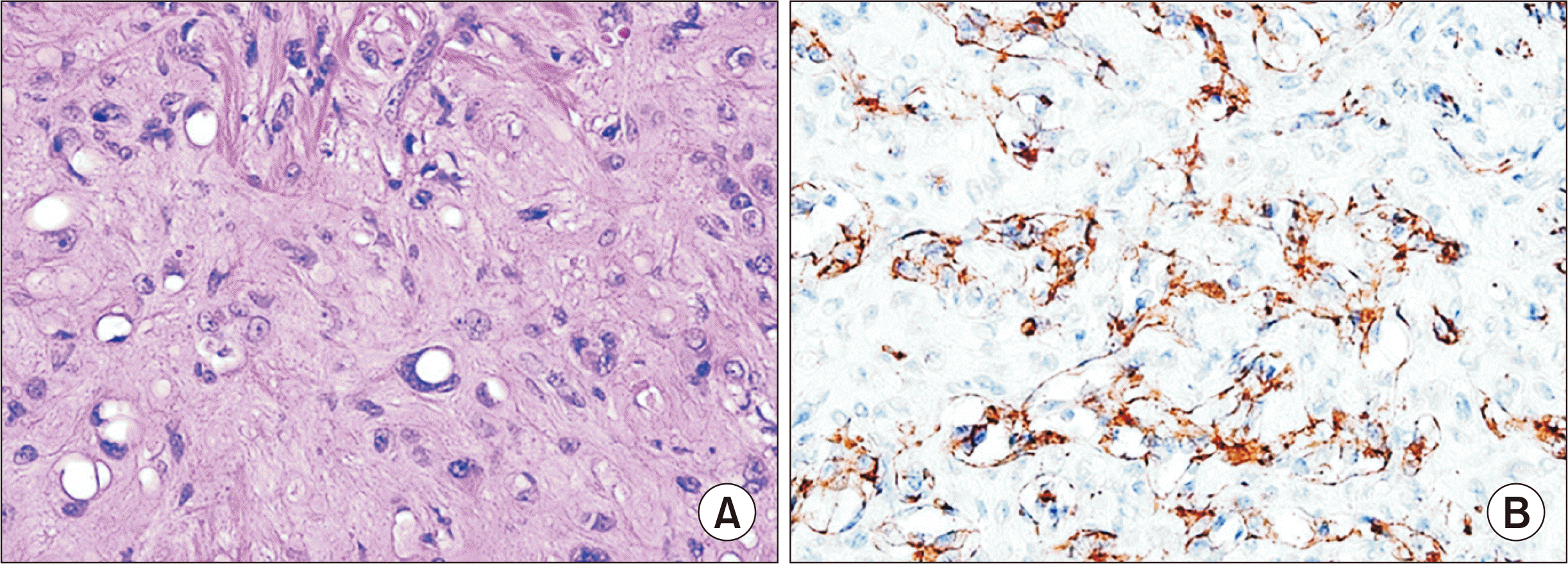
The pathological features of the explanted livers are summarized in Table 2. All four patients had multiple tumors scattered over the entire liver (Fig. 2). Two patients (case No. 1 and 2) had portal vein tumor thrombi, and one (case No. 1) had lymph node metastases. Microscopic examination showed low cellular epithelioid or spindle tumor cells on a background of fibromyxoid stroma, with the epithelioid tumor cells containing vacuolated cytoplasm (Fig. 3A). Immunohistochemical staining showed that the tumor cells in all four patients were diffusely immune-positive for CD31, CD34, and FVIII:Ag (Fig. 3B). Each one patient had hepatic EHE-LT scores of 7 and 5, whereas two had hepatic EHE-LT scores of 0 (Table 1).
Posttransplant Outcomes
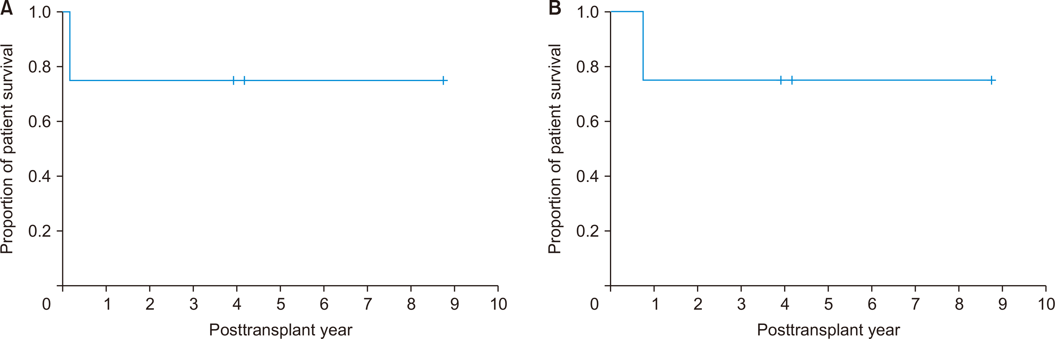
One patient, who had a large-sized tumor, 15 cm in diameter, and 13 small liver masses with portal vein tumor thrombosis and lymph node metastasis, was positive for lung metastases 2 months after LT. Pulmonary metastasectomy and systemic chemotherapy were performed, but this patient died due to tumor progression 9 months after transplantation (Fig. 1). The other three patients have done well without tumor recurrence. The 1-, 3-, and 5-year disease-free and overall patient survival rates were all 75% (Fig. 4).
Go to :
DISCUSSION
Treatments for hepatic EHE include hepatic resection, LT, chemotherapy, radiotherapy, hormone therapy, radiofrequency ablation, and surveillance alone. The 5-year patient survival rates have been reported to be 75% in 22 patients who underwent hepatic resection, 20% in 60 patients treated with chemotherapy/radiotherapy, and 4.5% in 70 patients who underwent surveillance alone [5]. Another study found that the 5-year patient survival rates were 86% in 11 patients who underwent hepatic resection and 73% in 11 LT recipients [22]. The 3-year patient survival rates have been reported to be 74.1% in 17 patients who underwent hepatic resection patients and 81.6% in 12 patients who underwent transarterial chemoembolization (TACE) [6]. Although these studies reported similar survival rates in patients who underwent hepatic resection, LT and TACE, the indications for each treatment modality were different. Hepatic resection is indicated for resectable intrahepatic lesions, whereas LT and TACE are indicated for unresectable lesions. Extrahepatic involvement, including lymph node and distant metastasis, is a contraindication for surgical treatment. The roles of nonsurgical therapies, including systemic/regional chemotherapy, radiotherapy, hormone therapy, and immunotherapy, have been investigated in only a few small case series [23-25].
The Europe Liver Transplant Registry (ELTR) reported a 5-year survival rate of 83% in 59 LT recipients with hepatic EHE, whereas the United Network for Organ Sharing (UNOS) registry reported a 5-year survival rate of 64% in 110 LT recipients, respectively [11,12]. LT is regarded as primary or salvage therapy for patients with multiple unresectable tumors not responsive to other nonsurgical treatments. However, DDLT in patients with hepatic EHE is limited by various factors, including donor shortage, high medical costs, the need for lifelong immunosuppressant therapy, patient willingness, and risk of tumor recurrence.
The indications of LT for hepatic EHE have not been well-defined. A prognostic score based on analysis of the ELTR-ELITA (European Liver Intestine Transplant Association) registry suggested that macrovascular invasion, short waiting time (≤120 days), and lymph node involvement are risk factors for post-LT tumor recurrence, whereas extrahepatic disease was not a formal contraindication to LT [20].
Hepatic EHE is currently a formally recognized indication for MELD score exception point priority in the United States under the new National Liver Review Board. A study investigated the role of LT and exception point waitlist priority using the UNOS database, in which exception point applications were submitted for 91.6% (120/131) of patients [26]. The 88 patients who received transplants had a median MELD score at LT of 7 and had waited 78.5 days. Unadjusted 1-, 3-, and 5-year post-LT survival rates of hepatic EHE recipients were found to be 88.6%, 78.9%, and 77.2%, respectively. Unadjusted post-LT patient and graft survival rates of hepatic EHE patients did not differ significantly from those rates in patients with hepatocellular carcinoma within the Milan criteria receiving exception point priority. That study concluded that most hepatic EHE recipients receive exception points at a universal approval rate, allowing them to promptly undergo DDLT.
Patients with hepatic EHE may not have priority for liver allocation in countries with limited numbers of deceased donors. In Korea, an exception point priority is given only to patients with hepatocellular carcinoma within the Milan criteria [27]. These patients may be eligible for LDLT if a living donor is available [14,15,23,28,29]. All four patients in the present study underwent LDLT because the MELD scores were too low for liver allocation [27,30]. Because patients who experience posttransplant tumor recurrence have a poor prognosis, LT candidates should be selected prudently.
The hepatic EHE-LT scoring system described above and based on three risk factors appears to be invalid for LDLT because the concept of prolonged waiting time is usually not included in LDLT. The primary reason to include waiting time as a factor in calculating hepatic EHE-LT score may be to have an observation period to exclude rapid tumor progression. Therefore, we suggest that the modified hepatic EHE-LT scores for LDLT should include only two risk factors, macrovascular invasion and regional lymph node metastasis. Patients with no, one, and two risk factors can be classified as being at low, intermediate, and high risk, respectively. This suggestion requires further validation to apply to clinical LDLT. The four patients with hepatic EHE who underwent LDLT in the literature may have been at low risk because there were no comments about macrovascular invasion and lymph node metastasis [14,23,28,29]. None of them showed tumor recurrence during a follow-up period of 8 months to 3 years.
Although extrahepatic disease was not a significant risk factor for tumor recurrence in the ELTR-ELITA registry study [20], another collective review study revealed that extrahepatic disease was significantly associated with higher tumor recurrence rates [29]. Our limited experience [15] and a review of the literature suggest that extrahepatic disease, including regional lymph node metastasis, may not be an eligible indication for LT, especially LDLT. The present study had several limitations, including its retrospective design and its inclusion of a small number of patients treated at a single center. Multi-center studies that include larger numbers of patients are necessary to confirm these results.
In conclusion, LDLT can be an effective treatment for patients with unresectable hepatic EHEs that are confined within the liver and absence of macrovascular invasion and lymph node metastasis.
Go to :
 XML Download