

 PDF
PDF Citation
 Print
Print


Introduction
There is an increase in the number of lumbar pathologies most common being disc prolapse and nerve compression, mostly in the adult age group between 30–50 years, which require surgical interventions in this region. With the advances in minimally invasive procedures in the lumbar region, there is a growing need to study various options related to surgical approaches. However, for safe execution of the procedure, the surgeons need to require thorough anatomical knowledge of the neural canal and adjoining structures to avoid complications like paraplegia, sensory-motor deficit, psoas hematoma etc. [1, 2].
Kambin described a triangular area for less invasive endoscopic surgery which was later known as the Kambin’s triangle (KT) [3]. KT is considered as safest corridor for passing the cannula or any other endoscopic instrument. It allows better view and avoids manipulation of the surrounding structures and minimises post-operative complications. Surgical procedures have been satisfactorily accomplished by approaching through this area [3-5]. It is also a useful approach for interventional radiologists and pain management physicians to reach the disc and surrounding neural elements.
KT is defined as a three-dimensional (3D) right angled triangle over dorsolateral aspect of the lumbar intervertebral disc which is bounded by the superior endplate of inferior vertebral body (base), dura/traversing nerve root (height) and the exiting nerve root- hypotenuse (Fig. 1A) [3, 6]. While Fanous et al. [7] termed this three dimensional area as ‘Kambin’s Prism’, Pairaiturkar et al. [8] mentioned two types of KTs-neural and bony. Some authors also describe it as a trapezoidal area [9]. As no important structure reportedly pass through KT, hence, surgeons and radiologists have satisfactorily approached the spinal canal through this area [5]. Moreover, it is mentioned that due to various advanced techniques, this area is better visualised during any surgical or radiological procedures and is a strategic site to approach for nerve root of interest [3, 4]. It is considered a preferable passage for correcting central disc herniation and a safe route to ventral epidural space.
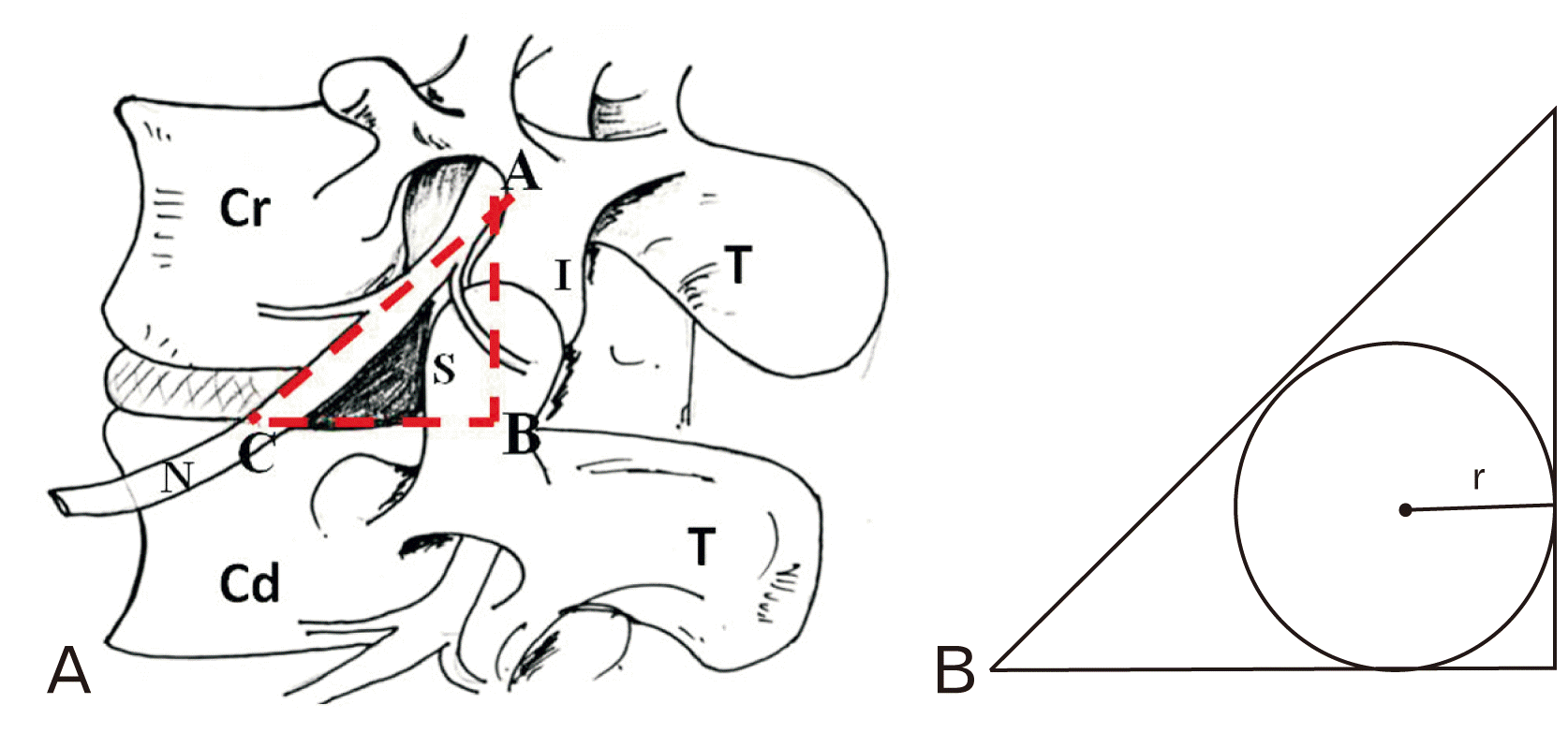 | Fig. 1(A, B) Schematic diagram of the Kambin’s triangle. I-Inferior articular process of Cr, S-Superior articular process of Cd. A, point of intersection of the vertical plane with the exiting nerve root; AB, height; B, point of intersection of vertical and horizontal plane; BC, base, C, point of intersection of the horizontal plane with the exiting nerve root; CA, hypotenuse, Cd, caudal vertebra; Cr, cranial vertebra; r, radius of inscribed circle.
|
Size of the cannula/any surgical instrument to be passed through the KT will depend on the area of the triangle approached. Studies have shown that cannula diameter can vary from 6.3 mm to 7.5 mm and some authors have even performed posterolateral discectomy and decompression with 6.9 mm cannula [2, 10].
The aim of the present study was to calculate the total area of bony KT in the lumbar region at four intervertebral spaces bilaterally in North West Indian cadavers. The calculated total area will provide information of the optimal size of cannula to be inserted through KT. The information will be a reference for the neurosurgeons, spine surgeons, interventional radiologists and pain management physicians for North-Western Indian population.
Go to : 
Materials and Methods
The present study was conducted on five randomly chosen adult donated human cadavers (one female and four male), age ranging from 52–70 years (Mean, 60±7.97), in the Department of Anatomy, Post Graduate Institute of Medical Education and Research, Chandigarh, India. Since the study was done upon the donated cadaver, approval from the ethical committee was not applicable. The average height of the cadavers was 160 cm (range, 154–168 cm). The cadavers having history of previous spine surgery or any spine pathology were excluded from the study. The area around the vertebral column was carefully cleaned to expose the exiting nerve root. Intertransverse ligament was removed to have a clearer view of the area. Nikon D7000 camera (Nikon Co., Tokyo, Japan) was used for taking the required photos. Keeping in view that removing the adjacent structures might alter the normal anatomy of the exiting nerve or the structures of our interest, minimal handling was ensured and sharp scalpel blade was used to cut out the ligaments so that minimal manipulation was required to expose the area of interest.
The boundary of bony KT (Fig. 1A) was demarcated by identifying the superior articular process and superior border of immediate caudal vertebra [6]. The dimensions of the KT at all lumbar intervertebral disc levels (L1–L2, L2–L3, L3–L4, and L4–L5) were measured bilaterally. This accounted for measurements of 40 triangles. Measurements were taken with the help of digital Vernier calipers (Mitutoyo digital calipers/573-291-30) (0.02 mm accuracy). At each level, the measurements were taken three times and average of the respective values was considered.
Two planes were considered for the measurements:
a) Vertical plane along the superior articular process at the level of the articular facet
b) Horizontal plane along upper margin of immediate caudal vertebra
The following measurements were made (Figs. 1B, 2):
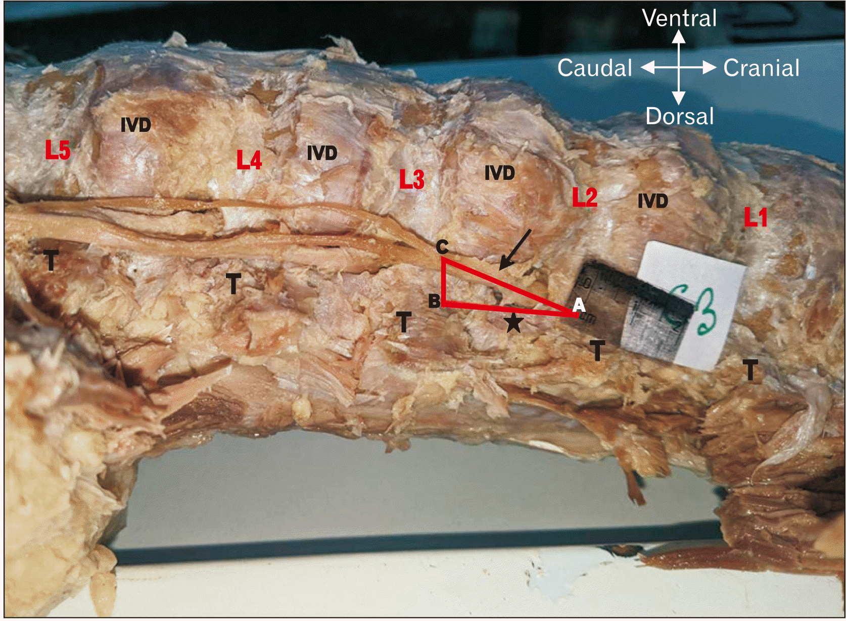 | Fig. 2Kambin’s triangle in a dissected cadaver (left lateral view) (black arrow). A, point of intersection of the vertical plane with the exiting nerve root; AB, plane along the superior articular process (star, black) of caudal lumbar vertebra (L3); B, point of intersection of vertical and horizontal plane; BC, Plane along superior surface of body of caudal lumbar vertebra (L3); C, point of intersection of the horizontal plane with the exiting nerve root; IVD, Intervertebral disc; L1,2,3,4,5, Lumbar vertebrae from cranial to caudal direction; T, Transverse process.
|
-
Height: AB, where A-point of intersection of the vertical plane with the exiting nerve root
B-Point of intersection of vertical and horizontal plane. -
Base: BC, where B-Point of intersection between vertical and horizontal plane.
C-Point of intersection of the horizontal plane with the exiting nerve root Hypotenuse: AC, where A and C are the points as described above.
Based on the measurements, a representative triangular area was determined at each lumbar intervertebral level on both sides. The area of the triangle was calculated by geometric formula: area=0.5 (base×height) [6].
Diameter (twice of the radius) and the area of circle inscribed within the triangle (Figs. 1B, 2) using the formula Πr2 were calculated at each lumbar level bilaterally. Radius was measured using the formula [(s–a)(s–b)(s–c)/s]1/2 where ‘s’ is the semiperimeter of the triangle and a, b and c are the dimensions of the sides of the triangle.
Means and standard deviations were calculated. Paired t-test was used to compare right and left sides. The P<0.05 was considered to be statistically significant. Statistical analysis was performed using Graph pad 6.0 software (GraphPad Software, Inc., La Jolla, CA, USA).
Go to :
Results
On the right side, the mean height of the triangle increased from 13.15 mm at L1-2 level to 16.37 mm at L2-3 level. Again there was a jump from 16.85 mm (L3-4) to 20.31 mm (L4-5). These measurements on the left side were slightly different from that on the right side (Table 1). However, statistically the difference between the right and the left side was insignificant (P>0.05). The mean value of the base of the triangle ranged from 8.19 mm to 9.30 mm on the right side with hardly any significant increase in the caudal spaces. Similar trend was observed on the left side (Table 2, Fig. 3). The calculated mean areas of the triangle at each intervertebral space from L1-2 to L4-5 were 54.01, 66.84, 74.59, and 94.44 mm2 respectively on the right side while on the left side these were 53.81, 73.42, 83.91, and 115.84 mm2 respectively (Table 2). The table also gives the mean diameter of the circle inscribed within KTs at all intervertebral spaces craniocaudally on both sides. The mean diameter (mm) increased from 5.83 at L1-2 level to 7.27 at L4-5 level on the right side while on the left side it increased from 5.67 at L1-2 level to 8.16 at L4-5 level. The difference between right and left side was not significant (P>0.05).
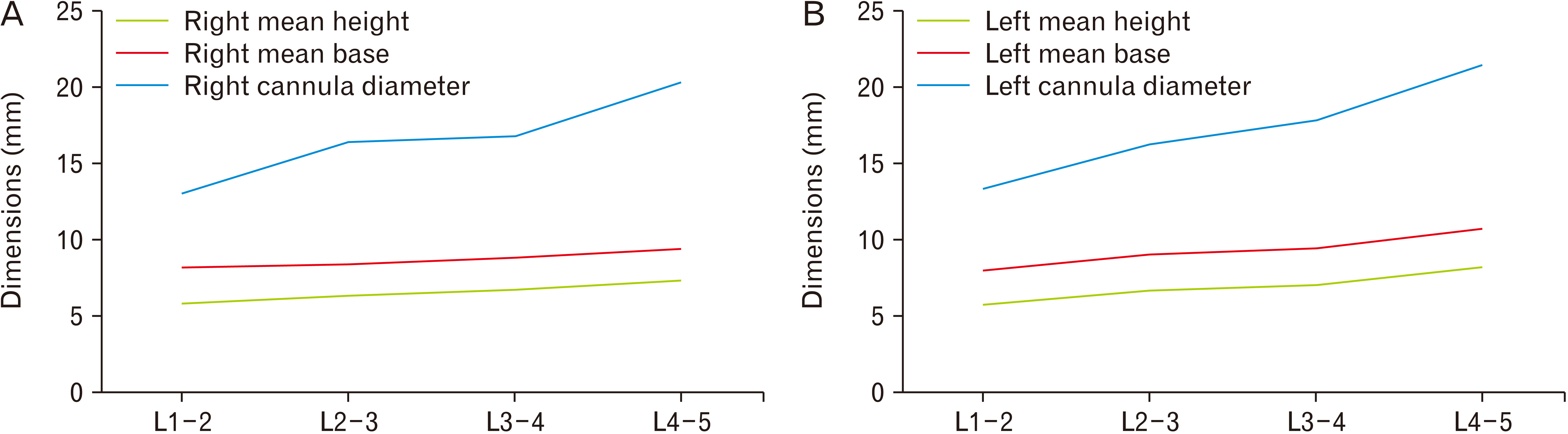 | Fig. 3(A, B) Line diagram showing the correlation of parameters between the lumbar levels on the right (A) and the left (B) side. L1-2, L2-3, L3-4, L4-5, lumbar intervertebral levels.
|
Table 1
Table shows the various dimensions measured and calculated in 40 Kambin’s triangles of five human cadavers on the right and the left side
![]()
Table 2
Mean dimensions of observed Kambin’s Triangles
![]()
On the right side, the mean diameter of the inscribed circle within the triangle was found to be 5.83 mm at L1-2 level and on further analyzing the data, it was observed that only 60% of the studied spaces at this level permitted insertion of cannula or any other instrument, having mean diameter of 5.83 mm. The maximum permissible diameter in all spaces at this level was found to be 5.24 mm in the present study (Fig. 4). In the subsequent three caudal levels only 40% of spaces admitted the corresponding mean diameters of cannula which were 6.29 mm, 6.66 mm, and 7.27 mm respectively. Further analysis revealed that the maximum permissible diameters which worked in accordance with all the spaces at the corresponding levels were 5.78 mm, 6.02 mm, and 7.20 mm respectively.
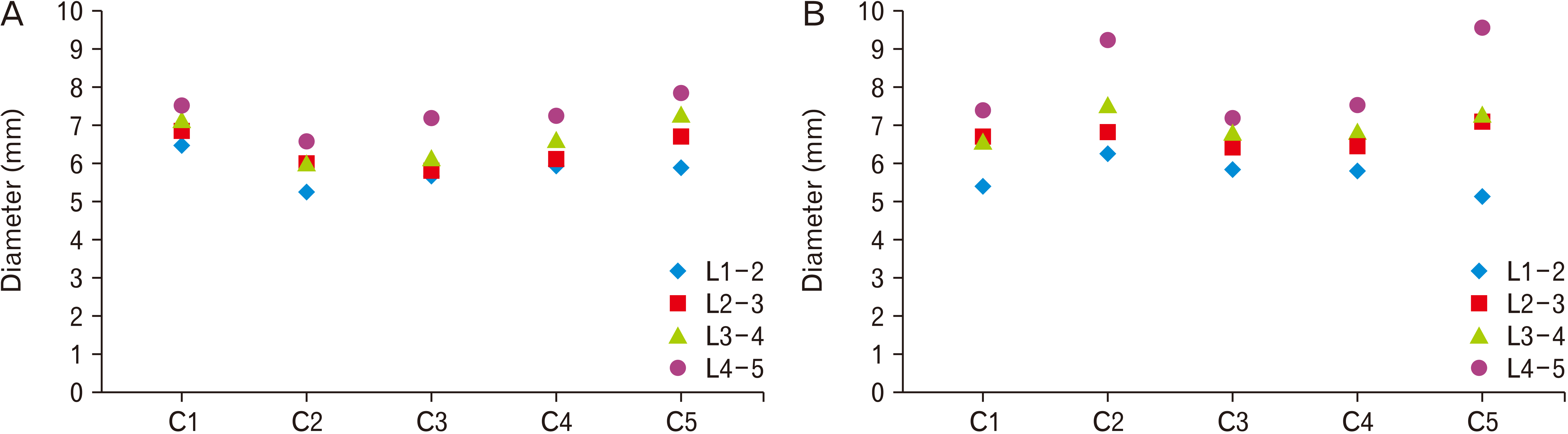
Analysis on the left side also shows that the calculated means do not completely represent every space. At L1-2 level, on the left side, it was observed that 60% cases had the permissible area to allow an instrument of mean diameter of 5.67 mm to pass through them while cannula of the diameter of 5.12 mm could be passed easily through all the spaces at this level. On the same side, the mean diameter at L2-3 level was found to be 6.69 mm which on further analysis shows that 60% spaces will easily permit an instrument of this diameter while the spaces in rest 40% are comparatively smaller with a minimum dimension of 6.42 mm. On the other hand, observation at L3-4 level reveals that 40% spaces had the adequate area to permit an instrument of the mean diameter of 7.05 mm while rest 60% had smaller spaces with minimum diameter of 6.62 mm. At L4-5 level, the mean diameter was found to be 8.16 mm on the left side and on detailed analysis; it was found that 40% of the cases had areas which could easily permit an instrument of diameter 8.16 mm, while rest 60% spaces have smaller areas with minimum diameter of 7.5 mm.
Go to :
Discussion
A triangular safe area to approach the spinal canal was described by Kambin which was later named as the KT [2, 3]. Since bony KT has become clinically relevant with the development of transforaminal procedures [5], this triangle represents a safe corridor to reach neural foramina, epidural space and further into disc spaces. Targeted delivery of drugs at clinically relevant neural foramina has become more effective with transforaminal approach compared to the traditional caudal epidural injections. Similarly, for nerve root blocks, surgeons can use KT to reach specific nerve root of choice [5]. Diagnostic procedures like discography also approach KT to reach the desired area. Presence of well defined bony landmarks of Kambin’s helps to navigate it intraoperatively using fluoroscopy which makes it even more clinically relevant to operating surgeons. Transforaminal endoscopy is now well established surgical procedure using KT and this procedure can be very helpful for surgeries like foraminotomy, foraminoplasty, fragmentectomy, pediculotomy, annuloplasty etc. [11-13].
Zhang et al. [14] in a retrospective CT based study in 110 Chinese patients with low back pain found that the mean area for the safe working zone gradually increased from L2-3 to L5-S1. However, they considered the safe zone to be a trapezoid area bounded by exiting nerve root and inferior pedicle. Ozer et al. [15] performed both cadaveric measurements and surgical observations of Kambin’s safety zone. They considered the angle between the edge of the facet joint and the exiting nerve root. They found wide angle (similar to KT) only in 17.6% patients and 20.8% cadavers, in rest of the cases there was narrow or no space.
To the best of our knowledge, no cadaveric study has been done on this triangle previously except by Hoshide et al. [6], who measured sixteen KTs in two cadavers. They dissected the bony KT after assessing it with a Kirschner wire. The present authors also followed the definition of KT as described by Hoshide et al. [6]. We dissected out the bony KT and measured the height and base (width) and then calculated the area of the right angled triangle. We found that the maximum area of the triangle was 94.44±8.09 mm2 on the right (L4-5) and 115.84±23.82 mm2 on the left side (L4-5). From the dimensions of the triangular spaces we calculated the diameter of the circle inscribed within each triangle and found that the maximum diameter was 7.27±0.48 mm on the right and 8.16±1.13 mm on the left side. We conclude that the maximum outer diameter of cannula or any other surgical instrument which can be passed through the triangular area should not exceed the average maximum diameter of the circle which in our case was found to be 7.26 mm on the right and 8.16 mm on the left side. Although a difference in measurement has been observed in the right and left sides, the difference was statistically insignificant (P>0.05). The differences might be due to slight change in the position of the vertebral column during measurement on both sides or due to bilateral variation in bony components or intervertebral discs, although proper care had been taken to avoid any confounding factors and mean of three values were considered to avoid possible error. Zhang et al. [14] observed difference in the safe working zone between both the sides at L4-5 and L5-S1 level which they opined was due to differences they found in the vertical distance between middle of the pedicle and exiting nerve root. However, we have not considered this parameter in our study. In another Indian study, on 3D magnetic resonance imaging (MRI) (oblique, axial and sagittal views) done in 50 patients, authors found that the mean maximum cannula diameter permissible through the bony KT increased from 5.67±1.38 mm at L1-2 level to 9.7±3.82 mm at L5-S1 [8]. The minimum diameter as given by the above authors is comparable to the observations of the present authors but the maximum diameter is slightly less in the present study compared to that observed by Pairaiturkar et al. [8]. The difference may be due to the difference in methodology used for the study, i.e. cadaveric versus MRI. The above authors also mentioned about difference in parameters between right and left sides but the differences were not significant [8]. Larger cannula diameter may lead to injury to the related nerve root with consequent postoperative complications [10]. Wimmer and Maurer [16] observed cannula diameter of 8 mm from L1-4 and 7 mm from L4-S1. In our study, we found that the areas of the caudal triangles are larger compared to the cranial triangles, the L4-5 having the maximum area on both sides.
We found that the area gradually increased from L1-2 space to L4-5 space. The observations by the present authors are in accordance with that of Zhang et al. [14] but do not correlate with that of Ozer et al. [15] and Wimmer and Maurer [16]. The reason for differences in dimensions may be due to the different methodologies used by the later. Most of the earlier authors concluded that the maximum area is at the L4-5 level [6, 8, 14].
From our observations, we also found that on the right side an instrument having outer diameter of 5 mm will be admissible in 100% cases at L1-2 level while in the subsequent caudal spaces the maximum diameter admissible through the respective spaces in all the cases were observed to be 5.5 mm, 6 mm, and 6.5 mm. Similarly, on the left side, an instrument having outer diameter of 5 mm will be admissible in 100% cases at L1-2 level while in the subsequent caudal spaces the maximum diameter admissible through the respective spaces in 100% cases were observed to be 6 mm, 6.5 mm, and 7 mm. Overall, we propose that the maximum diameter of an instrument which can be successfully inserted through the Kambin’s spaces of any side in all the cases at L1-2, L2-3, L3-4 and L4-5 level should be 5 mm, 5.5 mm, 6 mm, and 6.5 mm respectively (Fig. 4).
In general, a range of 7–11 mm canula is available for any transforaminal approach to the spinal canal. Our observations suggest that a surgeon approaching these lumbar spaces may come across areas of smaller dimensions which may require some amount of bony debridement for ease of entering any instrument. Based on our results, it is suggested that smaller diameter instruments can be used for approaching the desired area without unnecessarily disturbing the surrounding bony components.
Limitations in the study
Formalin induces 10% to 15% shrinkage of tissue which may mask the original dimensions [17, 18]. In contrast, another study done on breast cancer specimen, reported that in 96% cases there was no significant change between fresh and fixed states [19]. Also a study done on bone, did not show much shrinkage by formalin [20].
Go to :
 XML Download