

 PDF
PDF Citation
 Print
Print


Introduction
Incidences of ligament injuries are on the rise over the decades due to increased involvement in sports activities. Reconstructive surgeries using autografts have altered the scenario in regards to the management of ligament injuries. Unsatisfactory graft diameters often lead to failure [1-3] especially in situations like multiligament knee injuries [4]. A thorough knowledge about the graft options becomes extremely vital in preventing such situations and knowledge regarding the alternate options other than the conventional pes anserinus tendons would lead to a less painstaking experience.
The autografts used in knee ligament reconstructive surgery includes pes anserinus tendons, bone patella tendon bone (BPTB) graft, quadriceps tendon (QT) and peroneus longus. It’s difficult to predict the size of these tendons prior to harvest. The pes anserinus tendons have a great variability in their length and diameters [5]. This variability often percolates to a situation in which one finds the graft either small in diameter or shorter in length. Of the studies present pertaining the prediction of tendon size, a recent systematic review found out that only 20% of pes anserinus tendon size can be predicted by knowing patient’s height [1, 6-9].
Various studies have assessed the cross-sectional area (CSA) and diameters of pes anserinus tendons preoperatively using magnetic resonance imaging and correlated it with intraoperative diameters [10-15]. However, there is a paucity of literature in the Indian scenario regarding the thickness of the pes anserinus tendons, the QT and the patellar tendon (PT) [16-18]. We did an magnetic resonance imaging (MRI) based study to find out the CSA and diameters of pes anserinus tendons, thickness of QT, thickness of the PT and the relation of the these anatomical structures with patient’s height, weight, sex, patellar thickness and anterior cruciate ligament (ACL) foot print sagittal diameter. This study hopes to offer a normal baseline in Indian population from which future studies could be developed.
Go to : 
Materials and Methods
The study enrolled MRI of 114 consecutive patients with suspected knee injury who presented to our tertiary care centre (All India Institute of Medical Sciences, Rishikesh) after approval from institutional ethics committee (No. 253/IEC/PGM/2018). Exclusion criteria included knees with osteoarthritic changes, abnormalities of extensor mechanism, history of previous surgery and history of patellar dislocation or subluxation. A total of 104 patients were selected for the study using the exclusion criteria. The study period was September 2018 to August 2019. The average age of the subject group was 28 years (standard deviation 4.8) with 58 males and 46 females. The height, weight and body mass index (BMI) were recorded for the enrolled patients. The MRIs were performed with a Sigma 1.5 T MRI System (GE Healthcare, Milwaukee, WI, USA) with the patients’ knee in extended position and a slice thickness of 3 mm.
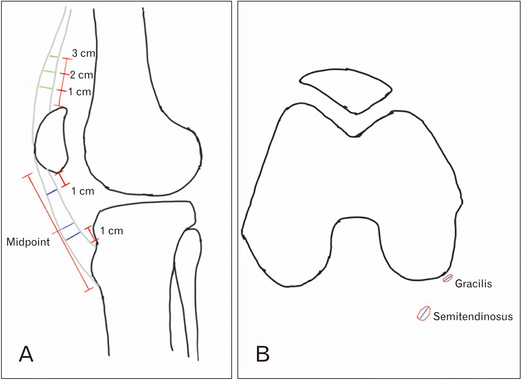
Sagittal proton density T1- or T2-weighted slice in which the patellar and QT appears to have the maximum diameters are chosen for measurement of tendon thickness. PT thickness is measured as the mean of three measurements done at 1cm from the lower pole of the patella, midpoint of the PT, and 1 cm above the upper border of the lower attachment of tendon (Fig. 1A) [19]. The QT thickness is calculated as the mean of three measurements at 10 mm, 20 mm, and 30 mm from the upper border of patella (Fig. 1A) [11]. Axial T2W slice at the level of the joint was used to calculate the CSA and the tendon diameter of the pes anserinus tendons [14, 15]. The method of measurement is illustrated in Fig. 2. A schematic representation of patellar and QT thickness measurement has been shown in Fig. 3A with measurement of pes anserinus tendon diameter and CSA in Fig. 3B. The free hand tool of the Osirix software (Osirix 9.5; Pixmeo Sarl, Bernex, Switzerland) was used for the measurement of CSA (Fig. 2A). The axial T2 image showing the thickest patella (including the cartilage) of all the images is used to assess the thickness of the patella. The thickness of the patella is measured perpendicular to the axis of patella obtained by joining the medial and lateral most ends (Fig. 1C) [20]. The tibial foot print sagittal diameter of the ACL was assessed in the sagittal T2 image showing the maximum length of the foot print (Fig. 1B) [21].
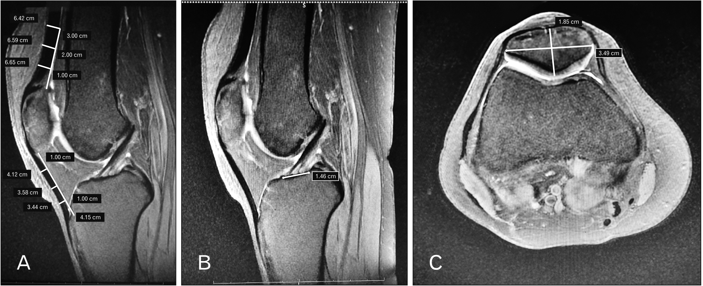 | Fig. 1T2 weighted sagittal MRI image showing the largest diameter of patella and QT taken (A) to measure patellar and QT thickness at three points as described. T2 weighted sagittal MRI slice showing largest diameter of ACL tibial footprint taken (B) for ACL footprint measurement. T2 axial image showing thickest patella (C) used to measure patella thickness as described. ACL, anterior cruciate ligament; MRI, magnetic resonance imaging; QT, quadriceps tendon.
|
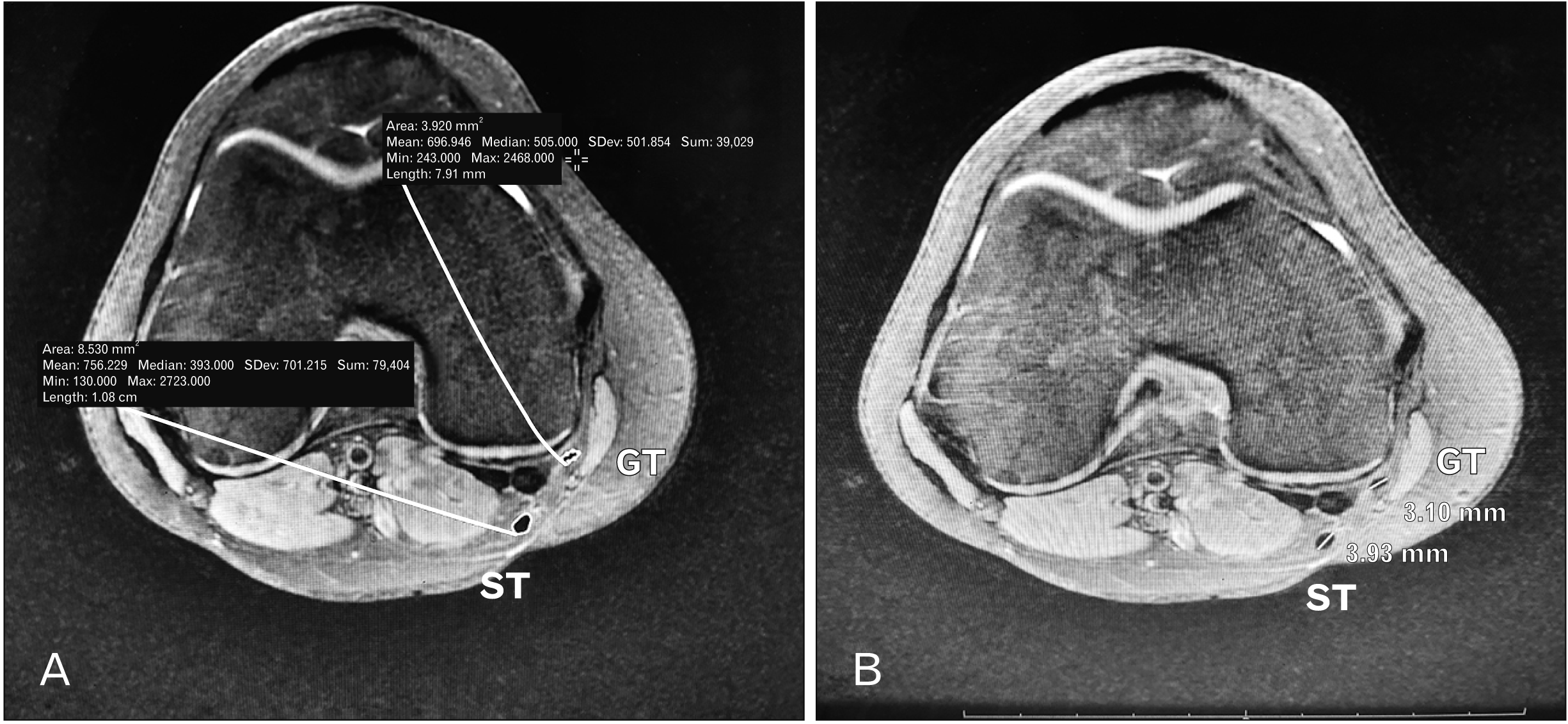 | Fig. 2Axial T2 weighted MRI slice at the level of the joint used to calculate the cross-sectional area and the diameter of pes anserinus tendons. The free hand tool of the Osirix software used to measure the CSA of pes anserinus tendons (A). Largest diameter at the chosen slice taken as the diameter of pes anserinus tendons (B). CSA, cross-sectional area; GT, gracilis tendon; MRI, magnetic resonance imaging; ST, semitendinosus.
|
The values are expressed as mean±standard deviation. The statistical analysis was done with SPSS software and Student’s t-test/Mann Whitney test was employed after checking the normality with Shapiro-Wilk test. A value of P<0.05 is considered as significant. Ten MRI measurements were assessed by a single examiner (first author) 2 times and by two different observers (first and second authors) to assess the intra- and inter-observer reliability. The relationship between the pes anserinus tendon diameter and CSA, PT, and QT thickness with anthropometric data, thickness of the patella and ACL tibial foot were calculated using the Pearson’s correlation coefficient.
Go to :
Results
The mean diameters of the semitendinosus tendon (ST) and gracilis tendon (GT) were 3.77±0.49 mm and 2.87±0.27 mm respectively. QT and PT thicknesses were 7.36±0.87 mm and 4.50±0.62 mm respectively. The mean CSA of the pes anserinus tendons were 11.62±1.62 mm2 and 6.64±1.18 mm2 for ST and GT respectively. The mean patella thickness was 21.05±0.89 mm and the mean ACL tibial footprint sagittal diameter was 15.29±1.7 mm. The sex wise comparison of these anatomical structures is expressed in Table 1. The mean patient’s height, weight and BMI were 166.9±6.55 cm, 73.4±7.41 kg and 22.7±2.71 kg/m2 respectively. Assessment of correlation coefficients revealed a significant but moderate positive association of height with STmeasurements (diameter and CSA) and PT thickness.
Table 1
Sex wise comparison of various anatomical knee structures as measured on preoperative MRIs
| Variable | Mean (n=104) | Male (n=58) | Female (n=46) | P-value |
|---|---|---|---|---|
| Diametera) | ||||
| ST | 3.77±0.49 | 3.98±0.39 | 3.51±0.48 | < 0.05 |
| GT | 2.87±0.27 | 2.90±0.30 | 2.84±0.22 | >0.05 |
| Thicknessa) | ||||
| QT | 7.36±0.87 | 7.45±0.95 | 7.23±0.76 | >0.05 |
| PT | 4.50±0.62 | 4.64±0.71 | 4.29±0.41 | <0.05 |
| CSAb) | ||||
| ST | 11.62±1.62 | 12.03±1.53 | 11.10±1.75 | <0.01 |
| GT | 6.64±1.18 | 6.66±1.06 | 6.62±1.30 | >0.05 |
![]()
Patient’s height was also found to have a weak association with gracillis tendon measurements (diameter and CSA) and QT thickness. Patellar thickness was seen to have moderate correlation with ST CSA and PT thickness while a significant, moderate correlation was seen between ACL tibial foot print sagittal diameter with semitendinosus CSA. No significant correlation was found between patient’s weight or BMI and thickness of tendons. A detailed explanation of the relationships beween tendon thickness and the variable assessed has been given in Table 2. An excellent inter and intra-observer reliability was seen with the techniques of measurement of tendon thickness in MRI as depicted in Table 3.
Table 2
Assessment of coefficient of correlation of different anatomical structures with anthropometric variables, patellar thickness and ACL footprint diameter
![]()
Table 3
Inter and intra-observer reliability in measurement of various anatomical structures
![]()
Go to :
Discussion
A rising concern among surgeons is the association of graft failures with smaller pes anserinus tendon grafts. A recent systematic review revealed a relative risk of failure of 6.8 times for pes anserinus tendon ACL reconstructions with a graft diameter less than 8 mm [1]. Pes anserinus grafts often have the disadvantage of having a smaller diameter intraoperatively. Hence one would be wise to understand the available autografts in surgeon’s armamentarium and it would help even more if one can predict the graft sufficiency preoperatively.
Variations in the CSA and diameter of the pes anserinus tendons is often seen in consecutive MR images owing to the myotendinous bulk, irregular shapes and variable orientations of the tendons. Various literatures quote different values of these parameters owing to the difference in their methodology (Table 4) [11, 13-15, 19, 23, 27]. Hodges et al. [22] revealed that the MRI measurement at the medial joint level corresponds to most accurate intra-operative graft thickness values. The minimum combined CSA for the average person on MRI to achieve a graft size of 8 mm was 17.168 mm2 (P<0.001) by Hanna et al. [23]. Our study revealed that height has moderate correlation with the pes anserinus tendon thickness (Table 2). Patient’s height is the only anthropometric variable which showed a significant positive correlation with the graft tendon thickness. The finding is comparable to literature on Indian population on preoperative prediction of graft assessment where they find correlation of height with the tendon or graft sizes [16-18]. Goyal et al. [17] reported in their study that Indian patients with height less than 147 cm were at risk of yielding final quadrupled autograft of less than 7 mm. Our study further fortifies the fact that patient’s height is the best predictor of all graft tendon diameters. Various previous studies have also correlated patient’s anthropometric measurements with pes anserinus tendon diameter. Boisvert et al. [6] evaluated 132 patients undergoing hamstring autograft ACL reconstruction. Intraoperative graft diameter was correlated with anthropometric measurements revealing a positive correlation with of graft diameter with height in women only. Zakko et al. [11] did an MRI based study in 62 patients and found a weak correlation between anthropometric measurements and MRI based PT and QT thickness. Previous studies also showed a smaller graft diameter in the female population. Treme el al. [24] conducted a cohort study in 50 patients undergoing ACL reconstruction using hamstring graft and concluded that small graft diameters are most likely in older, short, female subjects with small thigh circumferences. Our study showed significantly smaller graft thickness of ST and PT (Table 1). One should be cautious while utilizing pes anserinus and patellar tendon in ACL reconstruction in females and a use of other autograft options should be considered. The current study also correlated the patellar thickness and the ACL tibial footprint diameter with the available tendon graft sizes. Both these variables showed moderate correlation with the semitendinosus CSA while patellar thickness also showed a positive association with PT thickness.The current study is the first to study relation of the ACL tibial foot print diameter with the tendon sizes including patellar and QTs.
Table 4
Summary of literature on preoperative tendon measurements using MRI
| Studies | Country | Sample size (n) | Diametera) | CSAb) | Thicknessa) | |||||
|---|---|---|---|---|---|---|---|---|---|---|
|
|
|
|
||||||||
| ST | GT | ST | GT | PT | QT | |||||
| Our study | Indian | 114 | 3.77±0.49 | 2.87±0.27 | 11.62±1.62 | 6.64±1.18 | 4.50±0.62 | 7.36±0.87 | ||
| Zakko et al. [11] | USA | 62 | 8.60±1.60 | 23.30±5.60 | 4.30±0.80 | 8.40±1.50 | ||||
| Camarda et al. [13] | Italy | 77 | 4.20±0.40 | 3.30±0.40 | 4.50±0.60 | 7.30±1.10 | ||||
| Beyzadeglou et al [14] | Turkey | 51 | 4.20±0.40 | 3.10±0.30 | ||||||
| Hanna et al. [23] | USA | 30 | 17.168 | |||||||
| Hamada et al. [15] | Japan | 79 | 10.10±2.10 | |||||||
| Chang et al. [19] | Korea | 147 | Proximal-4.30±0.80 | |||||||
| Middle-3.70±0.80 | ||||||||||
| Distal-4.40±0.80 | ||||||||||
| Cobanoglu et al. [27] | Turkey | 70 | AP at JL-4.07±0.89 | At JL-7.38±0.90 | ||||||
| ML at JL-2.95±0.79 | At PL-8.58±3.12 | |||||||||
| AP at PL-3.65±0.90 | ||||||||||
| ML at PL-3.8±0.86 | ||||||||||
Values are presented as number only or mean±SD. MRI, magnetic resonance imaging; ST, semitendinosus tendon; GT, gracilis tendon; QT, quadriceps tendon; AP, antero-posterior diameter; ML, medio-lateral diameter; JL, joint line; PL, physeal line. a)Measurements in millimmeter (mm); b)Measurement in millimetre square (mm2).
![]()
Studies have attempted to show relationship between the pes anserinus tendon sizes with anthropometric data [11]. Zakko et al. [11] revealed moderate-to-good accuracy and high reliability of measurements of tendon sizes in MRI. Our study is the first to assess the tendon sizes of pes anserinus tendons (ST and GT) along with PT thickness and quadriceps thickness in the Indian population using MRI to the best of our knowledge and the first one to assess the relationship of ACL tibial foot print diameter with the tendon sizes.
QT and bone PT bone graft are alternate graft options in multi ligamentous knee reconstructions. Literature on the thickness of the patellar and QT is pretty rare [11, 13, 19]. Only a few of these looked into the relationship of anthropometric data with the PT thickness and QT thickness [11, 13]. Our study revealed the mean quadriceps and PT thicknesses to be 7.36±0.87 mm and 4.50±0.62 mm respectively with males having significantly larger PT diameter than female (P<0.05). Zakko et al. [11] recorded QT thickness of 8.4 mm and a PT thickness of 4.3 mm in Caucasian population. The height and thickness of the patella were seen to have moderate association with the PT thickness. Weight and height had weak relationship with the quadriceps thickness.
The study has its own limitations. First, relation of the MR findings with the intraoperative tendon sizes couldn’t be measured as majority of the cases included in the study were meniscal injuries. A study focussing on the relationship of MRI size and intra-operative tendon sizes would have been ideal but, the current study is the only one to truly assess the tendon autograft sizes in MRI in Indian population and correlate with the anthropometric data. Second, the length of the pes anserinus tendons couldn’t be measured in the study. Short tendons would lead to inadequate graft sizes intra-operatively. A long tendon would be adequate for single bundle reconstruction as one can use it either in tripled or quadrupled form. This was another shortcoming of the study. Third, the patellar bone block and the tibial tuberosity bone block thickness which is important in a BPTB graft were not measured. Age and activity levels were not included as these were extensively researched and proven to be insignificant[6, 8, 24-26]. Despite these short comings, this study holds significance in that it’s the first study in Indian population to show moderate positive correlation of height and patellar thickness with ST size and PT thickness. Moreover, this study is the first to analyse the relationship of ACL tibial foot print morphology with the auto graft sizes.
The study found moderate correlation of height and thickness of the patella with ST CSA and PT thickness. Weak correlation was seen between the patient’s anthropometric data (height, weight), ACL tibial foot print morphology and thickness of patella with gracillis tendon thickness and quadriceps thickness. The study provides new variables of patellar thickness and ACL tibial footprint diameter for the assessment of autograft thickness. These values may be taken for preoperative assessment, planning and counselling of patients especially in cases of multi ligamentous knee injuries wherein it would help the surgeon to navigate the surgery without enduring intraoperative difficulties and postoperative failures with autografts.
Go to :
 XML Download