

 PDF
PDF Citation
 Print
Print


Introduction
People subjected to excessive impact of physical effort at work are susceptible to knee damages while others complain of anterior knee pain due to conditions like arthritis or simple wear-and-tear with age [1]. Patellofemoral pain syndrome (PFPS) is one of the most frequent causes of anterior knee pain in adolescents and adults [2, 3]. This illness can much affect patients’ capability and quality of life and walk [4, 5]. PFPS is often caused by imbalances in the muscles surrounding the knee, which affect the kneecap (patella) and cartilage within the joint. This syndrome most frequently caused by quadriceps weakness or tight lateral structures such as iliotibial band and vastus lateralis (VL). PFPS sometimes gets better on its own without any treatment, though you may have symptoms for several years [6].
Clinicians know that injuries to a single part of the musculoskeletal system necessarily impinge on the workings of other (even remotely distant) parts. Thus, weak quadriceps can lead to an unstable knee joint. And an unstable knee joint can lead to weak quadriceps. It’s a vicious cycle [7]. Such information would be clinically important because it could improve the design of rehabilitation protocols [8].
The most distal fibers of vastus medialis (VM) are usually described as a separate part of the muscle, the vastus medialis oblique (VMO) muscle. The primary function of the VMO was medial stabilization of the patella throughout extension. This view has been supported by several authors [9-11]. The quadriceps heads (specifically the VM) are suspected to play a role in pain and degenerative changes in the femoropatellar and femorotibial joints [10]. Painful knees frequently had 5.2% lower quadriceps muscle cross-sectional areas than contralateral pain-free knees, whereas thigh flexor and adductor muscles anatomical cross-sectional area (ACSA) didn’t differ significantly between painful and pain-free knees [12]. Numerous studies have examined the connection amongst muscle strength, structural pathology (e.g. radiographic stage, cartilage loss) and knee symptoms (pain and physical function) [12, 13]. Villafañe et al. [14] found that muscle strengthening addressed to the symptomatic knee reduced pain.
Magnetic resonance imaging (MRI) of skeletal muscle, allowed investigators to accurately assess muscle mass at an individual time point and its changes over time. However, usage of ultrasound (U/S) has been promoted as a possibly reliable means for the measurement of skeletal muscle mass in young and older healthy volunteers [15] and in experimental populations, such as intensive care patients [16]. Imaging procedures such as computed tomography (CT) and MRI are expensive means and are not available to the majority of clinicians and researchers. Nevertheless, the literature has not established consensus on the ‘best’ technique to measure cross sectional muscle area. Because of the need for consensus and standardization for both clinicians and researchers, VMO was investigated in anatomical and morphometric aspects by widely used imaging techniques and was investigated for how VMO participates in anterior knee pain. Within this broader context, we focused on the challenge of early detection of knee problems through radiological measurements of VMO.
Go to : 
Materials and Methods
Cadaveric study
Morphological investigations were performed on ten cadaveric lower limbs, which were provided by the Department of Anatomy, Ain Shams University following the ethical guidelines “On the use of cadavers and parts of cadavers in medical research and for pre-, postgraduate and continued education and research with human subjects”. Ten cadaveric specimens (fresh or formalin-based cadaver) were examined. The cadaver parts were investigated using macro-dissection techniques based on a standardized dissection protocol. Each lower limb was placed supine on the dissection table. The femoral nerve and artery were localized and traced distally. With the aid of these neurovascular structures, the rectus femoris, VL, vastus intermedius, and VM were identified. VM was traced from proximal to distal till its insertion. The different parts of VM were studied with special emphasis on the direction of the muscle fibers [17].
Participant characteristics
Forty subjects were selected by authors for the present study and divided into two groups. Group (I) (control group); 20 young, healthy males (age=24±3 years, body mass index [BMI]=24.1±2.7 kg/m2) with no any previous anterior knee pain, trauma, surgery, or other lower extremity disease. The subjects in this group were recruited from the staff and students from Faculty of Medicine, Ain Shams University. Group (II) (study group); 20 males recruited in Ain Shams University Hospital, from 20 to 24 years old (BMI=25.1±1.8 kg/m2) who presented unilateral PFPS. They were recruited from the waiting list in Rheumatology and Rehabilitation Department. The initial diagnosis was established combining medical records by CT or MRI and the presence of anterior knee pain [14, 18]. Patients were included in this study if the following criteria were fulfilled: onset of pain longer than 3 months; positive clinical signs of PFPS (i.e., retro patellar pain, crepitation, pain in patellar grinding, direct patellar compression); no history of physical therapy. The exclusion criteria were previous knee surgery, previous meniscal or ligamentous injuries, or musculoskeletal disorders. Subjects with a lower limbs dissymmetry, with cardiac or vision dysfunctions, or neurologic (whether central or peripheral) which can influence quadriceps circumference, were also excluded from the present study [14]. All participants were subjected to MRI and U/S studies of the anterior compartment of the thigh with all experimental procedures conducted in the physical therapy research lab of the hospital from 2017 to 2019 (Retrospective study). Ethical approval for the present study was obtained from the Ethics Committee of Ain Shams University, Faculty of medicine institutional research board (ASU-IRB).
Anatomical Cross-Sectional Area assessments
Magnetic resonance imaging measurements
By means of a MRI scanner (Signa 1.5T; GE Healthcare, Madison, WI, USA), a sequence of axial, sagittal and coronal MRI pictures (echo time: 10 ms, repetition time: 520 ms, matrix: 256×192, field of view: 24 cm, slice thickness: 1 cm) of the entire right thigh was attained after the subject had been lying supine for minimum 20 minutes [19]. The subjects lay supine with their legs completely extended and muscles relaxed throughout MRI. From the MRI pictures, the outlines of VMO were manually digitized, and ACSA of the muscles were determined using ImageJ2 software [20]. Attention was done to eliminate observable intermuscular adipose and connective tissue. Each image was digitized two times, and the mean values were used for further analysis.
Ultrasonography
ACSA was assessed via the same investigator from images obtained in vivo at rest using US images recorded by a Mindray Z6 Digital Ultrasonic Diagnostic System (Shenzhen Mindray Bio-Medical Electronics Co., Ltd., Shenzhen, China) by using a linear transducer type L4-P with a frequency bandwidth range of 5–10 MHz. ACSA has been assessed by placing the U/S probe transversally relative to the patella and evaluated as the perpendicular space between the skeletal muscle interfaces [21]. The transducer was located on the anteromedial thigh along the sagittal axis of the VMO, and clearly visualizes fascicles on the U/S screen. VMO-ACSA was assessed at one fingerbreadth above the patella, upper border of patella and at mid-patellar level (level of femoral condyles). Three images were acquired and stored for offline analysis.
Statistical analysis
The statistical analyses were performed using IBM SPSS Statistics for Windows, Version 20.0 (IBM Co., Armonk, NY, USA). Descriptive statistics (mean and standard deviation [SD]) were used to describe the VMO muscle ACSA in the study groups. Differences between painless and painful knees in addition to differences between MRI and U/S measurements were assessed by two-way analysis of variance (ANOVA) with post-hoc Tukey tests. Furthermore, the difference % in VMO-ACSA (in MRI measurements) for painful vs. painless knee was calculated and expressed [10]. Results were considered statistically significant when P-value was<0.05.
Go to :
Results
Origin, insertion, innervation and architecture of the VMO muscle
VM has a proximal part called the vastus medialis longus (VML) that has vertical fibers. The VMO represents the distal portion of the VM and it is defined by an increased obliquity of the muscle fibers, which originate largely from the same inserting points of pubic part of adductor magnus, the medial lip of linea aspera (Fig. 1) and medial supracondylar line (Figs. 1, 2). The strong muscle belly of VMO finally inserted into the medial proximal margin of the patella and capsule of the knee joint.by fleshy fibers (Figs. 1–3). VMO further has a small region of direct continuity with the patellar tendon (Fig. 4). At the patellar insertion, VMO reached its maximal obliquity. Nine of the 10 limbs analyzed (90%) exhibited a distinct change in muscle fiber direction in VMO. Separate innervation (i.e., a branch that was given off from the posterior division of the femoral nerve was observed in 9 specimens; the VM branch, coursing distally along the anteromedial border of the muscle in a separate fibrotic tunnel (Fig. 1). VMO showed a variable architecture. Seven specimens of the examined cadaveric lower extremities demonstrated fibro-fascial plane of epimysium that would clearly separate the two portions of the VM muscle from one another (Figs. 1, 3). Three specimens showed no separation of VM muscle into two independent parts. Through serial transverse cuts at lower third of femur, VMO covered the anteromedial surface of medial condyle of femur totally at epicondylar level; consequently, the knee joint space was compressed medially (Fig. 2). In addition, in MRI coronal sections of fully extended knee, the medial condyle was pushed posteriorly than the lateral condyle (Fig. 4). To illustrate the VMO muscle in sonogram, we used a diagram from Anatomage. The lower horizontal fibers of VMO were observed to insert in the medial margin of patella as it wraps around the medial femoral condyle. VMO fascicles were transversely oriented near the point of insertion (Fig. 5).
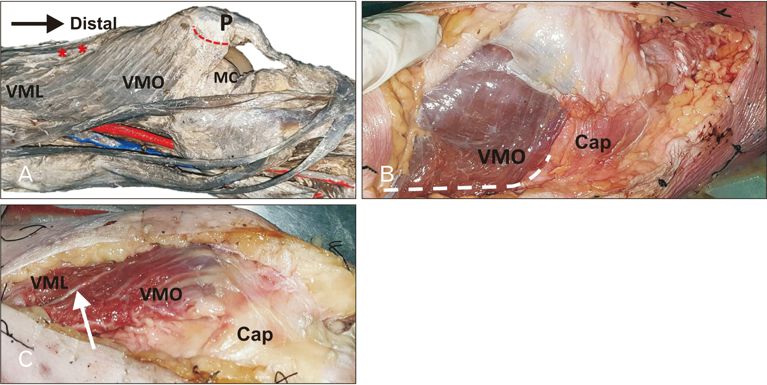 | Fig. 1VM muscle dissection showing (A) VML has vertical fibers and VMO with increased obliquity of muscle fibers. VMO inserted into the medial proximal margin of the patella with fleshy fibers (dotted red line). Lowermost fibers of VMO covered the medial aspect of medial condyle of femur. Nerve to VM runs in a separate fibrotic tunnel (red asterisks). (B) The dotted white line demonstrates the origin of VMO. VMO muscle fibers merge with fibrous capsule of the knee joint. (C) A fascial plane (arrow) separates the VM into two independent parts. Also, the VMO overlaps and merges with the joint capsule. cap, capsule of the knee joint; MC, medial condyle of femur; P, patella; VM, vastus medialis; VML, vastus medialis longus; VMO, vastus medialis oblique.
|
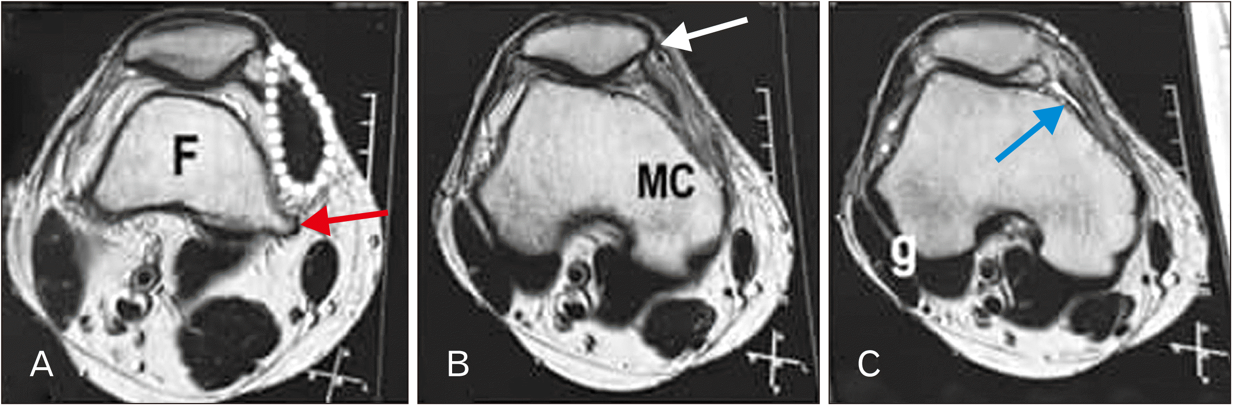 | Fig. 2MRI cross sections of the knee joint through the lower third of the right thigh showing: (A) at lower end of shaft of femur, VMO (marked by dotted white line) originates from medial supracondylar line (red arrow). (B) at medial epicondyle, VMO totally covered the medial aspect of medial condyle. VMO inserted in patella by fleshy fibers (white arrow) (C) at level of the popliteal groove, VMO was well defined that compressed the knee joint space (blue arrow) medially. F, lower end of shaft of femur; g, popliteal groove; MC, medial condyle of femur; MRI, magnetic resonance imaging; VMO, vastus medialis oblique.
|
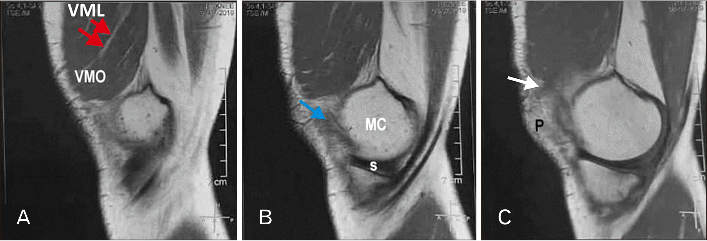 | Fig. 3MRI sagittal sections through the extensor apparatus of the knee showing: (A) a fascial plane (red arrows) separates VM into two parts. (B) Lowermost fibers of VMO (blue arrow) overlap the anterior aspect of medial condyle of femur. (C) VMO inserted into the medial proximal margin of the patella by fleshy fibers (white arrow). MC, medial condyle of femur; MRI, magnetic resonance imaging; P, patella; VM, vastus medialis; VML, vastus medialis longus; VMO, vastus medialis oblique.
|
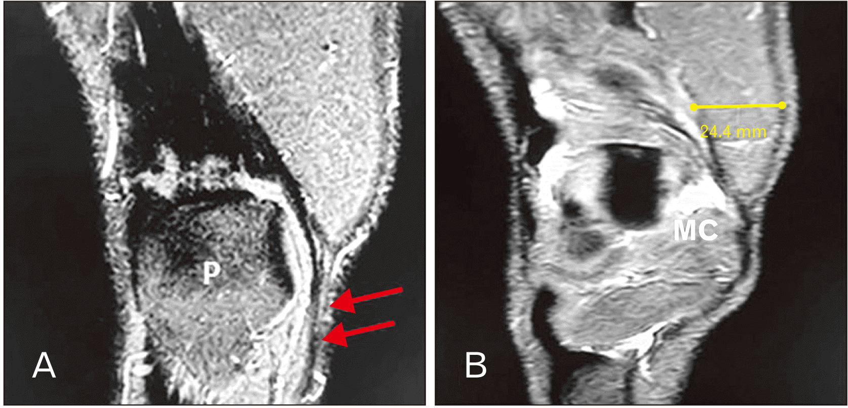 | Fig. 4MRI coronal sections through the extensor apparatus of the knee showing: (A) VMO inserted into the medial proximal margin of the patella and has a small region of direct continuity with the patellar tendon (red arrows) (B) Lowermost fibers of VMO overlapped the anterior aspect of the medial condyle of femur. In fully extended knee, the medial condyle was pushed posteriorly than the lateral condyle. MC, medial condyle of femur; MRI, magnetic resonance imaging; P, patella; VMO, vastus medialis oblique.
|
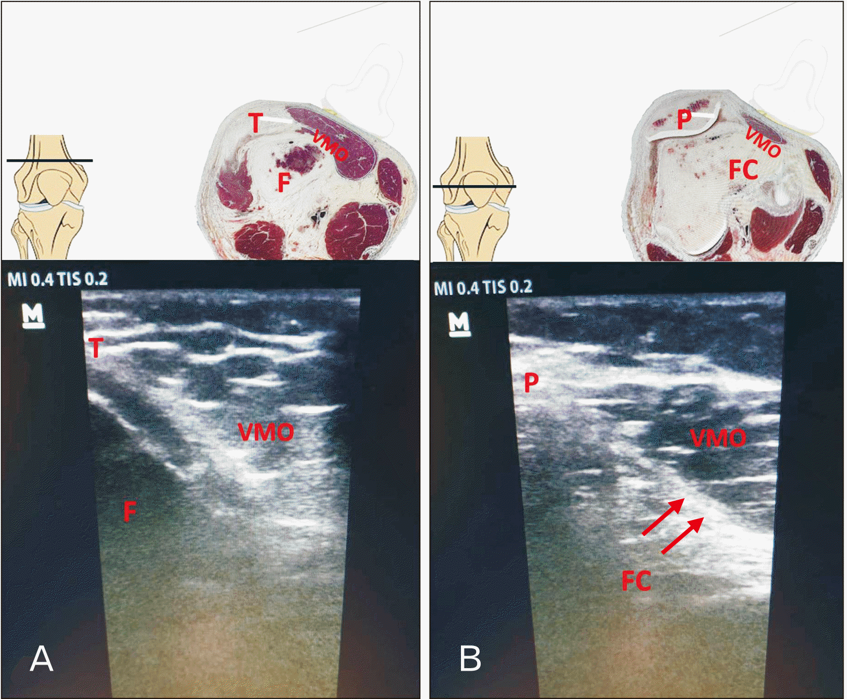 | Fig. 5U/S images of the VMO from a representative participant made by linear probe. (A) Crosssection showing anatomical structures immediately above the patella (B) Crosssection at midpatellar level. arrow, the VMO overlaps and merges with the joint capsule; F, femur; Fc, femoral condyle; P, patella; T, quadriceps tendon; U/S, ultrasound; VMO, vastus medialis oblique.
|
VMO anatomical cross-sectional area (ACSA) in painful vs. painless limbs
The mean values of VMO-ACSA in painful vs. painless knees are summarized in Table 1. In painless knees, VMO cross sectional area was measured on MRI image from proximal to distal; at lower end of shaft of femur it was 23.2±1.9 cm2 (mean±SD), 16.7±1.7 cm2 at upper border of patella and, 14.1±1.1 cm2 at mid-patellar level (level of femoral condyles). However, no statistically significant difference was found between MRI and U/S measurements in the corresponding levels. In painful knee, all parameters were significantly decreased as compared to painless knee. The noticed decrease in VMO-ACSA in painful knee was expressed as a percentage decrease in relation to painless knee. The differences % in VMO-ACSA were calculated in painful knee vs. painless knee; –17.2%±11.0% at lower end of shaft of femur, –21.1%±6.0% at upper border of patella, –36.7%±11.0% at mid-patellar level.
Table 1
A comparison between vastus medialis oblique anatomical cross-sectional area (cm2) measured by MRI and U/S in painless and painful knee
| Anatomical level | Painless knee | Painful knee | Difference % in MRI measurements (painful vs. painless knee) | |
|---|---|---|---|---|
| 1- Lower end of shaft of femur | MRI measurement | 23.2±1.9 | 19.2±1.6* | –17.2±11.0% |
| U/S measurement | 22.5±0.58 | 19.5±0.04* | ||
| 2- Upper border of patella | MRI measurement | 16.7±1.7 | 13.02±1.9* | –21.1±6.0% |
| U/S measurement | 16.5±0.8 | 12.7±0.62* | ||
| 3- Mid-patellar level | MRI measurement | 14.1±1.1 | 8.66±0.6* | –36.7±11.0% |
| U/S measurement | 13.5±1.14 | 9.4±0.08* |
![]()
Go to :
Discussion
The quadriceps muscle is a primary contributor to both functional knee joint stability and knee joint loading [22]. Dynamic knee joint stability may be compromised when weak quadriceps muscles are unable to provide adequate control of tibial translation during ambulation, increasing risk of damage to joint structures [23, 24]. Kim et al. [25] found that with respect to knee extensor muscle strength, the more painful knees showed significantly lower strength than the less painful knees at angular velocities and there is a significant association between dynamic balance measurements and quadriceps strength at angular velocity. The current study provides an insight for the morphology of the VMO and its participation in anterior knee pain.
A closer look at the VM reveals that the uppermost fibers (VML) are more vertically oriented while the lower fibers (VMO) are more obliquely oriented [11]. VMO has received moderate attention as potentially being more responsible for medial tension on the patella.
Muscles with a larger number of insertion and origin points have a greater impact on the musculoskeletal system when perturbed than muscles with few insertion and origin points [26]. The present study showed that VMO originates from medial lip of linea aspera and has attachments to the medial supracondylar line as it wraps around the femur. VMO inserted directly to the upper interior corner of the patella by fleshy fibers. Such finding might reflect the force of movement (applied to the patella) produced by this muscle. Lefebvre et al. [9]; Grob et al. [17] and Grob et al. [27] have demonstrated similar attachment. Motor point stimulation and anatomical dissection clearly showed that VM distal fibers were attached directly to the medial aspect of the patella that contributes to dynamic medial patellar stabilization and opposing its lateral dislocation [28]. Altered muscle activity and weaknesses in VMO cause mal-tracking of the patella and subsequent damage to surrounding structures and aching pain [29]. In the current research, the oblique fibers of VM are not only involved in the medial border of the patella, but additionally, they have a minor area of direct continuity with the patellar tendon. Also, VMO muscle fibers merge with the knee joint capsule. These observations were in accordance with a previous study [17]. Recently, Castanov et al. [11] stated that VMO attached to the mid-portion of the patella, the medial patellofemoral ligament and adductor magnus tendon. Its attachment and oblique alignment provide a mechanical advantage to promote medial stabilizing force to the patella. Also, Muhamed et al. [30] reported that the patellar thickness is important variable to be considered, while assessing the South Indian female for patellofemoral disorders.
Some of the examined cadavers clearly had a nerve branch entering the distal portion of VM muscle separately from the proximal portion. Although most muscles are innervated from their deep surface, it is not unusual for a nerve to travel superficially in a fascial plane prior to entering a muscle belly. The branch to VM from femoral nerve was the furthermost distally situated, almost half-way down the thigh. The branch was well distinct running lengthwise the antero-medial surface of the muscle with the saphenous nerve and was possibly mobilized well proximally. The nerve to VM is designated as giving a short branch entering the muscle proximally, and a longer nerve fiber wandering in the adductor canal before inflowing the muscle belly in its mid-portion [31]. Our nerve branch corresponds to the longer medial branch noted by Page et al. [31]. Separate nerve innervation is a sufficient evidence to support the concept that VMO is an anatomically and functionally separate muscle from VML in the quadriceps femoris muscle group.
VM is more powerful and extends more distally than VL and its relative predominance is traditionally described as medial patellar stabilizer [10]. The findings of this study indicate that VMO is sufficient to rotate the medial condyle of femur internally and the muscle could act independently. “Proof of medial rotation” has been provided by VMO spiral fiber arrangement that covered the anteromedial aspect of the medial condyle. Moreover, the joint space is more compressed medially. Medial rotation of the femur (locking) occurs during the last 30 degrees of extension and locking is produced by the continued action of the same muscles that produce extension i.e., the quadriceps femoris muscle [32]. Moreover, Toumi et al. [33] stated that the medial fusing points of the quadriceps tendon in the intermediate layers violates the insertion of the VM with potential consequences on the terminal phase of extension. Recently, Chang et al. [34] recorded that an appropriate VMO: VL ratio had beneficial effects on PFPS.
Several different clinical tools have been utilized to objectively report the muscle tension in the clinical and operative settings. The use of MRI is a reasonable and more logical approach, but care must be taken to ensure VMO attachment to the patella is accurate. In the present study, the VMO-ACSA of painless knees was 23.2±1.9 cm2 at lower end of shaft of femur, 16.7±1.7 cm2 at medial epicondyle, 14.1±1.1 cm2 at popliteal groove with no statistically significant difference between MRI and U/S measurements. The relative ACSA taken by VMO varies substantially with anatomical location. In the current investigation, we made an attempt to overcome this limitation by selecting a variable slice level among the lower end of the femur. Sattler et al. [10] recorded that in the distal third of the quadriceps femoris muscle, VM took the largest (36%) proportion of the total quadriceps volume. The relative contribution of VM to the total quadriceps increased from proximal to distal. A sum of researches have long-established the trustworthiness of U/S procedure for assessing the dimensions of the quadriceps muscle in health [35]. U/S has the benefit of being movable and encompasses no ionizing radiation and can conclude breadth and cross-sectional areas of superficial muscles. It is potential to quantity basic parameters of muscle construction with U/S investigation, like muscle volume, fascicle length (muscle fiber length that indicates the variety of lengths over which the muscle is capable of actively producing force), and pennation angle (muscle fiber angle comparative to the force-generating alliance) [36].
ACSA was used to represent muscle size as it has the benefit of being a good predictor of both isometric and isokinetic muscle force [37, 38]. In the current work, the VMO-ACSA decreased significantly and similarly in all the experimental groups comparing painless to painful knee (decrements ranged from 17.2% to 36.7%). Painful limbs displayed significantly smaller VMO-ACSA. Therefore, impaired VMO muscle function may lead to injurious loading of articular structures, including menisci, ligaments, cartilage, and bone. Wang et al. [39] reported that a low VM-ACSA was associated with pain and with medial tibial cartilage volume loss. Providing support for this hypothesis are data from human studies in which quadriceps muscle dysfunction may precede degenerative changes [40]. Patients with PFPS were found to have a lower muscle activity ratio between VM and VL muscles than healthy subjects [29]. Engelina et al. [2] added that VMO angle may be a contributory factor in a subset of PFPS, especially in patients who have patellar instability. Previous studies have reported a reduction in anterior knee pain following an increase in muscle strength with exercise training in people with symptomatic knee osteoarthritis [41, 42]. Further, significant increase in VM-ACSA in response to training over time was found to be associated with a concurrent reduction in anterior knee pain, a reduced medial tibial cartilage loss, and a reduced danger of performing knee replacement [39]. So, the current study supports training intervention as a successful measure to induce VMO muscle strength, even at advanced age. This may effectively increase joint health and protect against knee degenerative changes.
Changes in VMO-ACSA as a percentage over a certain time duration using U/S is not widely used for screening and staging. In the near future, it may become a valid method to assess muscle in different settings. Also, further study is recommended to explore the differences in VMO-ACSA as regards age, sex and differences in body weight and to assess whether these correlate to functional findings.
In conclusion, VMO is a distinct muscle within the quadriceps femoris group. VMO would track the patella medially and participating in last phase of knee extension. The assessment of the VMO-ACSA by ultrasonography may constitute promising and reliable tool to evaluate PFPS staging.
Go to :
 XML Download