

 PDF
PDF Citation
 Print
Print


Introduction
The femoral nerve arises from the dorsal branch of the second to fourth lumbar ventral rami. This nerve, after passing behind the inguinal ligament into the thigh, is separated into anterior and posterior divisions by the lateral circumflex femoral artery. The branches of the anterior division are the medial and the intermediate cutaneous femoral nerve and the nerve to the sartorius. Meanwhile, the posterior division is composed of the saphenous nerve and the nerves to the quadriceps femoris, which are related to the sensory profile of the medial aspect of the leg and extension of the knee joint, respectively [1].
The lateral circumflex femoral artery, mostly branching from the root of the deep femoral artery, passes between divisions of the femoral nerve. This artery then divides into the ascending, transverse, and descending branches. The ascending branch courses superiorly behind the tensor fasciae latae muscle to the hip joint and supplies the greater trochanter, the femoral neck, and the femoral head with its anastomosing arteries [1].
Recognition of the normal anatomy and understanding possible variations of the neurovascular structure inside the femoral triangle are important for regional anesthesiologists performing the femoral nerve block with minimal complications [2]. Also, some variations may be associated with pathologic conditions in the lower extremities. Herein, we report a case of an ascending branch of the lateral circumflex femoral artery showing an anomalous course passing through the nervous structure inside the femoral triangle.
Go to : 
Case Report
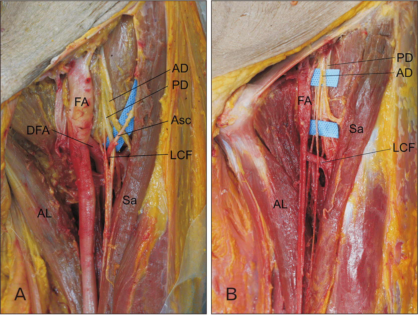
During routine dissection of the thigh, unilateral variation of the femoral nerve penetrated by the ascending branch of the lateral circumflex femoral artery was identified in an 80-year-old Korean male cadaver. No signs of previous surgery or injury were observed in this region. On the left side, the femoral nerve was already split into the anterior and the posterior divisions at a point higher than the location of the lateral circumflex femoral artery. The lateral circumflex femoral artery branched from the deep femoral artery behind the femoral nerve and spit off into the ascending branch as usual. On its way to the hip joint, the ascending branch passed through the neighboring femoral nerve, particularly the posterior division. The distance from the inguinal ligament to the point where the ascending branch pierced the posterior division of the femoral nerve was about 8.4 cm.
The further courses of the ascending branch and the femoral nerve were similar to those previously described. The ascending branch coursed superiorly and laterally to the hip joint behind the tensor fascia latae. Meanwhile, the posterior division of the femoral nerve was divided into the saphenous nerve and the nerves to the quadriceps femoris. The tributaries of the lateral circumflex femoral vein coursed independently from the artery, not penetrating the femoral nerve. There was no corresponding variation on the right side.
Go to :
Discussion
There have been multiple reports of partial splitting and reunion of the femoral nerve at different locations. Anloague and Huijbregts [3] identified bifurcated or trifurcated femoral nerves, separated by the psoas major muscle fibers at the posterior abdominal wall, that rejoined before passing behind the inguinal ligament. Variations of the femoral nerve pierced by muscular slips from the iliacus or the psoas major in the iliac fossa have also been commented on [4]. Wong et al. [5] identified multi-split femoral nerve by a variant psoas quartus muscle. However, no cases of femoral nerve split and reunification in the femoral triangle have been reported before, to the best of our knowledge.
Anatomy of the ascending branch of the lateral circumflex femoral artery was investigated, particularly in regard to its origin, course, and distributions, for the clinical conduct of flap surgery [6]. However, to the best of our knowledge, the ascending branch of the lateral circumflex femoral artery coursing through the femoral nerve with penetration has not been described yet.
The hind-limb buds are formed in the fifth week of gestation and, during this period, muscles bring associated nerves from the torso into the limb buds [5]. This is also true in the case of the femoral nerve. The arterial system of the lower extremity begins to develop from the fifth week of gestation and is completed in the third month with a pattern corresponding to that of a typical adult [7, 8]. Both the sciatic artery and the external iliac artery, during the embryogenic development, develop from the umbilical artery and form the rete femorale together, which supplies the lower-limb bud [8]. The femoral artery and the deep femoral arteries develop from the rete femorale with regression of the sciatic artery [8]. Senior [8] previously described the origin of the circumflex arteries from the rete femorale, whereas Ciftcioglu et al. [9] presumed the circumflex branches of the deep femoral artery might develop independently from the abovementioned rete femorale depending on their blood-flow distribution territory. Regarding these facts, variation in the femoral nerve and the lateral circumflex femoral artery of the type observed in this study occurs during the early stage of prenatal development.
Pathologic conditions with penetration of a nerve by a neighboring vessel have been identified at different locations. Patrick [10] reported a case of tic douloureux caused by the anterior inferior cerebellar artery passing through the trigeminal nerve. In rare cases of trigeminal neuralgia, intraneural vessels penetrating the trigeminal nerve have been identified [11, 12]. Hemifacial spasm patients whose symptoms were caused by intraneural vessels passing through the facial nerve were also reported [13, 14]. Considering the abovementioned cases in which the intraneural vessels resulted in neuropathies, penetration of the posterior division of the femoral nerve by the arterial branch might also have caused pain or paresthesia, particularly of the medial aspect of the leg in the distribution of the saphenous nerve derived from the posterior division. However, it was not possible to identify the preterm pathologies of the donor used in this study because of the limited available medical records.
Awareness of this anatomic variation is important for regional anesthesiologists performing the femoral nerve block [2]. The inguinal crease has been recommended for transducer placement or needle insertion site in ultrasound-guided regional anesthesia for the femoral nerve block [15]. According to Lechner et al. [16], the inguinal creases lies distal to the inguinal ligament by about 6.7 cm. Considering that the point where the ascending branch of the lateral circumflex femoral artery pierced the posterior division of the femoral nerve was 8.4 cm below the inguinal ligament in this case, this point was located near the inguinal crease. Therefore, displacement of the needle into the arterial branch could happen due to the intimate anatomical relationship between the femoral nerve and the arterial branch of this case, which can cause unexpected complications such as hematoma formation or local anesthetic systemic toxicity [17]. Conducting ultrasound visualization with recognition of possible vascular variations could reduce the complication rate [2].
Flap surgeons should also consider this possible variation when harvesting the arterial pedicle for the tensor fascia latae flap. The ascending branch of the lateral circumflex femoral artery forms the major pedicle of the tensor fasciae latae musculocutaneous flap [6]. In a patient with this variation, the risk of accidental nerve injury would increase while approaching the arterial pedicle because of the intimate anatomical relationship between the ascending branch of the lateral circumflex femoral artery and the femoral nerve.
This is solely based on anatomical dissection, thus the information of depth dimension is lacked. Also, frequency of the variation cannot be suggested.
Despite the abovementioned limitations, an understanding of the anatomic variation identified in this study will be helpful for regional anesthesiologists to minimize unexpected serious complications during the femoral nerve block. In addition, the intraneural vessels identified in the femoral triangle may pose possible pathogeneses of refractory leg pain.
Go to :
 XML Download