

 PDF
PDF Citation
 Print
Print


INTRODUCTION
Aortoenteric fistula is defined as an abnormal communication between any portion of the gastrointestinal tract and the aorta.1 Primary aortoenteric fistula (PAEF) is the spontaneous development of direct communication between the gastrointestinal tract and native aorta, unlike secondary aortoenteric fistula that occurs after surgery, such as a vascular graft.2 PAEF is an extremely rare cause of upper gastrointestinal bleeding.3,4 This paper reports the case of a 75-year-old man who presented with massive melena and was diagnosed with primary aortoduodenal fistula (PADF). This case is unique because the diagnosis was made by endoscopy rather than CT or autopsy.
CASE REPORT
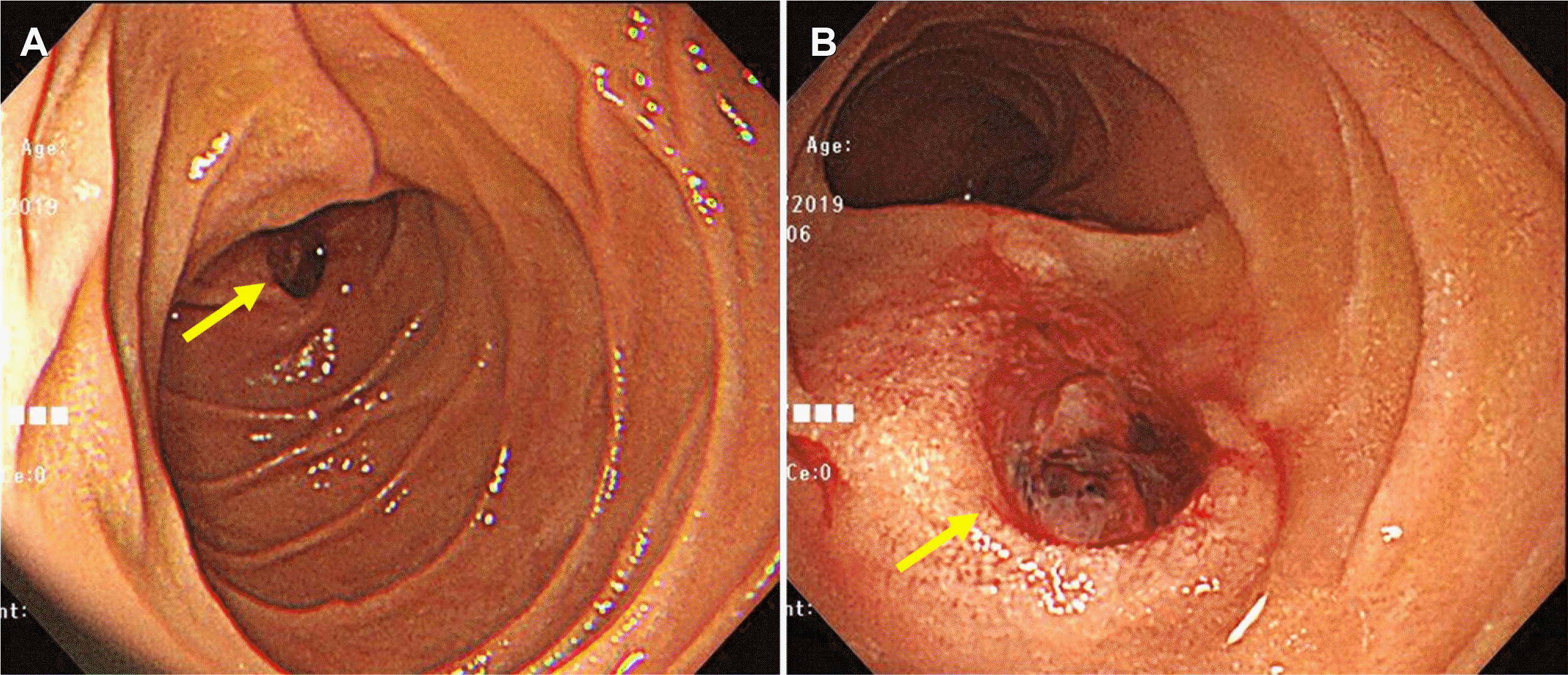
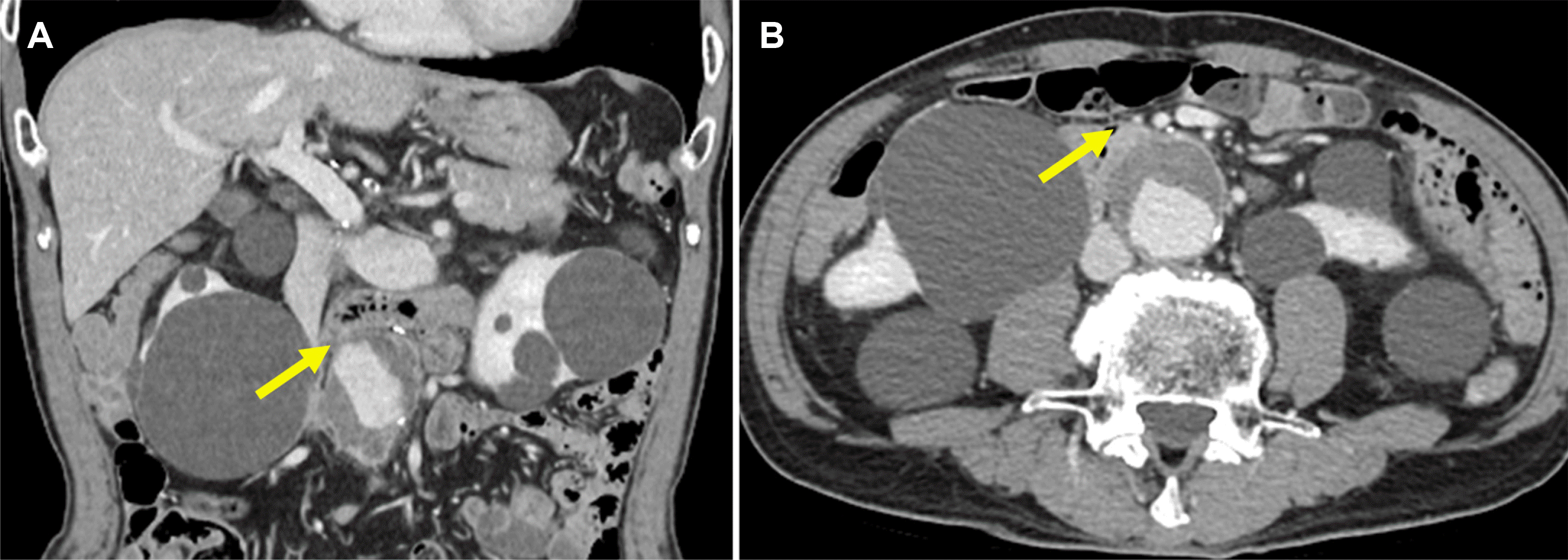
A 75-year-old man visited the emergency department due to a large amount of melena that started 4 hours earlier. He had experienced abdominal pain after dinner 12 hours earlier. At the time of admission, there was no abdominal pain, and his blood pressure, heart rate, respiratory rate, and body temperature were 123/72 mmHg, 63 beats/min, 20 breaths/min, and 36.2℃, respectively. The hemoglobin level and hematocrit were 11.3 g/dL and 33.1%, respectively. He had hypertension and was being followed-up for an abdominal aortic aneurysm at another hospital. No abdominal tenderness was observed during the physical examination at the time of admission. Esophagogastroduodenoscopy revealed an ulcer measuring approximately 1.3 cm with a huge pulsating vessel in the proximal third portion of the duodenum (Fig. 1). An abdominal CT scan revealed an infrarenal abdominal aortic aneurysm measuring 5 cm with adjacent fat stranding abutting to the small bowel (Fig. 2). These observations confirmed the clinical diagnosis of PADF. The patient was prepared for emergency surgery, but the massive bleeding occurred again. Unfortunately, he died in the preoperative stage.
DISCUSSION
PAEF is an extremely rare cause of upper gastrointestinal bleeding, with an incidence of 0.04-0.07% in autopsy studies.3,4 The annual incidence rate of PAEF has been estimated to be 0.007 per million population.3-5 PADF is prevalent in the third and fourth portions of the duodenum because of its location close to the aorta.6,7 A report published in 2008 reviewed 366 cases of PAEF reported over the last 165 years since Salmon reported the first case of PAEF in 1843. Among 366 cases of PAEF, 267 cases (72.9%) were PADF.6 Approximately 80% of PAEFs are associated with atherosclerotic aortic aneurysms, while the other 20% are associated with infections, foreign bodies, post-traumatic pseudoaneurysms, tumors, and radiation therapy.8
The classic triad of PAEF is abdominal pain, upper gastrointestinal bleeding, and a pulsating abdominal mass. On the other hand, these symptoms have been reported to appear in only 11% of cases. Other known symptoms include hematemesis, pain in the back with or without the abdomen, melena, shock, hematochezia, and syncope.4 PAEF usually presents with spontaneous intermittent gastrointestinal bleeding before massive bleeding. Intermittent gastrointestinal bleeding is usually mild and self-limiting and is probably due to low blood pressure and the formation of thrombus.4 Thrombus may plug the fistula and temporarily stop bleeding. On the other hand, upper endoscopy and excessive volume supply can promote massive gastrointestinal bleeding.7 The time interval between intermittent herald gastrointestinal bleeding and massive bleeding can range from a few hours to months.9,10 In this case, a patient visits the hospital because of a small amount of gastrointestinal bleeding and eventually dies from massive bleeding; it is sometimes mistaken for a treatment error.
Upper endoscopy is the diagnostic modality of choice for acute upper gastrointestinal bleeding.11 On the other hand, stable patients often do not have current bleeding, and endoscopy rarely reveals confirmatory evidence of PAEF. In addition, endoscopic visualization of the lower third part of the duodenum, the most common area of PAEF, is complicated and is generally not observed by the endoscopist.4,7 Owing to the aforementioned reasons, the diagnosis of PAEF is achieved mainly by an abdominal CT scan or autopsy and is rarely diagnosed with endoscopy.
Currently, the standard for a diagnosis is CT with intravenous contrast.12 Compared to endoscopy or angiography, CT is less invasive, easier to use, and has no risk of promoting massive gastrointestinal bleeding. The presence of air in the retroperitoneum and thrombus, along with the loss of the fat plane, is probably an indicator of PADF.6 Arterial angiography is useful only when there is active bleeding. If there is no active bleeding or a small amount of bleeding, the condition is impossible to diagnose, even with an examination. As a result, a re-examination may be required to confirm the diagnosis of PADF.6
The treatment for PAEF is the same as that for a secondary aortoenteric fistula and involves an aortic reconstruction and duodenal repair with an exploratory laparotomy.1 Multivariate analysis of 791 patients with primary and secondary aortoduodenal fistula suggested that an aortic reconstruction is an independent predictor of survival.13 Recently, successful endovascular treatment of PADF was reported. Endovascular repair is becoming an attractive treatment option. On the other hand, endovascular stent removal is required in patients with PADF months after successful initial endovascular treatment because of the high risk of infection with endovascular stent grafts.14 To the best of the authors’ knowledge, there are no reports of success with PAEF treatment through endoscopy. A case of attempting endoscopic treatment of PAEF was reported. In that case, the patient had a large amount of bleeding several hours after the endoscopy and died while being prepared for emergency surgery.15 In the authors’ opinion, endoscopic devices, such as clips used for endoscopic hemostasis, can damage the aneurysm wall. A damaged aneurysm causes massive bleeding and is associated with a poor prognosis.
Early diagnosis of PADF is difficult because of its non-specificity and subtle clinical features.15,16 Moreover, it is often overlooked as the cause of upper gastrointestinal bleeding owing to its rarity. The mortality rate of untreated PAEF is almost 100%.2,4,16 The key to an early diagnosis of PAEF is the suspicion of the endoscopist. The doctors should recognize PAEF as a potential cause of gastrointestinal bleeding. This is particularly true for patients with a history of abdominal aortic aneurysms. Finally, if a duodenal ulcer has a pulsating nature at a location other than a common peptic ulcer in the endoscope, endoscopic hemostasis should not be attempted, and an abdominal CT scan should be performed for confirmation.
 XML Download