

 PDF
PDF Citation
 Print
Print


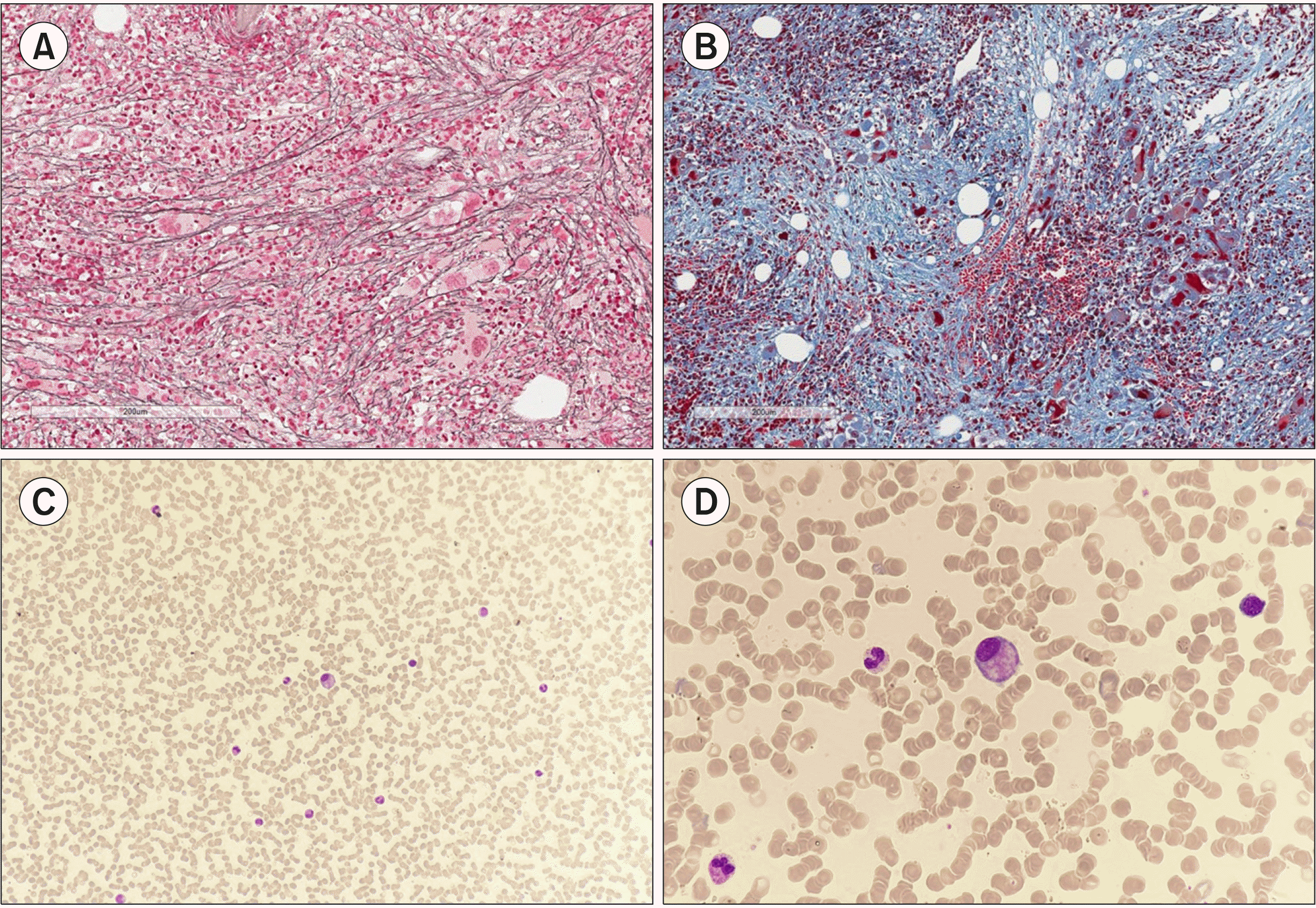
A 57-year-old man diagnosed with JAK2+ prefibrotic/early primary myelofibrosis (MF) progressed during the one-year follow-up to the overt fibrotic stage (A, reticulin stain, ×100 and B, Masson’s trichrome stain, ×100). Complete blood count showed no cytopenia (hemoglobin 14.4 g/dL, 12.79×10⁹ leukocytes/L, and 148×109 platelets/L). Initial blood-smear examination with low amplification objectives (C, Giemsa stain, ×200) showed the leukoerythroblastic morphology frequently seen in MF, with circulating myeloid precursors, erythroblasts, and anisopoikilocytosis. Surprisingly, a megakaryocyte was found in the body of the slide. Higher amplification (D, Giemsa stain, ×600) enabled better characterization of this atypical small megakaryocyte with a non-lobated nucleus. Isolated (bare or naked) megakaryocyte nuclei are typical findings in MF. Intact megakaryocytes have rarely been reported in peripheral blood smears. If present, the feathered edge of the smear is the predilected location of these cells as they are among the largest cells in the sample and tend to be dragged when extending the sample, whereby their morphology may be altered. This finding emphasizes the importance of a systemic approach to blood-smear examination, starting scanning at low magnification for full coverage of the sample and identification of the best regions for higher magnification.
 XML Download