

 PDF
PDF Citation
 Print
Print


INTRODUCTION
Chromosomal aberrations (CA) in hematologic malignancies can be commonly examined using conventional cytogenetics, fluorescence in situ hybridization (FISH), and multiplex reverse transcriptase polymerase chain reaction (multiplex RT-PCR). A commercially available multiplex RT-PCR system has been introduced to detect 28 fusion genes in patients with acute leukemia (AL) [1].
CA develops in almost 56% of adult de novo acute myeloid leukemia (AML) and slightly more often in childhood cases [2-4]. Racial differences in hematological malignancies between Asian and Western countries have been reported [5, 6]. The prevalence of CA in AL might change over time, possibly due to changes in various risk factors, such as environmental pollutants and lifestyle, as mentioned in a previous study [7, 8]. Therefore, we compared the frequency and spectra of CA in patients with AL during two time periods (2006–2009 vs. 2010–2015) in a single institute and with other previous reports.
MATERIALS AND METHODS
Patients
In total, 717 patients with AL were enrolled during a 6-year period (2010–2015). This study included 125 children (median age, 8 yr; range, 0–18 yr) and 592 adults (median age, 57 yr; range, 19–88 yr). A total of 66% (83/125) of the children were male and 34% (42/125) were female; 58% (341/592) of the adults were male and 42% (251/592) were female; 73% (521/717) were diagnosed with primary (84%) or secondary (16%) AML (40 children, 481 adults), 26% (187/717) were diagnosed with acute lymphoid leukemia (ALL) (80 children, 107 adults), and 1% (9/717) were diagnosed with mixed phenotype acute leukemia (MPAL) (Supplementary Table 1). Clinical and laboratory data, such as sex, age, and the results of multiplex RT-PCR, FISH, and conventional cytogenetics were obtained from electronic medical records (EMR). This study was approved by the institutional review board (CNUHH No. 2009-35). Informed consent was obtained from all participating patients.
Multiplex RT-PCR system
In total, 89% (637/717) of AL patients were analyzed using the multiplex RT-PCR system. Briefly, RNA was extracted from the peripheral blood (PB) or bone marrow (BM) using a commercial kit (RNAqueous kit; Ambion, Austin, TX, USA), in accordance with the manufacturer’s protocol. The multiplex RT-PCR test kit (DNA Technology, Aarhus, Denmark) was used to detect 28 of the most common leukemic fusion genes and more than 80 splice variants (Supplementary Fig. 1).
Fluorescence in situ hybridization (FISH)
FISH was performed in appropriate BM or PB specimens available from 609 patients, in addition to the conventional cytogenetic method. It was performed using an appropriate probe (Abbott Molecular/Vysis, Des Plaines, IL, USA), in accordance with the manufacturer’s instructions.
Conventional cytogenetics
Conventional cytogenetic analysis was performed at the initial diagnosis phase using short-term cultures of BM cells. At least 20 metaphases were analyzed in each case, and the clonal CA was described according to the International System for Cytogenetic Nomenclature 2009 and 2013 (Supplementary Fig. 2).
RESULTS
Detection of CA using a multiplex RT-PCR system
Sixteen CA types were detected in 36% (228/637) of the patients using a multiplex RT-PCR system (Supplementary Table 2). The multiplex RT-PCR system detected fusion transcripts in 35% (159/460) of AML patients, 38% (64/168) of ALL patients, and 56% (5/9) of MPAL patients (Table 1).
Table 1
Frequency and spectra of chromosomal aberrations according to the type of all acute leukemia patients using the multiplex RT-PCR system.
![]()
Detection of CA using conventional cytogenetics, including FISH
In total, conventional cytogenetic methods were performed in 97% (506/521) of the AML patients, 94% (175/187) of the ALL patients, and 100% (9/9) of the MPAL patients. Among these, 93% of AML, 87% of ALL, and 100% of MPAL were successfully analyzed. Successful analyses were achieved in 92% (634/690) of the patients. Among the successfully analyzed cases, 59% (372/634) had detectable CA and 41% (262/634) were considered cytogenetically normal (Table 2).
Table 2
Frequency and spectra of chromosomal aberrations according to the type of acute leukemia and detection method in all patients using conventional cytogenetics, including FISH.
| Chromosomal aberration by conventional cytogenetics | Fusion transcript | AML | ALL | MPAL | Total | ||||||
|---|---|---|---|---|---|---|---|---|---|---|---|
|
|
|
|
|||||||||
| K | F | K | F | K | F | ||||||
| t(15;17)(q24;q21) | PML/RARA | 54 | 13 | 67 | |||||||
| t(9;22)(q34;q11) | BCR/ABL1 | 3 | 4 | 22 | 8 | 4 | 41 | ||||
| t(8;21)(q22;q22) | RUNX1/RUNX1T1 | 36 | 36 | ||||||||
| 11q23 | |||||||||||
| t(4;11)(q21;q23) | KMT2A /AFF1 | 1 | 1 | ||||||||
| t(6;11)(q27;q23) | KMT2A /AFDN | 5 | 5 | ||||||||
| t(11;19)(q23;p13.3) | KMT2A /ELL | 0 | |||||||||
| t(9;11)(p22;q23) | KMT2A /MLLT3 | 6 | 1 | 7 | |||||||
| t(10;11)(p12;q23) | KMT2A /MLLT10 | 4 | 1 | 5 | |||||||
| t(11;19)(q23;p13.3) | KMT2A /MLLT1 | 1 | 1 | ||||||||
| t(12;21)(p13;q22) | ETV6/RUNX1 | 5 | 7 | 12 | |||||||
| t(1;19)(q23;p13) | TCF3/PBX1 | 1 | 1 | ||||||||
| inv(16)(p13;q22) | CBFB/MYH11 | 21 | 1 | 22 | |||||||
| t(9;9)(q34;q34) | SET/NUP214 | 0 | |||||||||
| del(1p32) | STIL/TAL1 | 0 | |||||||||
| t(16;21)(p11;q22) | FUS/ERG | 2 | 2 | ||||||||
| t(6;9)(p23;q34) | DEK/NUP214 | 2 | 2 | ||||||||
| Extra-aberrationa) | 113 | 5 | 43 | 7 | 2 | 170 | |||||
|
|
|||||||||||
| Positive cases excluding extra-aberration | 151 | 46 | 5 | 202 | |||||||
| Positive cases | 269 | 96 | 7 | 372 | |||||||
|
|
|||||||||||
| Total cases | 521 | 187 | 9 | 717 | |||||||
| Cases of ‘NT’ | 15 | 12 | 0 | 27 | |||||||
| Cases excluding ‘NT’ | 506 | 175 | 9 | 690 | |||||||
| Positive cases excluding ‘extra-aberration’ & ‘NT’ (%) | 30 | 26 | 56 | 29 | |||||||
| Positive cases excluding ‘NT’ (%) | 53 | 55 | 78 | 54 | |||||||
![]()
Thirteen types of fusion transcripts, excluding extra-aberrations, were detected in 29% (202/690) of patients using conventional cytogenetics. Meanwhile, the extra-aberration indicated numerical abnormalities or structural rearrangements, excluding 28 fusion transcripts, in the multiplex RT-PCR system. The most frequent CA was t(15;17)(q24;q21) in 9.7% (67/690), followed by t(9;22) (q34;q11) in 5.9% (41/690), t(8;21)(q22;q22) in 5.2% (36/690), inv(16)(p13;q22) in 3.2% (22/690), and KMT2A gene rearrangements in 2.8% (19/690) of patients (Table 2, Supplementary Table 3). Within a group of 59% (23/39) of childhood AML patients, the most common CA was t(15;17)(q24;q21) (N=8), followed by t(8;21)(q22;q22) (N=6) (Supplementary Table 4). Within a group of 25% (19/75) of childhood ALL patients, the CA was t(12;21)(p13;q22) in 12 cases, followed by t(9;22)(q34;q11) in five cases, and one case each of t(10;11)(p12;q23) and t(11;19)(q23;p13.3) (Supplementary Table 4). However, extra-aberrations were the majority in childhood ALL patients. Within a group of 27% (128/467) of adult AML patients, the most common CA was t(15;17)(q21;q22) (N=59), followed by t(8;21)(q22;q22) (N=30) (Supplementary Table 4). Within a group of 27% (27/100) of adult ALL patients, the common CA was t(9;22)(q34;q11) (N=25). Meanwhile, the majority of adult patients showed a normal karyotype (Supplementary Table 4).
Frequency and spectra of CA according to childhood and adult groups using multiplex RT-PCR
A total of 46% percent (54/117) of the childhood group subjects exhibited a positive result using the multiplex RT-PCR system. Within a group of 65% (24/37) of childhood AML patients, the common CA included eight PML/RARA, followed by five RUNX1/RUNX1T1, five KMT2A gene rearrangements (three KMT2A/MLLT3 and two KMT2A/MLLT10), three CBFB/MYH11, and one of each of BCR/ABL1, DEK/NUP214, and FUS/ERG. In a group of 37% (28/75) of childhood ALL patients, the following fusion scripts were detected: 15 ETV6/RUNX1, 5 BCR/ABL1, four KMT2A gene rearrangements (two KMT2A/AFF1, one each of KMT2A/ MLLT1 and KMT2A/MLLT10), three STIL/TAL1, and one TCF/PBX1. A total of 33% (174/520) of adult ALL patients had a positive result. Within a group of 32% (135/423) of adult AML patients, the common CA included PML/RARA (N=59), followed by RUNX1/RUNX1T1 (N=29), CBFB/MYH11 (N=20), KMT2A gene rearrangements (N=15) [KMT2A/AFDN (N=5), KMT2A/MLLT3 (N=5), KMT2A/MLLT10 (N=3), KMT2A/ELL (N=2)], BCR/ABL1 (N=8), FUS/ERG (N=2), DEK/NUP214 (N=1), and SET/NUP214 (N=1). The common CA in 39% (36/93) of adult ALL patients included BCR/ABL1 (N=29), followed by TCF3/PBX1 (N=4), KMT2A/AFF1 (N=1), SET/NUP214 (N=1), and STIL/TAL1 (N=1) (Supplementary Table 5).
Frequency and spectra of CA according to AML and ALL patient groups using multiplex RT-PCR
In a group of 35% (159/460) of AML patients, the following fusion transcripts were detected: 67 PML/RARA, 34 RUNX1/ RUNX1T1, 23 CBFB/MYH11, 20 KMT2A rearrangements, nine BCR/ABL1, three FUS/ERG, two DEK/NUP214, and one SET/NUP214. In a group of 38% (64/168) of ALL patients, fusion transcripts included 34 BCR/ABL1, 15 ETV6/RUNX1, five TCF3/PBX1, five KMT2A gene rearrangements (three KMT2A/AFF1, one of each of KMT2A/MLLT1 and KMT2A/ MLLT10), four STIL/TAL1, and one SET/NUP214. In a group of 56% (5/9) of MPAL patients, the following fusion transcripts were detected: four BCR/ABL1 and one KMT2A/ MLLT3 (Table 1).
Results from multiplex RT-PCR system and conventional cytogenetics
There was an agreement between the multiplex RT-PCR system and conventional cytogenetics in 71% (394/553) of patients. No CA was detected using either method in 41% (228/553) of patients. Thirteen types of fusion transcripts were detected using a multiplex RT-PCR system in 30% (166/553) of the patients, which was in agreement with conventional cytogenetics. Conventional cytogenetics disclosed additional structural rearrangements and/or numerical abnormalities in 11% (61/553) of the patients with specific aberrations, which were demonstrated using both methods. The results of the multiplex RT-PCR system did not correspond with the conventional cytogenetics in 29% (159/553) of the patients. Cytogenetically cryptic translocations solely detected using multiplex RT-PCR system were present in 5% (28/553) of the patients with a normal karyotype, numerical abnormalities, or structural rearrangements based on conventional cytogenetics. Detailed comparison data between the multiplex RT-PCR system and conventional cytogenetics are summarized in Supplementary Table 6. A total of 36% (131/359) of the patients with a negative result based on the multiplex RT-PCR system had CA, according to conventional cytogenetics results (Supplementary Table 6–8).
Considering both PCR methods and conventional cytogenetics, the most frequent CA of childhood and adult AML was PML/RARA, which was followed by RUNX1/RUNX1T1. The most frequent CA of childhood and adult ALL was ETV6/RUNX1 and BCR/ABL1, respectively (Supplementary Table 9, 10, Fig. 1, 2). The most frequent CA of the mixed phenotype AL was BCR/ABL1, which was followed by KMT2A/MLLT3 (Table 1, 2).
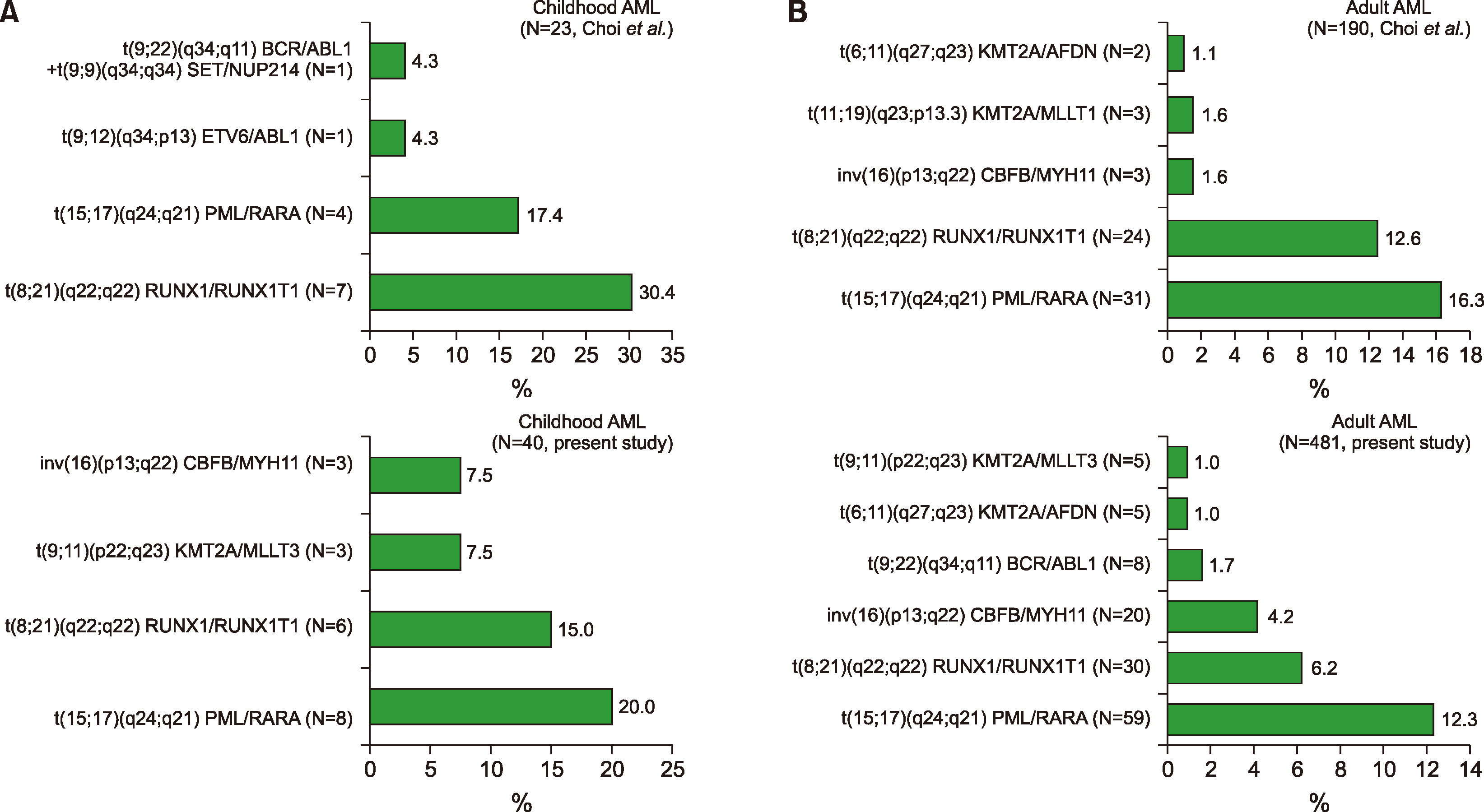
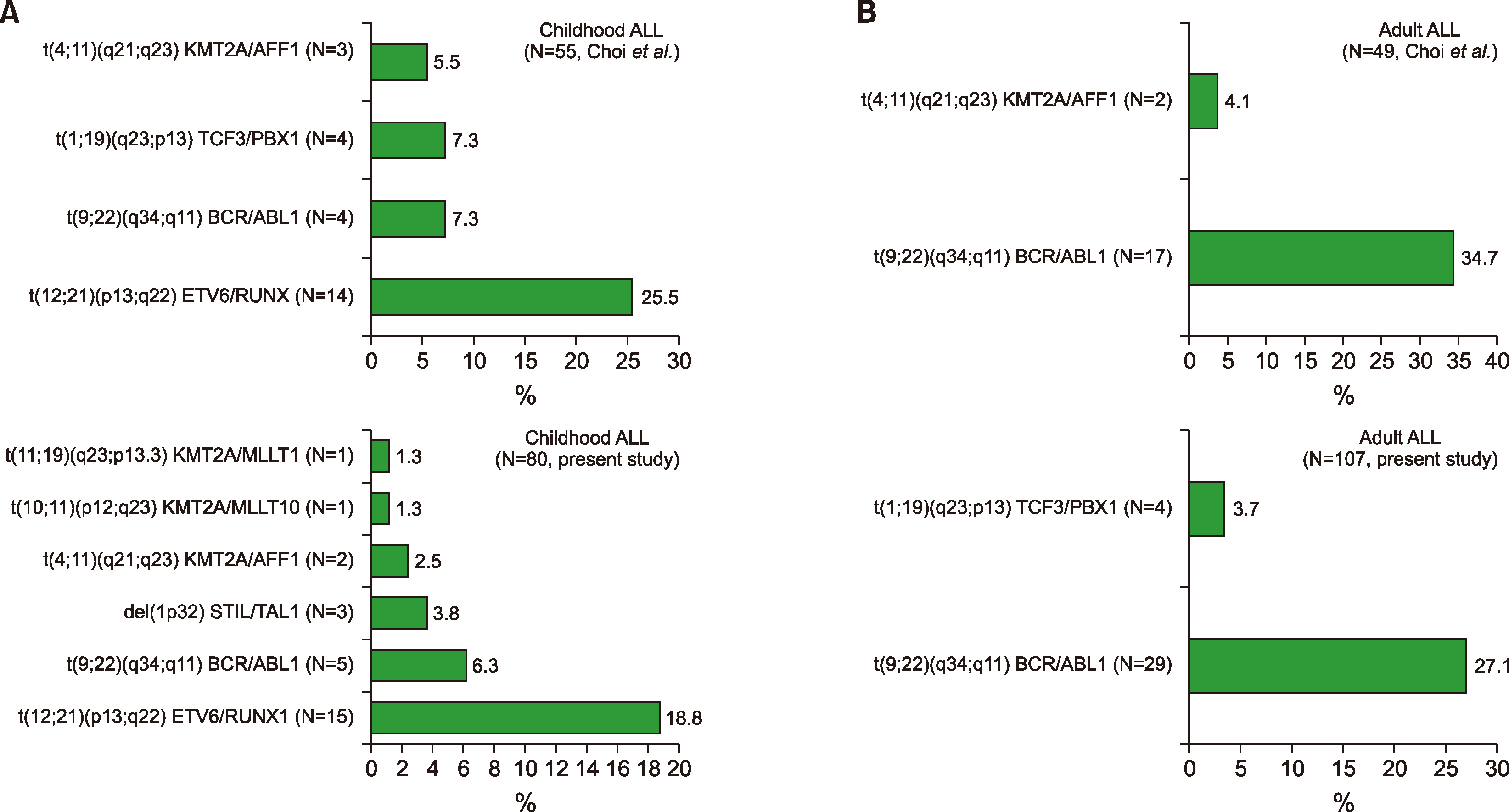
Fig. 1
The prevalence of major chromosomal aberrations, which were detected using conventional cytogenetics, including FISH and multiplex RT-PCR, were compared to the previous study [1] and described according to leukemia type and age. (A) Childhood AML, (B) adult AML.
![]()
Fig. 2
The prevalence of major chromosomal aberrations, which were detected using conventional cytogenetics, including FISH and multiplex RT-PCR, was compared to that of the previous study [1] and described according to leukemia type and age. (A) Childhood ALL, (B) adult ALL.
![]()
Important CA that were not covered by the multiplex RT-PCR system
In the current study, 98 types of CA were determined in 717 patients with hematological malignancies. The multiplex RT-PCR system for screening 28 fusion transcripts identified only 16 types among them. Eighty-two types of leukemic fusion could not be found using multiplex RT-PCR, excluding numerical abnormalities. Three important CAs, such as i(17)(q10), t(3;3)(q21;q26.2), and t(8;14)(q24;q32), were found using conventional cytogenetics (Supplementary Table 7, 8, Supplementary Fig. 2).
DISCUSSION
Considering the many fusion genes and breakpoint variants currently determined, more than 50 separate PCR reactions are required to screen an AL patient with a standard operation [9]. We adopted a commercially accessible multiplex-RT-PCR system (DNA Technology) to identify 28 common fusion genes and over 80 splice variants based on previous research [9]. Data from Denmark using this system, which was employed on specimens from 143 patients with a median age of 63 years (range, 0–85 yr; 132 adults, 11 children), revealed that CA was identified in only 15% (21/143) of the patients [10]. In contrast, this study showed a higher CA frequency of 36% (228/637), using the same multiplex RT-PCR system: 35% (159/460) of AML patients, 38% (64/168) of ALL patients, and 56% (5/9) of MPAL patients. The rate of occurrence and spread of leukemic fusion genes in AL also differed from earlier European data [10]. CA occurred in 65% (24/37) of childhood AML cases, whereas it only occurred in about 40% (24/60) of childhood AML cases based on the same multiplex RT-PCR system from Austria [11]. The frequency of leukemic fusion genes in our ALL patients (38%, 64/168) was analogous to an Italian dataset utilizing the same multiplex RT-PCR system: 37% (28/75) and 39% (36/93) of childhood and adult ALL patients, respectively. In the Italian study, 39% of 170 ALL patients carried leukemic fusion genes: 39% and 40% of the childhood and adult ALL patients, respectively [12].
The most common CA in AML patients was the t(15;17) abnormality (15%, 67/460). The occurrence rate was higher compared to data from other institutes of Korea (6.7%), Caucasian (6.5, 10%), Australian (12%), Japanese (11%), and Singapore-Chinese (11%), but it was analogous to the Chinese data (14.3%) [5, 6, 13]. This discrepancy may be due to the sample size and likely to ethnic differences. The geographic diversity of CA in leukemia needs further study and a better understanding of the genetic and environmental factors associated with leukemia. The frequency and range of CA in ALL were similar between this study and previous studies [6, 14]. A balanced translocation of the BCR/ABL1 fusion gene in ALL patients was the most common abnormality (20%, 34/168) in this study. The occurrence of the BCR/ABL1 fusion transcript was lower than in the Southwest Oncology Group data (26%) [15], while it was higher than those in Indian (6%) and Taiwanese data (8%) [16, 17]. The most common CA in childhood ALL patients was the ETV6/RUNX1 fusion transcript, which was followed by BCR/ABL1, KMT2A gene rearrangements, STIL/TAL, and TCF3/PBX1. In contrast, the BCR/ABL1 fusion transcript was the most common CA in adult ALL patients, which was followed by TCF3/PBX1, KMT2A/AFF1, SET/NUP214, and STIL/TAL1.
The discrepancy of 29% (159/553) between the multiplex RT-PCR system and conventional cytogenetics was mainly due to numerical and submicroscopic anomalies. The multiplex RT-PCR system used to screen 28 fusion transcripts identified 16 types in the group of 98 types of CA determined in 717 patients with leukemia. The concordance rate of 71% (394/553) between the multiplex RT-PCR system and conventional karyotyping was analogous to an earlier study (70%) [1].
Choi et al. [1] reported 20 fusion transcripts containing KMT2A/EPS15 (MLL1/AF-1p), ETV6/ABL1 (TEL/ABL), RUNX1/MDS1 (AML1/MDS1), ZBTB16/RARA (PLZF/RARA), and ETV6/MN1 (TEL/MN1) using a multiplex RT-PCR system. However, 16 fusion transcripts containing DEK/ NUP214 were determined using the multiplex RT-PCR system in this study, and the above-mentioned 5 fusion transcripts were not identified: KMT2A/EPS15 (MLL1/AF-1p), ETV6/ABL1 (TEL/ABL), RUNX1/MDS1 (AML1/MDS1), ZBTB16/ RARA (PLZF/RARA), and ETV6/MN1 (TEL/MN1).
In addition, the range or spectra of leukemic fusion genes in AL differed from a prior report in Korea (Fig. 1, 2) [1]. In a prior study, Choi et al. examined 325 leukemia patients during a 4-year period (2006–2009). Eighty-one children (median age 8 yr, range 0–18 yr) and 244 adults (median age 56 yr, range 19–86 yr) were included in their study. Of the 81 children, 35 (43%) were male and 46 (57%) were female; of the 244 adults, 123 (50%) were male. A total of 213 (66%) patients were diagnosed with de novo (92%) or secondary (8%) AML (23 children, 190 adults); 104 (32%) patients had ALL (55 children, 49 adults), and 8 had mixed phenotype acute leukemia (MPAL). Accordingly, these findings were somewhat heterogeneous and different from those of the present study, as mentioned in the Patients’ section of the materials in this study.
In addition, the most prevalent CA in childhood AML patients was PML/RARA, and was followed by RUNX1/ RUNX1T1 in this study (Supplementary Table 9), whereas the most prevalent CA was RUNX1/RUNX1T1, which was followed by PML/RARA in a previous report [1]. In addition, taking into account MPAL patients, the most prevalent CA was BCR/ABL1, followed by KMT2A/MLLT3, in this study (Supplementary Table 9, 10), while the most common CA was BCR/ABL1, followed by KMT2A/ELL in a previous report [1].
Interestingly, there were cases with significant CA, such as i(17)(q10), t(3;3)(q21;q26.2), and t(8;14)(q24;q32), that were not identified using the commercial multiplex RT-PCR system (Supplementary Table 8, Supplementary Fig. 2). These are very meaningful and typical CA in hematological malignancies, such as AL. t(8;14)(q24;q32) is a typical CA in patients with ALL (Burkitt type), and the t(3;3)(q21;q26.2) is a newly adopted recurrent CA in AML patients, in accordance with the 2008 WHO classification [1]. In addition, i(17)(q10) is repeatedly shown in the blast crisis of Philadelphia- positive CML patients. As a solitary CA, i(17q) has also been demonstrated in primary fibrosis, hypereosinophilic syndrome, and rare cases of myelodysplastic syndrome (MDS) and myeloproliferative neoplasm (MPN) that result in acute non-lymphocytic leukemia. In accordance with several studies, MDS/MPN, which contains the i(17q) as a single CA, is regarded as a distinctive entity with a frequency of 0.4–1.57% of MDS, or 1% of all myeloid cases, which features a male predominance, significant anemia, hyposegmented neutrophils, increased micromegakaryocytes, and an unfavorable prognosis [18, 19]. Thus, it is worth considering CA as an additional screening panel of leukemic fusion genes for improving the molecular detection system.
In conclusion, this study disclosed the frequency and range of CA in Korean AL patients, which differed from those of previous published reports. This change might be due to environmental and lifestyle changes [7, 8]. Our data were collected from a single center in Korea; thus, the findings may not be generalizable to other institutions. In addition, the limited number of patients might preclude definitive conclusions on the AL changes in Korea. Further studies to accumulate more evidence are needed to determine the change in spectra or range of AL. Nonetheless, this study might provide important information for improving the molecular detection system in Korean AL patients.
 XML Download