Journal List > J Korean Foot Ankle Soc > v.25(1) > 1146538

거골하관절 불안정성이 족관절 불안정성을 치료하는 데 중요한 요소로 작용할까?

Abstract

Subtalar joint instability (STI) is one of the causes of persistent hindfoot pain even after treatment for ankle instability. Diagnosis of STI is often neglected because it is a poorly understood concept. However, understanding of the anatomy and injury mechanism of the subtalar joint is a prerequisite of appropriate treatment. Patients with STI usually complain of several symptoms, such as recurrent sprains or a giving way sensation and rolling over. Recently, various diagnostic tools and treatment methods have been investigated. This study was undertaken with the aim of helping clinicians by providing a comprehensive understanding of STI.

서 론

만성 외측 후족부 불안정성은 족관절 염좌의 흔한 합병증이다. 해부학적으로 후족부 인대 불안정성은 족관절 불안정성(ankle joint instability)과 거골하관절 불안정성(subtalar joint instability)의 두 가지 형태로 구분된다.1) 족관절 불안정성과 거골하관절 불안정성은 아직도 임상적으로 분명하게 나누어 진단되지 않고 있다.2-5)
거골하관절 인대 손상과 불안정성은 족관절 불안정성과 비교하면 아직까지 임상적으로 잘 알려져 있지 않다. 거골하관절 불안정성은 1962년에 Rubin과 Witten6)에 의해 처음 임상적으로 언급되었다. 이 질병의 실제 유병률은 다양하게 보고되었다. Gillespie와 Boucher7)의 연구와 Brantigan 등8)의 연구에서는 거골하관절 불안정성이 만성 족관절 기능적 불안정성의 10%∼25% 정도에서 발생한다고 보고하고 있으며, Larsen9)은 외측 족관절 불안정성 환자의 25% 정도는 거골하관절 불안정성이 있다고 보고하였다. 또한 Meyer 등10)은 급성 족관절 외측 인대 파열 환자 40명을 대상으로 시행한 거골하관절 조영술 검사에서 80% 정도는 거골하관절 인대 손상이 동반되어 있다고 보고하였다.
정확히 진단되지 못한 거골하관절 불안정성은 단순히 족관절 외측 인대 봉합 및 재건술을 시행한 환자들의 지속적인 후족부 기능적 불안정성(functional instability)의 원인이 될 수도 있다. 따라서 족관절 염좌 발생 시 족관절뿐만 아니라 거골하관절에 대한 평가도 같이 이루어져야 한다. 때로는 거골하관절 인대들의 단독 손상도 발생할 수 있어 거골하관절의 해부학적 구조, 임상증상 및 치료 등에 대한 이해가 필요하다.

해부학적 구조

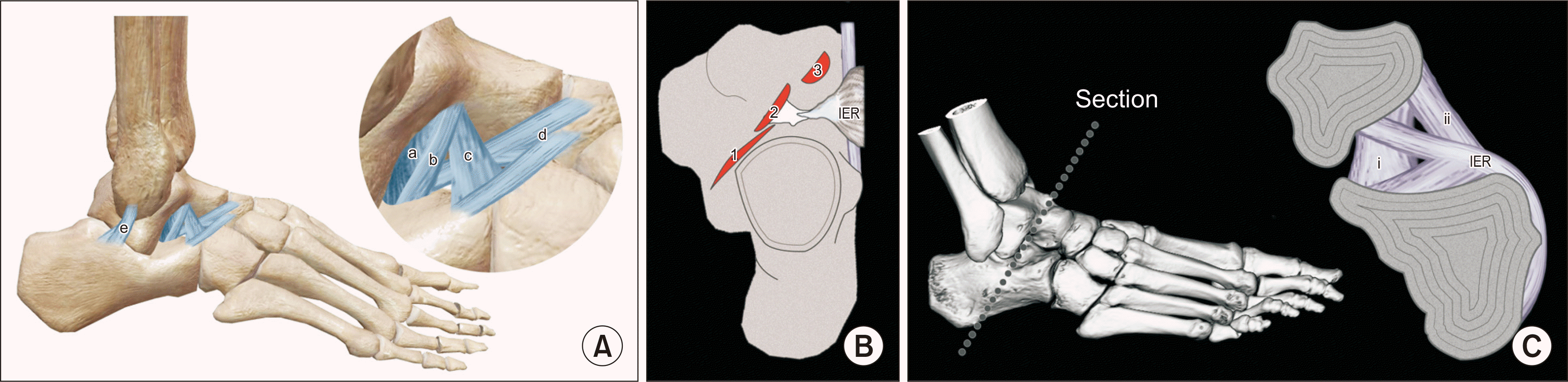
거골하관절의 해부학적 구조물은 종비인대(calcaneofibular ligament), 경부인대(cervical ligament), 전방 관절낭 인대(anterior capsular ligament), 거종골간인대(interosseous talocalcaneal ligament)로 알려져 있다.2,11,12) 종비인대는 경골거골관절(tibiotalar joint)과 거골하관절을 이어주는 유일한 인대로 거골하관절의 안정성에 중요한 구조물이다.13,14) 다른 구조물은 내재적(intrinsic) 거골하관절 인대에 해당된다(Table 1).15-21)

1. 경부인대

경부인대는 보통 하나 또는 두 개의 다발(bundle)로 이루어지지만 여러 개의 다발로 이루어진 경우도 있으며 전거비인대(anterior talofibular ligament)와 거종골간인대보다 넓고 길다.
거골의 경부인대 부착부(footprint)는 불규칙적이며 형태는 원형이거나 타원형이다. 길이는 12.7±2.1 mm이며, 두께는 7.7±2.4 mm 정도이다. 거골-입방골(talonavicular) 관절의 연골면에서 나와서 종골에 부착하는데 부착부는 종골의 전방 외측 상부에 있다.15)

2. 거종골간인대

거종골간인대는 전거비인대, 종비인대, 경부인대보다 얇고 짧다. 띠(band) 형태가 주를 이루며 거골 및 종골의 부착부는 거골동(tarsal canal)과 평행하게 주행하고 있다.15)

3. 전방 관절낭 인대

전방 관절낭 인대는 종골 후방 돌기(posterior facet)의 전방 관절막 두꺼운 부분에 해당한다. 종골의 부착부는 후방관절의 전방부에 위치하며 길이는 11.4±1.0 mm 정도이며, 거골하관절과 평행하게 주행한다. 내번력(inversion stress)을 받을 때 파열되는 인대로 생각된다.15)

4. 거골동인대

하부 신전 지대의 내측 띠와 거종골간인대에 의해 생기는 층과 중간 거종골관절 사이에 위치한 짧고 수직한 방향을 가진 인대이다. 거골과 종골 사이를 짧고 단단하게 연결하며 내측으로는 내측 거종골인대와 일부 연결되어 있다.18)
해부학적 관점에서 종비인대는 족관절 및 거골하관절 두 관절의 중요한 구조물이고 내재적 거골하관절 인대들 중에서는 거종골간인대가 중요한 구조물로 알려져 있었다. 최근에는 경부인대와 전방 관절낭 인대가 거골하관절 불안정성에 중요한 구조물로 인식되고 있으며 거종골간인대는 거골동 운동 축의 내측에 위치하기 때문에 외상 시 내번력을 저항하기에는 부족하다는 의견도 있다.15)
경부인대는 내번력과 전방 전위(anterior drawer)에 저항하는 인대이다. 거종골간인대는 족관절의 내측에 위치하고 있으므로 내번력에 대하여 저항하는 힘은 제한적이다(Fig. 1).

수상 기전

거골하관절의 불안정성은 족관절과 후족부의 고에너지 외전 외상(high-energy supination trauma)에 의하여 발생한다.22) 종비인대, 거종골간인대, 경부인대의 파열이 동반되고 거골하관절과 족관절이 동시에 손상되는 경우가 많지만 단독으로 손상될 수도 있다.
Meyer와 Lagier23)는 거골하관절 불안정성을 외상 기전(injury mechanism)과 인대 손상(ligament damage) 정도에 따라 네 가지 유형(type)으로 구분하였다(Table 2). 이 분류에서는 거골하관절 불안정성을 후족부의 외회전 힘과 족관절 위치에 의해 판단한다. Type I은 후족부의 강력한 외회전에 의해 발생된다. 족저굴곡 족관절 상태에서는 전거비인대와 경부인대가 손상된 후 종비인대와 외측 관절막이 손상된다. Type II는 경부인대 손상과 더불어 거종골간인대가 파열된 것을 말한다. Type III는 외상이 족관절이 족배굴곡 상태에서 종비인대, 경부인대, 거종골간인대의 파열이 동반되어 심한 연부조직의 손상이 발생한다. 전거비인대는 외력이 가해지지 않으므로 파열 없이 유지된다. Type IV는 족관절과 거골하관절 인대의 심한 손상이 있을 때를 말한다. 이는 족저굴곡 상태에서 후족부의 강한 외회전력으로 인한 회전 시에 발생한다. Pisani24)는 피겨 스케이트의 3회전 점프, 농구선수에서 발생하는 ‘편타성 기전(whiplash mechanism)’을 보고하였고, 이로 인해 거종골간인대 파열이 발생한다고 보고하였다. 신체 검사와 부하 방사선 검사가 흔하게 사용되고 있으나 아직까지 객관성에 대해서는 이견이 있다.

임상 증상

만성 거골하관절 불안정성 환자는 활동하는 동안 족관절의 힘이 빠지는 느낌(giving way)을 느끼면서 자주 겹질림, 동통, 부종, 뻣뻣함을 호소하며 증상은 명확하게 구분하기가 어렵다. 거골하 동통 증후군(sinus tarsi syndrome)의 증상처럼 거골동 부위를 만지면 압통이 있으며 족관절을 내번하면 동통이 발생하기도 한다. 또한 비골하 전방 부분의 동통과 묵직함, 과거에 비해 약 1/3만 보행하여도 불편함을 느끼는 증상(walking fatigue)을 호소하며 한발로 서있기가 힘들면 의심해야 할 것이다.25)
진단을 위해서는 족관절 염좌의 외상력과 초기치료, 반복적 염좌의 빈도, 과거 및 현재의 활동 정도와 더불어 전문적인 운동선수인지, 주된 운동 종목은 무엇인지, 증상을 악화시키는 자세 혹은 활동 인자가 있는지 등에 대한 문진도 필요하다.

검 사

거골하관절 불안정성의 진단을 위한 가장 큰 문제점은 질병의 정의에 대한 이해가 부족하다는 것이다. 정확한 진단과 치료를 위해서는 거골하관절 불안정성의 해부학적 이해와 정확한 연구가 필요하다.
일반적인 검사에 앞서서 환자의 문진과 정확한 신체 검사가 반드시 필요하고 일반적인 방사선 검사는 족관절과 후족부를 확인하여야 하며 거골하관절 조영술, 컴퓨터 단층촬영(computed tomography, CT), 자기공명영상(magnetic resonance imaging, MRI) 및 초음파촬영(sonography), 부하 단층촬영술(stress tomography) 등의 검사를 통해 외상 정도와 인대의 손상 정도를 명확하게 파악하고 정확히 진단하여야 한다.
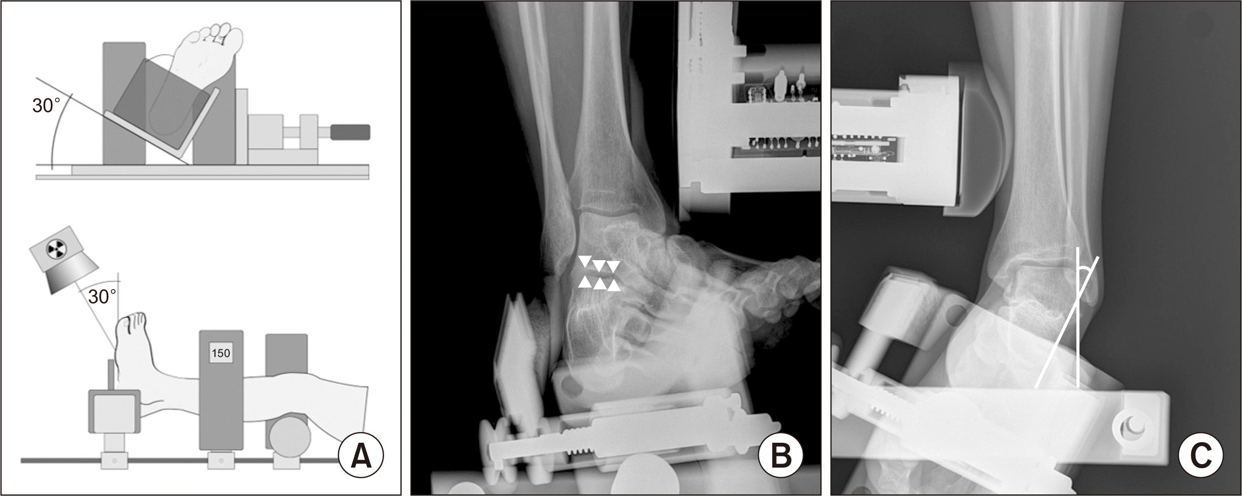
Karlsson 등26)은 족관절과 거골하관절이 동시에 보이는 일반적 족관절 전후방 부하 방사선을 사용하여 거종골관절면이 전후방 사진에서 건측에 비해 2 mm 이상 증가하는 것을 거종골간 불안정성이 있다고 정의하였다. Louwerens 등27)은 거골과 종골의 평행성이 무너지는 것을 거골하관절 불안정성의 진단으로 사용하였으며, Heilman 등28)은 Broden’s view 부하 검사에서 거골과 종골의 후돌기 사이 간격이 5 mm 이상 증가하는 것을 진단 기준으로 제시하였다. Broden’s view28,29)를 이용한 검사가 일반적인 전후방, 외측 단순 방사선 검사보다 거골하관절을 평가하기 좋다. 이 방법을 이용하면 평행한 후방관절을 명확하게 관찰할 수 있다(Fig. 2B).
Telos를 통한 부하 검사가 거골하관절 불안정성의 검사에 도움이 될 수 있는데, 거종골간 각이 5도 이상이거나, 거골의 내측 전위가 5 mm 이상이 확인되면 불안정성을 나타낸다는 보고가 있으며,30) 건측과 비교하였을 때 거종골간 각이 5도 이상 차이가 난다면 불안정성을 시사한다는 논문도 있다(Fig. 2).31,32)
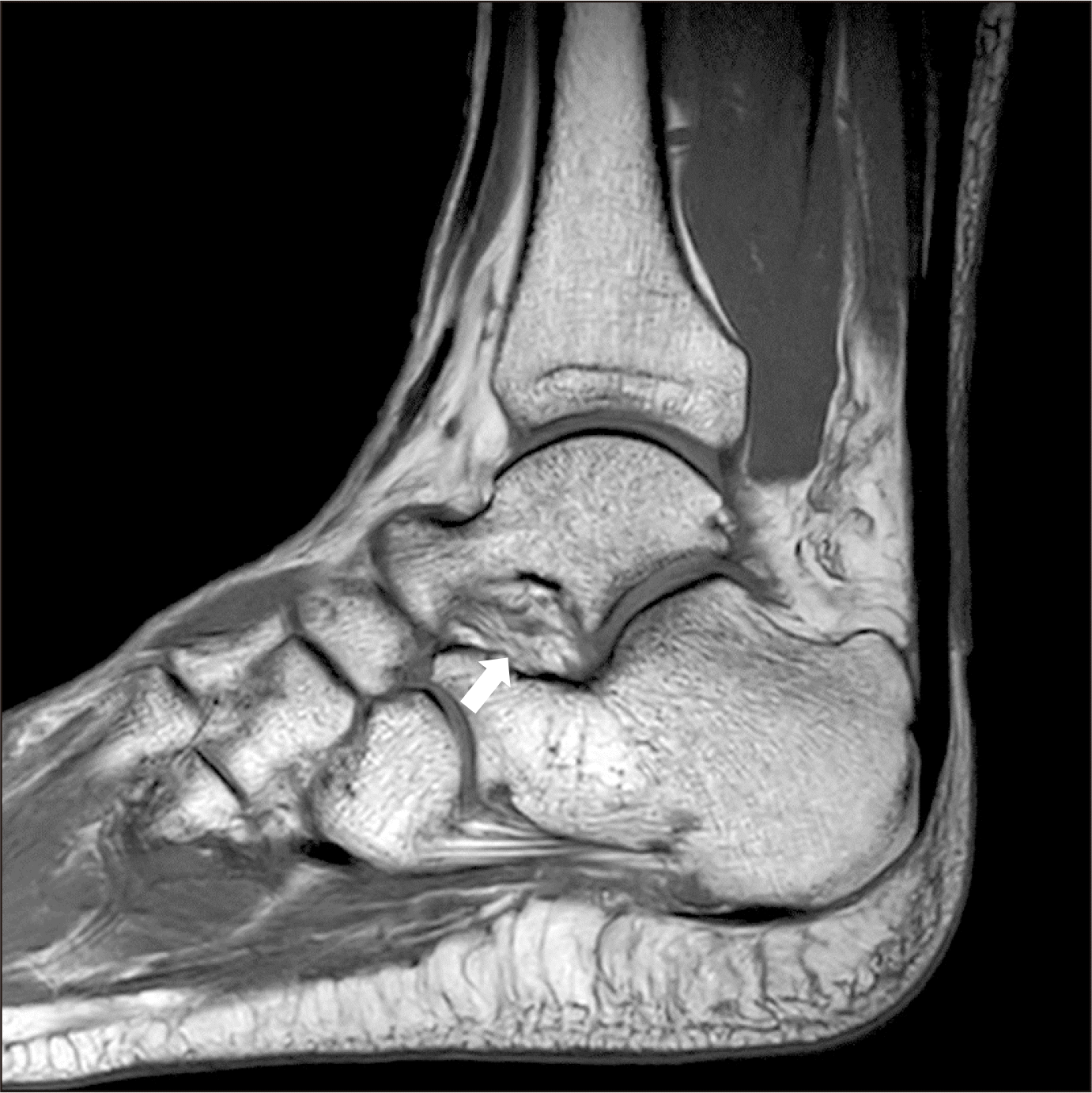
진단을 위한 검사에서는 MRI가 인대의 성상과 손상 정도, 구조를 확인하는 중요한 검사가 되며(Fig. 3) 거골하관절 불안정성이 의심되는 환자에서 전방 관절낭 인대와 경부인대의 손상 정도를 중점적으로 보아야 할 것이다. 거종골간인대 손상도 확인할 수 있으며 거골과 종골의 후방부위가 관절면에 따라 전방으로 전위되어 있는 것도 확인할 수 있다. 또한 리도카인을 거골동에 주사하여 동통의 변화를 알아보는 리도카인 검사 또한 통증의 유발 부위를 확인하고 수술 전 거골하관절 손상을 의심해볼 수 있는 중요한 검사 중에 하나이다.30) 거골하관절경 검사에서는 거종골간인대 손상 및 전방 관절낭 인대 및 경부인대 손상도 쉽게 발견된다.31-33)

치 료

1. 비수술적 치료

거골하관절 불안정성의 치료는 만성 족관절 불안정성의 치료에서와 마찬가지로 비골건 강화운동, 고유수용감각훈련, 뒤꿈치의 외측 쐐기(lateral heel wedge)와 같은 깔창이나 신발의 변형, 테이핑, 보조기 착용 등의 비수술적 치료가 우선된다.34) 특히, 인대의 결함에 의한 기계적 불안정성의 경우에는 수술적 치료를 고려할 수 있지만 수술 결정 이전에 6개월 이상의 비수술적 치료를 선행하는 것이 바람직하다. 비수술적 재활 치료운동은 관절가동운동, 근력강화운동, 균형감각 회복 및 기능적 운동 등이 있다. 이 중 균형감각 훈련이 환자 스스로 불안정성에 대해 평가하기에는 가장 효과적이며 염좌의 재발 방지에도 효과가 있다.35)

2. 수술적 치료

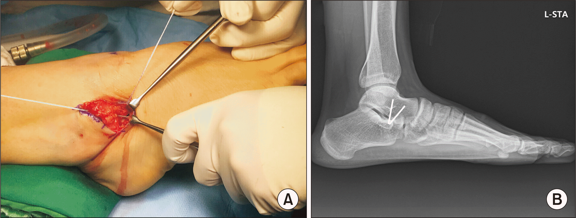
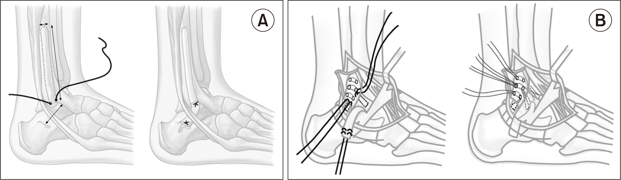
충분한 기간의 비수술적 치료를 시행하고도 거골하관절의 불안정성이 확인되며 증상이 경감되지 않고 거골동 리도카인 검사 양성인 경우, 관절의 안정성을 높이고 관절염의 예방하는 것을 목적으로 수술적 치료를 시행할 수 있다. 파열된 조직의 연속성을 회복하는 봉합(repair)과 결손조직을 기능적으로 보완하기 위해 원래의 조직이 아닌 다른 조직을 이용하는 재건(reconstruction)이 있다. 재건은 해부학적 재건(anatomical reconstruction)과 비해부학적 재건(non-anatomical reconstruction)으로 구분할 수 있다. 손상된 인대를 봉합하는 해부학적 봉합은 통상적으로 늘어난 인대를 단축시켜 봉합하는 것과 봉합나사(suture-anchor) 등을 이용하여 손상된 인대를 골에 재부착하는 것으로 근처의 국소조직과 인대를 같이 봉합하여 봉합 부위를 튼튼하게 만드는 비해부학적 보강의 개념이 포함된다(Fig. 4). 거골하관절 불안정성에도 Brostrom-Gould36) 술기가 사용될 수 있으며 인접한 신전 지대(extensor retinaculum)를 덮어 보강하는 술식으로 보강술에 해당한다.
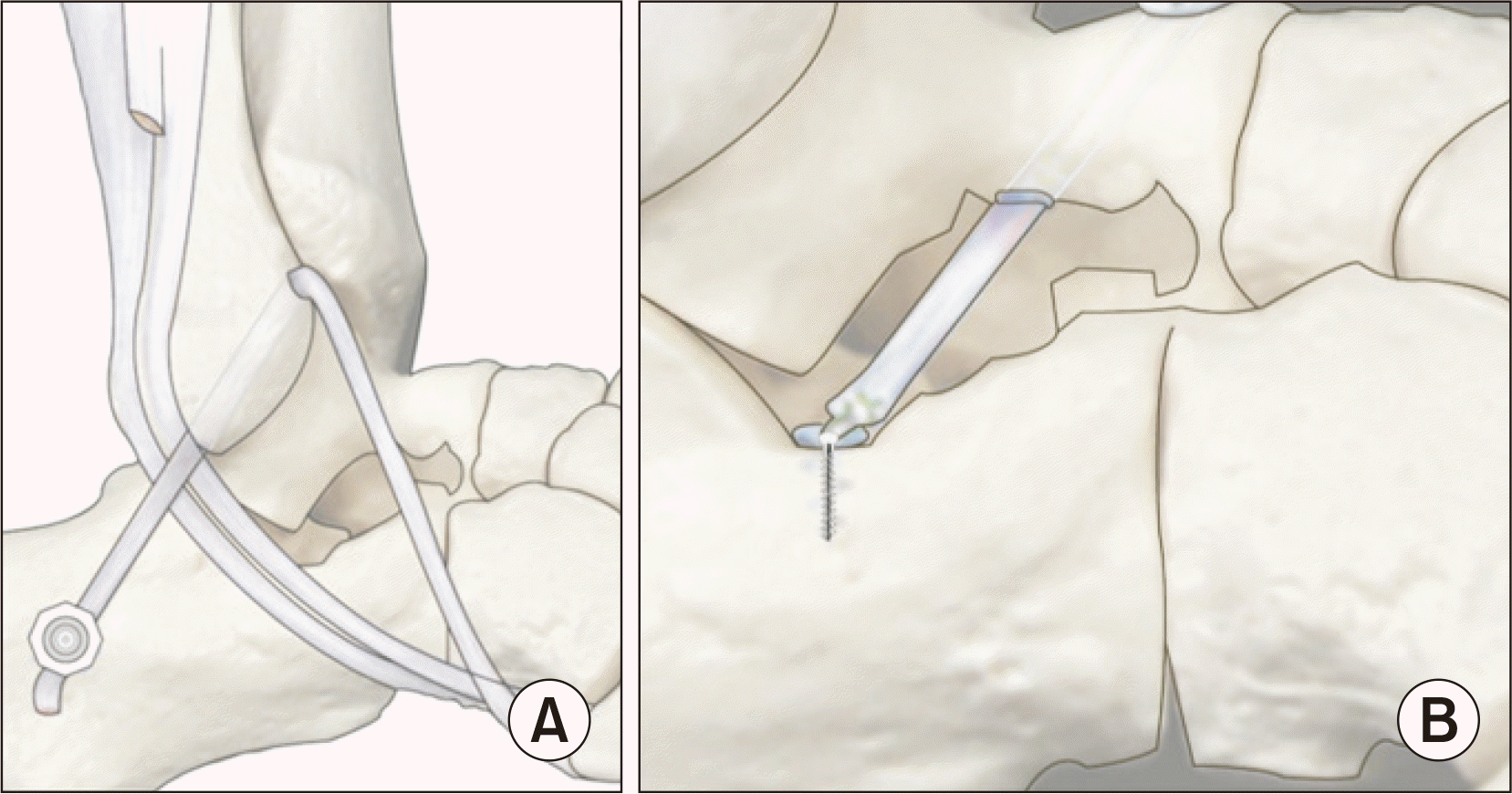
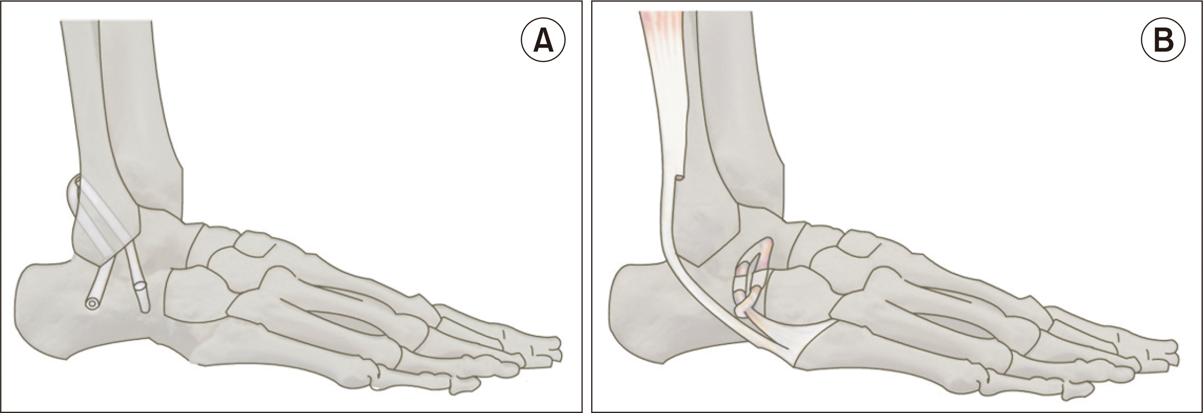
재건술은 조직의 이식을 동반하며 해부학적 재건은 이식건을 사용하여 거골과 종골의 해부학적 구조물을 종비인대, 전방 관절낭 인대, 경부인대의 부착부를 찾아 새로 만들어주는 수술로 골터널의 위치, 개수 등에 따라 여러 가지 방법이 시도되고 있다. 외측 인대 복합체는 해부학적 다양성이 큰 편이며, 이식건이 파열된 원래 인대 조직과 같은 방향과 모양으로 정확하게 이식되기는 어렵다. 비해부학적 재건은 주변의 건, 조직의 이전이나 이식을 통하여 손상된 인대의 기능과 유사한 역할을 해서 관절의 과도한 움직임을 제한하는 방법으로 아킬레스건이나 비골건 혹은 동종건 등을 이용하여 고정한다(Fig. 57). 일반적으로 해부학적 봉합술이 비해부학적 재건술보다 선호되나5,37,38) 해부학적 봉합술과 비해부학적 재건술 모두 좋은 결과를 보이고 있다(Table 3).9,24,26,30,32,39-42)
만성 족관절 불안정성과 동시에 거골하관절 불안정성이 있을 때는 종비인대의 재건이 안정성을 회복하는 데 중요한 요소이며 종비인대가 위축되거나 늘어났을 경우 종비인대를 적절하게 단축하거나 골 간 고정(transosseous fixation)을 시행하는 방법도 있다. 종비인대의 남은 조직이 빈약할 때는 비골 골막 피판을 이용하여 재건하기도 한다(Fig. 5A).

고 찰

거골하관절 인대의 해부학에 관한 연구는 아직 완전하지 않다.17,18,20) 여러 인대의 용어에 대해 아직까지 혼란이 있으며 이외에도 거골하관절의 안정성에 기여하는 여러 인대의 각각의 기능에 대한 연구가 더 필요하다.18,33)
종비인대는 족관절과 거골하관절의 안정에 중요한 역할을 한다.14,43) 그러나 거골하관절의 안정성에 대한 기여 정도는 여전히 논쟁의 여지가 있다. 일부 저자들은 거골하관절의 안정에 거종골간인대가 중요한 역할을 한다고 보고했지만12,14,44) 경부인대가 거골하관절 불안정성에 더 큰 기여를 한다는 의견도 있다.10,41) 전방 관절낭 인대는 처음에 후방관절의 전측 관절막의 두꺼워진 부분으로 설명되었다. Stephens와 Sammarco45)는 전방 관절낭 인대가 모든 자세에서 거골하관절의 안정성에 중요한 역할을 하고 있다고 밝혔다. 최근 MRI를 이용한 연구에서는 전방 관절낭 인대의 손상과 거골하관절 불안정성 사이의 상관관계가 확인되었다.33,46)
거골하관절의 기능은 본질적으로 족관절의 기능과 관련이 있으며 아직 잘 이해되지 않은 상호 보상 메커니즘이 있다. 내전 손상 후 높은 유병률을 가진다고 생각되나 거골하관절 불안정성은 종종 조기에 진단되지 않으며 만성 거골하관절 불안정성의 후기 단계에서 임상적으로 명백하게 나타난 이후에 진단된다.
적절한 치료를 결정하기 위해 임상적 지표, 기능적 지표 및 영상의학적 지표를 포함하는 알고리즘에 기반한 진단이 필요하다. 이를 위해서는 추후에 족관절과 거골하관절의 3차원적 동작 분석 및 두 관절의 상호 작용에 대한 연구가 필요하다.

결 론

거골하관절 불안정성은 후족부의 내번 및 외전 손상에 의해 발생하는 것으로 생각된다. 불안정성이 비골건의 약화 및 족관절의 균형 감각 이상에 의한 원인이 아니면 거골하관절 불안정성을 강력히 의심해 볼 수 있다. 종비인대가 일차적 중요한 인대로 생각되며 거골하 불안정성이 진단되었을 경우 종비인대의 파열 여부를 진단하고 치료해야 한다. 내재적 인대 중에서는 거종골간인대 외에도 경부인대 및 전방 관절낭 인대가 최근 중요하게 인식되고 있다. 불안정성의 검사는 부하 검사로 전후방 부하 검사, Broden’s 부하 검사 등이 도움될 것으로 생각된다. 치료는 일차적으로 비수술적 치료를 권장하며 비수술적 치료가 실패했을 시에 해부학적 및 비해부학적인 재건술 등을 시행할 수 있다. 추후 추가적인 진단 방법과 치료법에 대한 연구가 지속적으로 필요할 것으로 생각된다.

Notes

Financial support: None.

Conflict of interest: None.

REFERENCES

1. Usuelli FG, Mason L, Grassi M, Maccario C, Ballal M, Molloy A. 2014; Lateral ankle and hindfoot instability: a new clinical based classification. Foot Ankle Surg. 20:231–6. doi: 10.1016/j.fas.2014.05.003. DOI: 10.1016/j.fas.2014.05.003. PMID: 25457657.
crossref pmid
2. Michels F, Clockaerts S, Van Der Bauwhede J, Stockmans F, Matricali G. 2020; Does subtalar instability really exist? A systematic review. Foot Ankle Surg. 26:119–27. doi: 10.1016/j.fas.2019.02.001. DOI: 10.1016/j.fas.2019.02.001. PMID: 30827926.
crossref pmid
3. Michels F, Pereira H, Calder J, Matricali G, Glazebrook M, Guillo S, et al. 2018; Searching for consensus in the approach to patients with chronic lateral ankle instability: ask the expert. Knee Surg Sports Traumatol Arthrosc. 26:2095–102. doi: 10.1007/s00167-017-4556-0. DOI: 10.1007/s00167-017-4556-0. PMID: 28439639.
crossref pmid
4. Krähenbühl N, Weinberg MW, Davidson NP, Mills MK, Hintermann B, Saltzman CL, et al. 2019; Currently used imaging options cannot accurately predict subtalar joint instability. Knee Surg Sports Traumatol Arthrosc. 27:2818–30. doi: 10.1007/s00167-018-5232-8. DOI: 10.1007/s00167-018-5232-8. PMID: 30367197.
crossref pmid
5. Barg A, Tochigi Y, Amendola A, Phisitkul P, Hintermann B, Saltzman CL. 2012; Subtalar instability: diagnosis and treatment. Foot Ankle Int. 33:151–60. doi: 10.3113/FAI.2012.0151. DOI: 10.3113/FAI.2012.0151. PMID: 22381348.
crossref pmid
6. Rubin G, Witten M. 1962; The subtalar joint and the symptom of turning over on the ankle: a new method of evaluation utilizing tomography. Am J Orthop. 4:16–9.
7. Gillespie HS, Boucher P. 1971; Watson-Jones repair of lateral instability of the ankle. J Bone Joint Surg Am. 53:920–4. DOI: 10.2106/00004623-197153050-00007. PMID: 5557605.
crossref pmid
8. Brantigan JW, Pedegana LR, Lippert FG. 1977; Instability of the subtalar joint. Diagnosis by stress tomography in three cases. J Bone Joint Surg Am. 59:321–4. DOI: 10.2106/00004623-197759030-00005. PMID: 849942.
crossref pmid
9. Larsen E. 1988; Tendon transfer for lateral ankle and subtalar joint instability. Acta Orthop Scand. 59:168–72. doi: 10.1080/17453678809169701. DOI: 10.1080/17453678809169701. PMID: 3364187.
crossref pmid
10. Meyer JM, Garcia J, Hoffmeyer P, Fritschy D. 1988; The subtalar sprain. A roentgenographic study. Clin Orthop Relat Res. (226):169–73. DOI: 10.1097/00003086-198801000-00023. PMID: 3335091.
11. Ringleb SI, Dhakal A, Anderson CD, Bawab S, Paranjape R. 2011; Effects of lateral ligament sectioning on the stability of the ankle and subtalar joint. J Orthop Res. 29:1459–64. doi: 10.1002/jor.21407. DOI: 10.1002/jor.21407. PMID: 21445995.
crossref pmid
12. Tochigi Y, Amendola A, Rudert MJ, Baer TE, Brown TD, Hillis SL, et al. 2004; The role of the interosseous talocalcaneal ligament in subtalar joint stability. Foot Ankle Int. 25:588–96. doi: 10.1177/107110070402500813. DOI: 10.1177/107110070402500813. PMID: 15363382.
crossref pmid
13. Li L, Zhong S, Shen X, Li Q, Xu W, Tao Y, et al. 2019; Recent development on liquid chromatography-mass spectrometry analysis of oxidized lipids. Free Radic Biol Med. 144:16–34. doi: 10.1016/j.freeradbiomed.2019.06.006. DOI: 10.1016/j.freeradbiomed.2019.06.006. PMID: 31202785.
crossref pmid
14. Kjaersgaard-Andersen P, Wethelund JO, Nielsen S. 1987; Lateral talocalcaneal instability following section of the calcaneofibular ligament: a kinesiologic study. Foot Ankle. 7:355–61. doi: 10.1177/107110078700700612. DOI: 10.1177/107110078700700612. PMID: 3609987.
crossref
15. Michels F, Matricali G, Vereecke E, Dewilde M, Vanrietvelde F, Stockmans F. 2021; The intrinsic subtalar ligaments have a consistent presence, location and morphology. Foot Ankle Surg. 27:101–9. doi: 10.1016/j.fas.2020.03.002. DOI: 10.1016/j.fas.2020.03.002. PMID: 32169330.
crossref pmid
16. Cahill DR. 1965; The anatomy and function of the contents of the human tarsal sinus and canal. Anat Rec. 153:1–17. doi: 10.1002/ar.1091530102. DOI: 10.1002/ar.1091530102. PMID: 5885336.
crossref pmid
17. Jotoku T, Kinoshita M, Okuda R, Abe M. 2006; Anatomy of ligamentous structures in the tarsal sinus and canal. Foot Ankle Int. 27:533–8. doi: 10.1177/107110070602700709. DOI: 10.1177/107110070602700709. PMID: 16842721.
crossref pmid
18. Li SY, Hou ZD, Zhang P, Li HL, Ding ZH, Liu YJ. 2013; Ligament structures in the tarsal sinus and canal. Foot Ankle Int. 34:1729–36. doi: 10.1177/1071100713500653. DOI: 10.1177/1071100713500653. PMID: 23913369.
crossref pmid
19. Clanton TO, Campbell KJ, Wilson KJ, Michalski MP, Goldsmith MT, Wijdicks CA, et al. 2014; Qualitative and quantitative anatomic investigation of the lateral ankle ligaments for surgical reconstruction procedures. J Bone Joint Surg Am. 96:e98. doi: 10.2106/JBJS.M.00798. DOI: 10.2106/JBJS.M.00798. PMID: 24951749.
crossref
20. Poonja AJ, Hirano M, Khakimov D, Ojumah N, Tubbs RS, Loukas M, et al. 2017; Anatomical study of the cervical and interosseous talocalcaneal ligaments of the foot with surgical relevance. Cureus. 9:e1382. doi: 10.7759/cureus.1382. DOI: 10.7759/cureus.1382. PMID: 28775922. PMCID: PMC5522015.
crossref
21. Yamaguchi R, Nimura A, Amaha K, Yamaguchi K, Segawa Y, Okawa A, et al. 2018; Anatomy of the tarsal canal and sinus in relation to the subtalar joint capsule. Foot Ankle Int. 39:1360–9. doi: 10.1177/1071100718788038. DOI: 10.1177/1071100718788038. PMID: 30052059.
crossref pmid
22. Karlsson J, Eriksson BI, Renström PA. 1997; Subtalar ankle instability. A review. Sports Med. 24:337–46. doi: 10.2165/00007256-199724050-00005. DOI: 10.2165/00007256-199724050-00005. PMID: 9368279.
pmid
23. Meyer JM, Lagier R. 1977; Post-traumatic sinus tarsi syndrome. An anatomical and radiological study. Acta Orthop Scand. 48:121–8. doi: 10.3109/17453677708985122. DOI: 10.3109/17453677708985122. PMID: 868481.
crossref pmid
24. Pisani G. 1996; Chronic laxity of the subtalar joint. Orthopedics. 19:431–7. doi: 10.3928/0147-7447-19960501-13. PMID: 8727337.
pmid
25. Mittlmeier T, Rammelt S. 2018; Update on subtalar joint instability. Foot Ankle Clin. 23:397–413. doi: 10.1016/j.fcl.2018.04.005. DOI: 10.1016/j.fcl.2018.04.005. PMID: 30097081.
crossref pmid
26. Karlsson J, Eriksson BI, Renström P. 1998; Subtalar instability of the foot. A review and results after surgical treatment. Scand J Med Sci Sports. 8:191–7. doi: 10.1111/j.1600-0838.1998.tb00191.x. DOI: 10.1111/j.1600-0838.1998.tb00191.x. PMID: 9764439.
crossref pmid
27. Louwerens JW, Ginai AZ, van Linge B, Snijders CJ. 1995; Stress radiography of the talocrural and subtalar joints. Foot Ankle Int. 16:148–55. doi: 10.1177/107110079501600308. DOI: 10.1177/107110079501600308. PMID: 7599733.
crossref pmid
28. Heilman AE, Braly WG, Bishop JO, Noble PC, Tullos HS. 1990; An anatomic study of subtalar instability. Foot Ankle. 10:224–8. doi: 10.1177/107110079001000407. DOI: 10.1177/107110079001000407. PMID: 2307380.
crossref pmid
29. Broden B. 1949; Roentgen examination of the subtaloid joint in fractures of the calcaneus. Acta radiol. 31:85–91. doi: 10.3109/00016924909136638. DOI: 10.3109/00016924909136638. PMID: 18114628.
crossref pmid
30. Pisani G, Pisani PC, Parino E. 2005; Sinus tarsi syndrome and subtalar joint instability. Clin Podiatr Med Surg. 22:63–77. vii doi: 10.1016/j.cpm.2004.08.005. DOI: 10.1016/j.cpm.2004.08.005. PMID: 15555844.
crossref pmid
31. Sugimoto K, Isomoto S, Samoto N, Okahashi K, Araki M. 2017; Recent developments in the treatment of ankle and subtalar instability. Open Orthop J. 11:687–96. doi: 10.2174/1874325001711010687. DOI: 10.2174/1874325001711010687. PMID: 28979582. PMCID: PMC5620400.
crossref pmid pmc
32. Jung HG, Park JT, Shin MH, Lee SH, Eom JS, Lee DO. 2015; Outcome of subtalar instability reconstruction using the semitendinosus allograft tendon and biotenodesis screws. Knee Surg Sports Traumatol Arthrosc. 23:2376–83. doi: 10.1007/s00167-015-3504-0. DOI: 10.1007/s00167-015-3504-0. PMID: 25577222.
crossref pmid
33. Kim TH, Moon SG, Jung HG, Kim NR. 2017; Subtalar instability: imaging features of subtalar ligaments on 3D isotropic ankle MRI. BMC Musculoskelet Disord. 18:475. doi: 10.1186/s12891-017-1841-5. DOI: 10.1186/s12891-017-1841-5. PMID: 29162077. PMCID: PMC5696773.
crossref pmid pmc
34. Thermann H, Zwipp H, Tscherne H. 1997; Treatment algorithm of chronic ankle and subtalar instability. Foot Ankle Int. 18:163–9. doi: 10.1177/107110079701800309. DOI: 10.1177/107110079701800309. PMID: 9116897.
pmid
35. McKeon PO, Hertel J. 2008; Systematic review of postural control and lateral ankle instability, part II: is balance training clinically effective? J Athl Train. 43:305–15. doi: 10.4085/1062-6050-43.3.305. DOI: 10.4085/1062-6050-43.3.305. PMID: 18523567. PMCID: PMC2386424.
crossref pmid pmc
36. Gould N, Seligson D, Gassman J. 1980; Early and late repair of lateral ligament of the ankle. Foot Ankle. 1:84–9. doi: 10.1177/107110078000100206. DOI: 10.1177/107110078000100206. PMID: 7274903.
crossref pmid
37. Tourné Y, Mabit C. 2017; Lateral ligament reconstruction procedures for the ankle. Orthop Traumatol Surg Res. 103(1S):S171–81. doi: 10.1016/j.otsr.2016.06.026. DOI: 10.1016/j.otsr.2016.06.026. PMID: 27871968.
crossref
38. Tourné Y, Mabit C, Moroney PJ, Chaussard C, Saragaglia D. 2012; Long-term follow-up of lateral reconstruction with extensor retinaculum flap for chronic ankle instability. Foot Ankle Int. 33:1079–86. doi: 10.3113/FAI.2012.1079. DOI: 10.3113/FAI.2012.1079. PMID: 23199857.
crossref pmid
39. Chrisman OD, Snook GA. 1969; Reconstruction of lateral ligament tears of the ankle. An experimental study and clinical evaluation of seven patients treated by a new modification of the Elmslie procedure. J Bone Joint Surg Am. 51:904–12. DOI: 10.2106/00004623-196951050-00007. PMID: 4978936.
pmid
40. Schon LC, Clanton TO, Baxter DE. 1991; Reconstruction for subtalar instability: a review. Foot Ankle. 11:319–25. doi: 10.1177/107110079101100511. DOI: 10.1177/107110079101100511. PMID: 2037273.
crossref pmid
41. Kato T. 1995; The diagnosis and treatment of instability of the subtalar joint. J Bone Joint Surg Br. 77:400–6. doi: 10.1302/0301-620X.77B3.7744924. DOI: 10.1302/0301-620X.77B3.7744924. PMID: 7744924.
crossref pmid
42. Liu C, Jiao C, Hu Y, Guo QW, Wand C, Ao Y. 2011; Interosseous talocalcaneal ligament reconstruction with hamstring autograft under subtalar arthroscopy: case report. Foot Ankle Int. 32:1089–94. doi: 10.3113/FAI.2011.1089. DOI: 10.3113/FAI.2011.1089. PMID: 22338961.
crossref pmid
43. Li L, Gollhofer A, Lohrer H, Dorn-Lange N, Bonsignore G, Gehring D. 2019; Function of ankle ligaments for subtalar and talocrural joint stability during an inversion movement - an in vitro study. J Foot Ankle Res. 12:16. doi: 10.1186/s13047-019-0330-5. DOI: 10.1186/s13047-019-0330-5. PMID: 30923576. PMCID: PMC6421682.
crossref pmid pmc
44. Knudson GA, Kitaoka HB, Lu CL, Luo ZP, An KN. 1997; Subtalar joint stability. Talocalcaneal interosseous ligament function studied in cadaver specimens. Acta Orthop Scand. 68:442–6. doi: 10.3109/17453679708996259. DOI: 10.3109/17453679708996259. PMID: 9385243.
crossref pmid
45. Stephens MM, Sammarco GJ. 1992; The stabilizing role of the lateral ligament complex around the ankle and subtalar joints. Foot Ankle. 13:130–6. doi: 10.1177/107110079201300304. DOI: 10.1177/107110079201300304. PMID: 1601340.
crossref pmid
46. Yoon DY, Moon SG, Jung HG, Kim NR. 2018; Differences between subtalar instability and lateral ankle instability focusing on subtalar ligaments based on three dimensional isotropic magnetic resonance imaging. J Comput Assist Tomogr. 42:566–73. doi: 10.1097/RCT.0000000000000717. DOI: 10.1097/RCT.0000000000000717. PMID: 29727309.
crossref pmid

Figure. 1
(A) Lateral ligaments (a: ITCL, b: ATCL, c: OTCL, d: bifurcate ligament, e: CFL). (B) Insertion point of 3 components of IER (1: medial, 2: intermediate, 3: lateral). (C) Shows how IER blend with ITCL and OTCL (i: ITCL, ii: OTCL). IER: inferior external retinaculum, ITCL: interosseous talocalcaneal ligament, ATCL: anterior talocalcaneal ligament, OTCL: oblique talocalcaneal ligament, CFL: calcaneofibular ligament.
jkfas-25-1-38-f1.tif
Figure. 2
(A) Telos device standardized stress view of subtalar joint. Foot is internal rotated at 30°, X-ray beam is tilted plantarward 30°. Applied force is limited to 150 N. (B) Clinical example of patient with stable subtalar joint. Parallelism between talus and calcaneal posterior facet can be seen on Broden’s stress view (arrowheads). (C) Clinical example of a patient with chronic subtalar instability, in which a talocalcaneal tilt 25°.
jkfas-25-1-38-f2.tif
Figure. 3
Sagittal T1-weighted magnetic resonance images demonstrate the ruptured cervical ligament (white arrow).
jkfas-25-1-38-f3.tif
Figure. 4
(A) Suture-anchor inserted at lateral calcaneus for repair and augmentation of cervical ligament and inferior external retinaculum. (B) V-shape repair and augmentation of cervical ligament with suture anchor.
jkfas-25-1-38-f4.tif
Figure. 5
(A) Calcaneofibular ligament augmentation via autologous periosteal flap that is mobilized and pedicled at the distal fibula. (B) Anatomic repair including inferior external retinaculum reinforcement.
jkfas-25-1-38-f5.tif
Figure. 6
(A) Extra-anatomic reconstruction using pedicled tendon autograft from peroneus brevis tendon in a modified Elmslie ligamentoplasty. (B) Reconstruction of interosseous talocalcaneal ligament with free hamstrings tendon autograft.
jkfas-25-1-38-f6.tif
Figure. 7
(A) Reconstruction of OTCL, ITCL, and CFL with semitendinosus tendon allograft. (B) Reconstruction of ITCL via pedicled peroneus brevis tendon autograft. OTCL: oblique talocalcaneal ligament, ITCL: interosseous talocalcaneal ligament, CFL: calcaneofibular ligament.
jkfas-25-1-38-f7.tif
Table 1
Overview of Studies Assessing the Dimensions of ITCL, CL, and ACaL
Study No. of specimens ITCL (mm) CL (mm) ACaL (mm)
Cahill16) 5 embalmed Length: 15
Width: 5.6
Thickness: 1.6
Length: 19.6
Width: 11.6
Thickness: 2.8
NS
Jotoku et al.17) 40 embalmed Bandtype
Length: 10.0±3.5
Width: 8.5±2.7
Thickness: 2.3±0.7
NS Length: 8.3±1.6
Width: 8.3±1.7
Thickness: 1.4±0.4
Fantype
Length: 7.5±2.1
Width: 11.0±1.3
Thickness: 3.0
Multiple type
Length: 7.0
Width: 8.5
Thickness: 3.0
Li et al.18) 32 fresh-frozen Length: 10.6±1.2
Width: 7.5±1.2
Thickness: 1.5±0.2
Length: 13.3±1.9
Width: 9.5±1.6
Thickness: 2.6±0.5
Length: 13.6±2.0
Width: 10.1±1.4
Thickness: 2.4±0.3
Clanton et al.19) 14 fresh-frozen NS Length: 15.2 NS
Poonja et al.20) 26 formalin-fixed NS Anterior length: 8.27±2.52
Superior: 0.62±0.24
Inferior: 0.70±0.26
NS
Posterior length: 13.95±5.96
Superior: 1.05±0.30
Inferior: 1.20±0.34
Width: 9.15±2.45
Yamaguchi et al.21) 21 embalmed Footprint calcaneum included medial root IER
Width: 16.8±1.8
Footprint calcaneum
Width: 7.2±2.3
Length: 10.8±2.7
NS
Michels et al.15) 16 fresh-frozen Length
Lateral: 9.1±1.9
Medial: 7.5±1.7
Length
Anterior: 13.9±1.5
Posterior: 18.5±2.9
Length
Lateral: 11.4±1.0
Medial: 6.9±1.0
Width
Talar: 13.3±4.7
Calcaneal: 10.1±2.3
Thickness: 1.1±0.9
Width
Talar: 13.6±2.2
Calcaneal: 15.8±3.7
Thickness: 1.7±0.7
Width
Talar: 10.1±2.0
Calcaneal: 12.1±2.4

Values are presented as mean only or mean±standard deviation.

ITCL: interosseous talocalcaneal ligament, CL: cervical ligament, ACaL: anterior capsular ligament, NS: not stated, IER: inferior external retinaculum.

Table 2
Meyer and Lagier’s Classification of Subtalar Joint Instability
Type Ankle position Involved ligament
I Ankle plantarflexion CL+ACaL+CFL
II Ankle plantarflexion CL+ITCL
III Ankle dorsiflexion CFL+CL+ITCL
IV Ankle plantarflexion (severe injuries) Multiple ligaments

CL: cervical ligament, ACaL: anterior capsular ligament, CFL: cal­caneofibular ligament, ITCL: interosseous talocalcaneal ligament.

Table 3
Outcome Following Surgical Treatment of Subtalar Joint Instability
Author (year) Technique No. of patients Excellent or good results (%)
Larsen (1988)9) PB 25 93
Chrisman and Snook (1969)39) PB 3 100
Schon et al. (1991)40) Modified Elmslie NS NS
Pisani (1996)24); Pisani et al. (2005)30) ITCL anatomic 47 91
Karlsson et al. (1998)26) Anatomic+IER reinforcement 22 82
Kato (1995)41) ITCL or triligamentous 14 (2*/12) >90
Thermann et al. (1997)34) Chrisman–Snook 34 91
Liu et al. (2011)42) Hamstrings autograft 1 100
Jung et al. (2015)32) Anatomic ITCL, OTCL, and CFL 20 100

PB: pedicled peroneus brevis graft, NS: not stated, ITCL: interosseous talocalcaneal ligament, IER: inferior external retinaculum, OTCL: oblique talocalcaneal ligament, CFL: calcaneofibular ligament.

*ITCL reconstruction. Triligamentous reconstruction.

TOOLS
Similar articles