

 PDF
PDF Citation
 Print
Print


INTRODUCTION
Although perihilar cholangiocarcinoma (HCCA) represents 50-70% of all types of bile duct carcinomas, it is a very rare disease, with an annual incidence rate no greater than 1:100,000.1 Radical surgery with negative histological margins is needed for long-term survival.2 However, the adjacent location of HCCA to the hepatic artery, portal vein, and hepatic parenchyma complicates complete resection. Moreover, radical surgery for HCCA includes liver resection, bilio-enteric reconstruction, and radical lymph node dissection around the perihilar, retropancreatic, and para-aortic areas. Thus, infrequent occurrence and demanding surgical techniques may complicate the learning curve of surgical expertise for treatment of HCCA.3
Therefore, the current study set out to determine the learning curve of radical surgery for HCCA in a single surgeon working in a tertiary academic hospital and to assess the perioperative outcomes according to surgical proficiency.
MATERIALS AND METHODS
Patients
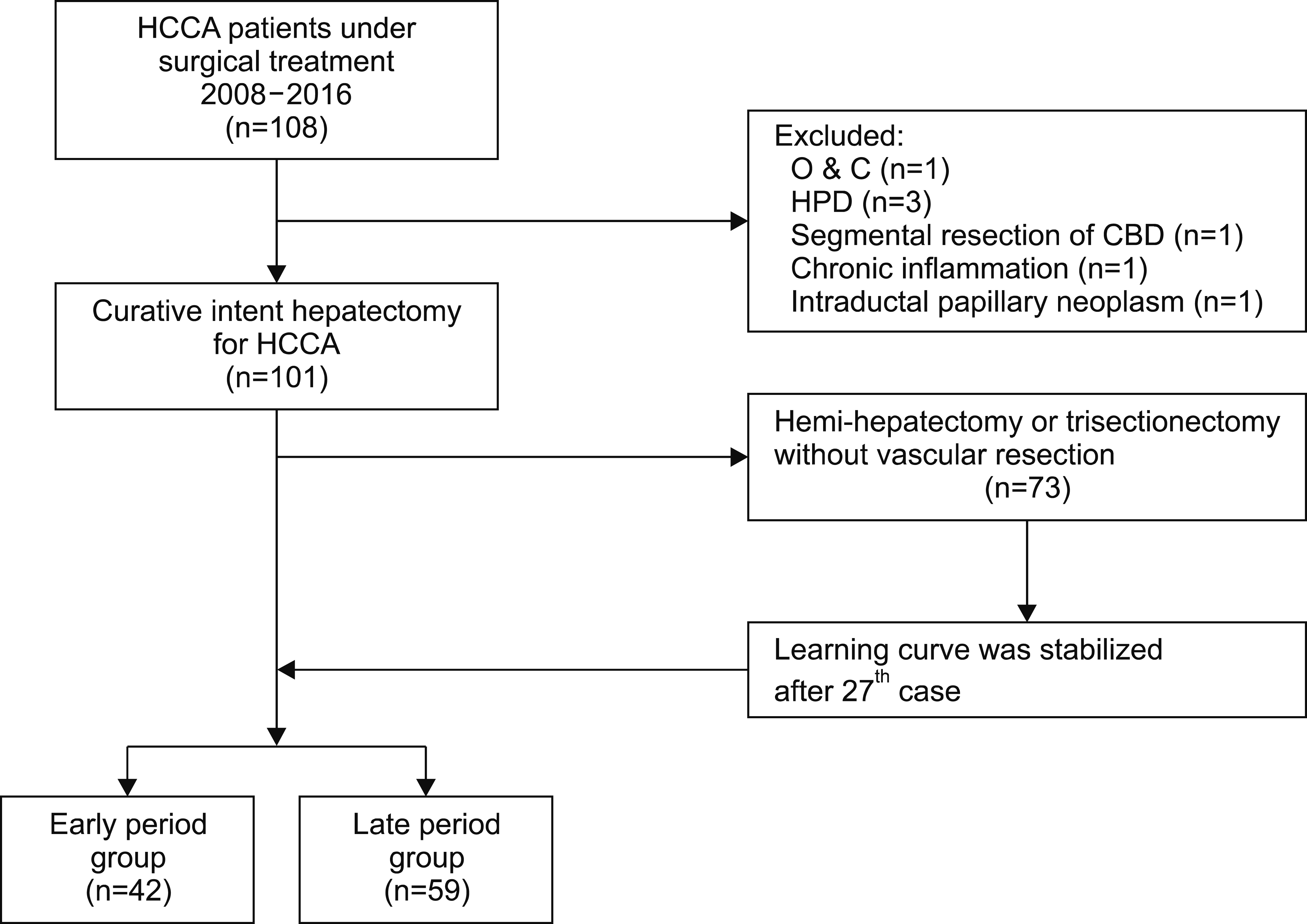
From January 2008 to December 2016, a total of 108 consecutive patients underwent curative-intent surgery for HCCA by a single surgeon (CGH) at Yonsei University College of Medicine, Seoul, Korea. Excluding seven cases (one case with only laparotomy, three cases with hepatopancreaticoduodenectomy, one case with segmental resection of the bile duct, and two cases with benign disease on final pathology report), 101 patients who underwent curative surgery consisting of liver resection, radical lymph node dissection, and Roux-en-Y hepaticojejunostomy for HCCA were included in this study. Therefore, a total of 101 patients was divided into two groups according to learning curve analysis: the early period group (EPG), consisting of 42 patients, and the late period group (LPG), consisting of 59 patients (Fig. 1).
Learning curve analysis
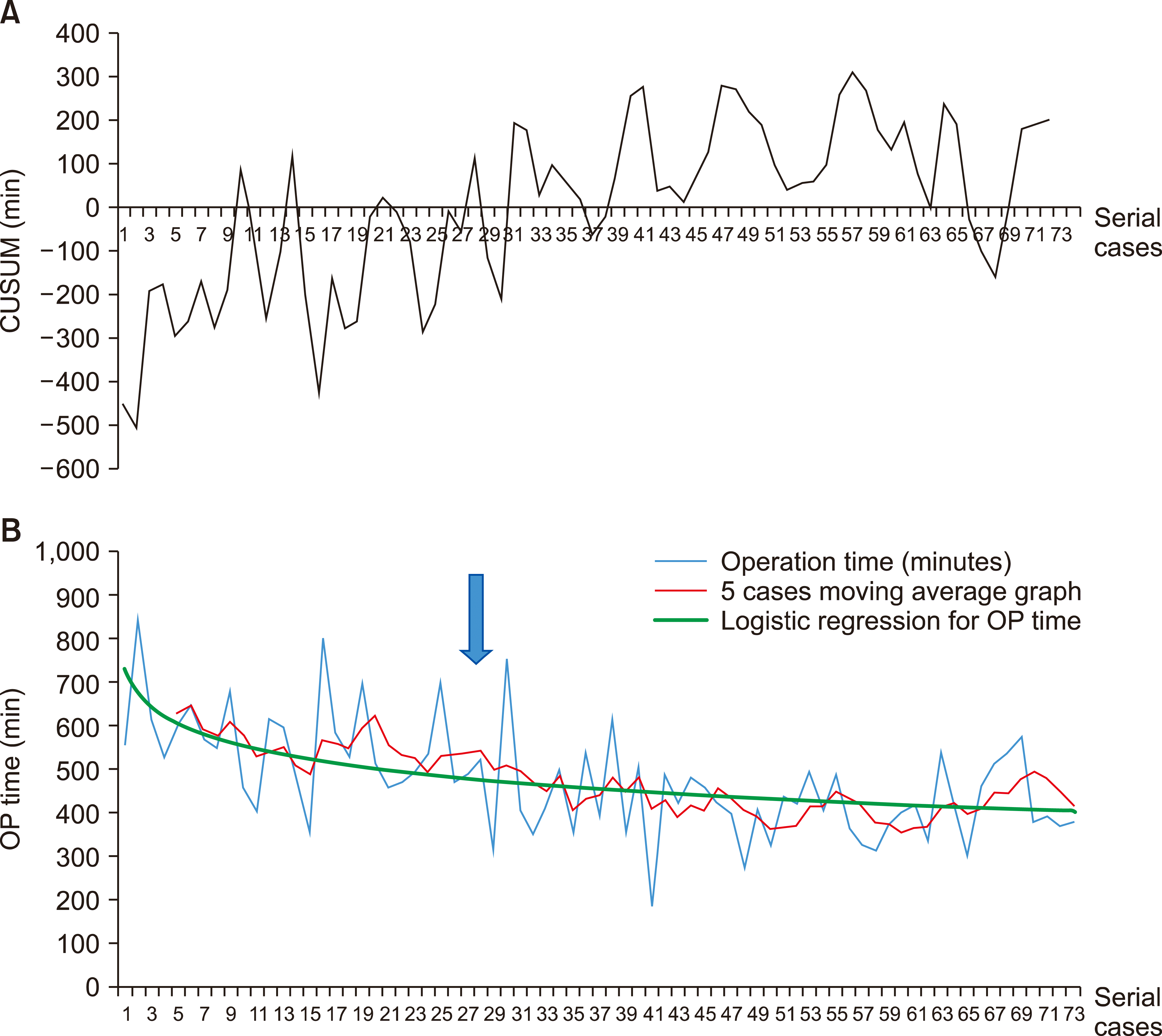
Operative time was used as a variable for learning outcome in this study and was defined as incision-to-closure time. Although the surgical procedure of HCCA was diverse and complex, it is composed of three essential processes: radical lymph node dissection, liver resection, and bilio-enteric anastomosis. Among these three steps, procedural time for liver resection may be affected by extent of the resection plane. Theoretically, central bisectionectomy or anterior sectionectomy might require more than twice the effort for parenchymal dissection compared to a right hemihepatectomy, left hemihepatectomy, right trisectionectomy, or left trisectionectomy, which only need one-side resection of the liver. Moreover, vascular resection and reconstruction to remove the tumor-invaded hepatic artery or portal vein may also prolong the operation time. Therefore, 71 patients who underwent one-side liver resection (hemihepatectomy, extended hemihepatectomy, and trisectionectomy) without vascular resection and anastomosis were selected for learning curve analysis. The learning curve was assessed with the cumulative sum (CUSUM) method and a moving average graph.
CUSUM method
The CUSUM method is a descriptive method that can represent data trends by calculating the serial differences between raw data and the mean value.4 In this study, 71 cases were ordered chronologically from the first to the last, and the CUSUM of the operation time (CUSUMOT) was defined as , where OTi is individual operation time, and OTmean is the mean operation time of the 71 cases. First, CUSUM was obtained by summing the differences between mean operative time and serial operative time. The learning curve was determined by plotting CUSUMOT (Fig. 2A); a CUSUMOT of zero meant that OTi was the same as OTmean. Thus, CUSUMOT greater than zero indicates competency in the procedure. The CUSUMOT was steadily over or near the zero baseline after the 27th case.
Verification of the learning curve with a moving average graph
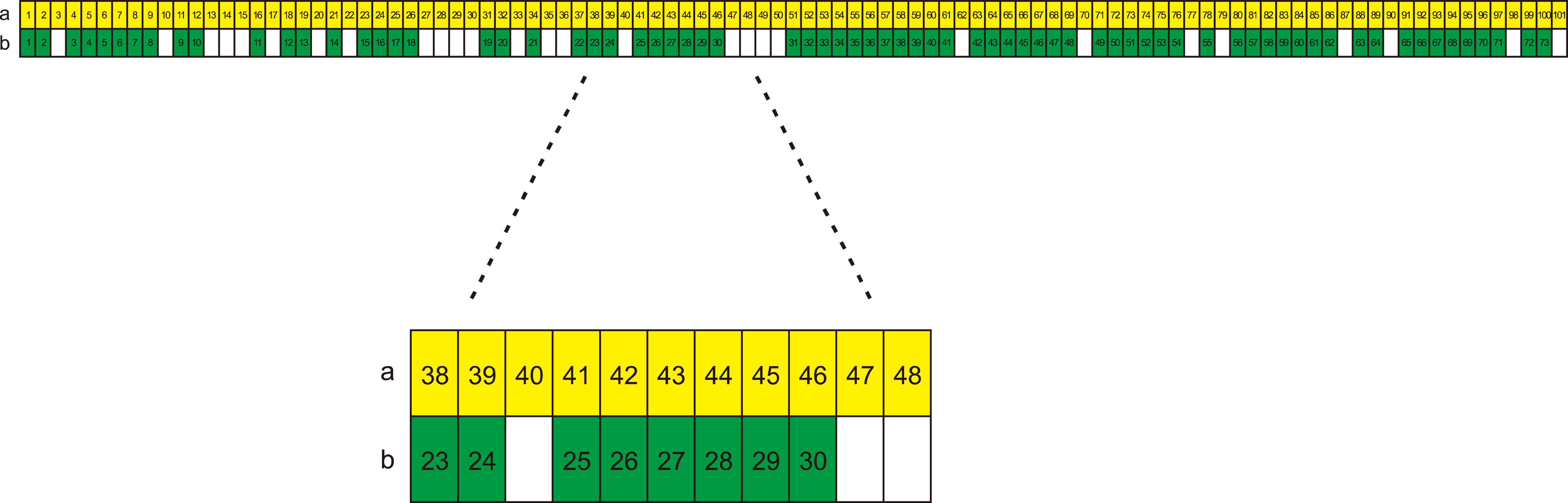
The mean operation time for each of five sequential cases was calculated and plotted. The moving average graph converged toward a plateau after the 27th case. A logarithm graph for serial operation time was created using Microsoft EXCEL 2016 (Microsoft Inc., Redmond, WA, USA) with the formula y=–93.43ln(x)+786.28, which also showed a plateau after 27 cases (Fig. 2B). Therefore, procedure competence was stabilized after the 27th of 71 cases selected for this learning curve analysis. The 27th case in these 71 selected patients was the 43rd of 101 sequential patients (Fig. 3). Therefore, the patients were dichotomized into two groups of EPG (n=42) and LPG (n=59).
Statistical analysis
Normality of continuous variables was tested using Levene’s test. Continuous variables with symmetrical distribution were compared using the independent t-test and presented as mean with standard deviation. Continuous data with asymmetrical distribution were compared using Mann-Whitney U test and described as median value with interquartile range. Categorical variables were compared with the χ2 or Fisher’s exact test as appropriate. Survival was calculated using the Kaplan–Meier method and compared between groups using the log-rank test. Perioperative mortality was included in overall survival analysis but excluded from disease-free survival analysis. Cox regression model with forward stepwise logistic regression analysis was applied for multivariate analysis using significant univariate variables affecting survival. Statistical analyses were performed using SPSS 25 for Windows (SPSS Inc., Chicago, IL, USA). A value of p<0.05 was considered statistically significant.
RESULTS
Patient characteristics
Of the 101 patients, 65 were men (64.4%), and the mean age at operation was 64.46±10.36 years. A total of 32 patients received neoadjuvant chemotherapy (31.7%), and 17 patients underwent preoperative portal vein embolization (16.8%). Bismuth-Corlette type IIIa was the most common type of HCCA (n=34, 33.7%). Right hemi-hepatectomy (n=41, 40.6%) was performed most frequently, followed by left hemihepatectomy (n=32, 31.7%). The most common pathologic stage according to AJCC 8th edition was stage II (n=48, 47.5%), followed by stage IIIB (n=35, 34.7%). Of all patients, 40 had LN metastasis (39.6%), and the R0 resection rate was 80.2% (n=81).
Perioperative outcomes of early and late period groups
There was no significant difference in sex, age, or pre- operative laboratory findings between the two groups. However, patients with complicated preoperative procedures such as neoadjuvant concurrent chemo-radiation therapy (CCRT) and portal vein embolization (PVE) were more common in the LPG (Table 1). Bismuth-Corlette type IV was most common among EPG patients (n=19, 45.2%), while type IIIa was most common in the LPG (n=26, 44.1%), though there was no statistically significant difference between the two groups. AJCC stage II was most common in both groups (EPG: n=20, 47.6%, LPG: n=28, 47.5%).
In terms of type of liver resection, left hemihepatectomy in the EPG (n=13, 31.0%) and right hemihepatectomy in the LPG (n=30, 50.8%) were most frequently performed. There was no significant difference in LN metastasis rate, R0 resection rates or number of retrieved lymph nodes during operation between the two groups (Table 2). However, perioperative outcomes were improved in the LPG, with significantly shorter average operation time (603.17±117.59 vs. 432.03±91.77, p<0.001), significantly decreased bleeding during operation (1127.86± 689.54 ml vs. 613.05±548.31 ml, p<0.001), and significantly decreased length of hospital stay (median hospital stay day: 23 days vs. 18 days, p=0.025). Severe complications greater than grade IIIA according to Clavien- Dindo classification were significantly more frequent in the EPG (47.6% vs. 27.1%, p=0.034). However, there was no significant difference in overall complications rate or perioperative mortality between the two groups (Table 2).
Regarding severe complications, cases of intra-abdominal fluid collection, pleural effusion, and bile leakage lead to percutaneous catheter drainage, and wound infection required secondary wound closure in grade IIIA complications. Patients with grade IV complications were admitted to intensive care units for post-operative bleeding, cholangitis, or intestinal obstruction. Biliary sepsis and aspiration pneumonia were the causes of perioperative mortality.
Survival outcomes
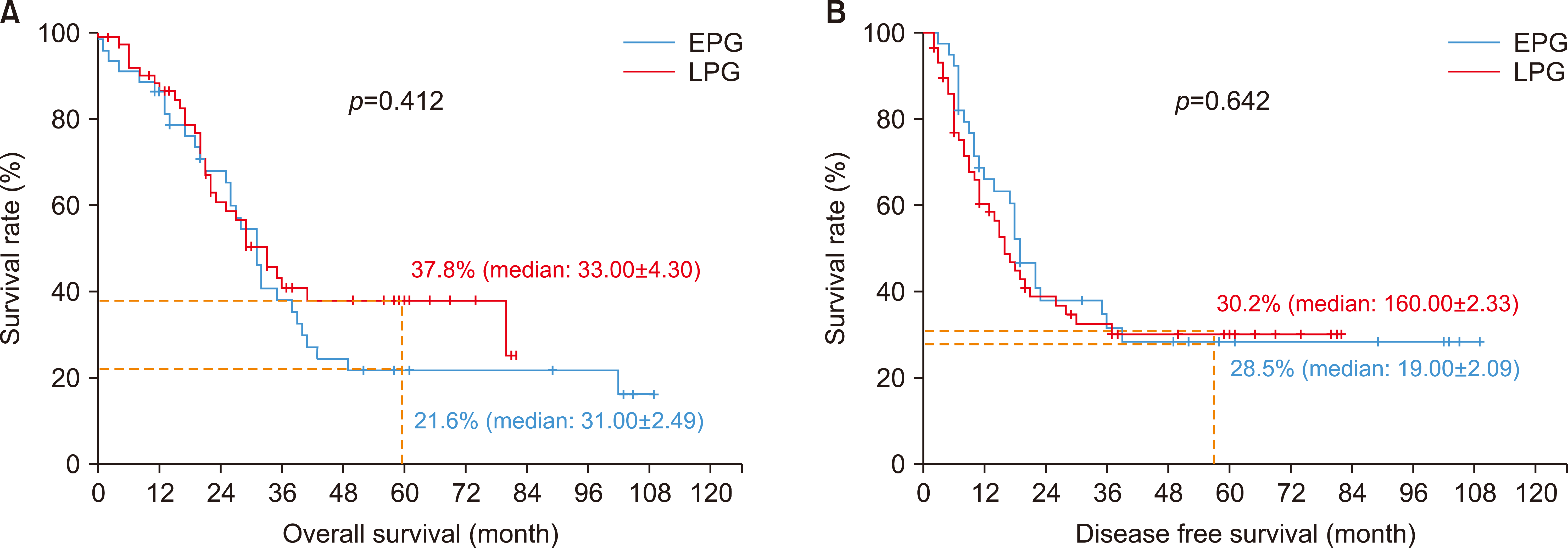
There was no significant difference in disease-free survival (DFS) or overall survival (OVS) rate between the two groups (Fig. 4). The 5-year DFS rate was 28.5% and 30.2% for the EPG and LPG, respectively (p=0.642). The 5-year OVS rate was 21.6% and 37.8% in the EPG and LPG, respectively (p=0.412). Preoperative serum carcinoembryonic antigen (CEA) level over 5 ng/ml and metastasis to lymph node were te significant risk factors for poor DFS rates according to multivariate analysis. The significant poor prognostic factors for OVS rates were high preoperative CEA level over 5 ng/ml and low serum albumin level under 3.5 g/dl (Table 3).
DISCUSSION
HCCA is a very rare disease, and only one-quarter of all patients are suitable for curative resection.5 Thus, it is very difficult to achieve surgical proficiency for HCCA treatment in a short period of time. Moreover, curative-intent operations for HCCA include various complicated surgical procedures.6,7 Along with dismal long-term oncologic outcomes even after curative surgery, perioperative outcomes are relatively poor.8,9 Therefore, such surgical treatment for patients with HCCA is typically performed in tertiary care hospitals. However, there is lack of information regarding the impact of surgical experience on surgical outcome for HCCA. To the best of our knowledge, the present study is the first learning curve study on surgical treatment of HCCA in a tertiary academic hospital.
Various surgical factors, such as operation time and intraoperative blood loss, and dichotomous surgical variables including postoperative complication or mortality may represent learning outcomes. Among these variables, operation time is the most commonly used variable for learning outcomes due to its easy accessibility even in retrospective studies. CUSUM analysis based on operation time may effectively reduce the inherent variability of surgical data and improve understanding of the learning curve.10 The current study also used operation time as a learning outcome. However, complex procedures such as central bisectionectomy or segmental resection of the portal vein or hepatic artery might affect the operation time.2 Therefore, the learning curve was created using data from patients undergoing similar procedures: one-side hepatectomy without vascular resection and anastomosis. Indeed, perioperative outcomes of the LPG were significantly improved compared to those of the EPG. Thus, the current learning curve study appears to reasonable to distinguish an inexperienced period from an experienced period. Indication of the operation was also extended after achieving learning curve. There were significantly more patients with neoadjuvant chemo-radiation therapy or portal vein embolization in the LPG (Table 1).2,11 However, severe complications, operation time, amount of bleeding during operation, and length of hospital stay were significantly reduced in the LPG.
Although the perioperative outcome was significantly improved in the LPG, there was no difference in long- term survival outcome between the LPG and EPG. There were no significant differences in disease-free and overall survival rates between the two groups, even after considering cancer stage in subgroup analyses. In general, the 5-year survival rates were around 30% even after curative resection due to the high chance of recurrence. Even though a few studies have reported that perioperative outcomes such as perioperative blood transfusion were poor prognostic factors for long-term survival,12 most studies have revealed tumor-related factors such as lymph node metastasis, R1 resection, high preoperative carcinoembryonic antigen level, poor differentiation, and vascular invasion as independent risk factors for long-term survival after curative treatment of HCCA.5,12-21 Therefore, improved perioperative outcomes might not necessarily improve the long-term survival outcomes.
This study has some limitations. This was a single-institutional, retrospective study involving a single surgeon. Although more than 100 cases were included, which might be considered a high volume given the rarity of HCCA,22 it is difficult to generalize the results that 40 cases are necessary to achieve surgical proficiency for treatment of HCCA. Therefore, prospective multi-center studies for novice surgeons are needed.
In conclusion, operation time, amount of bleeding during operation, length of hospital stay, and complication rates were improved after stabilization of the surgical learning curve. However, R0 resection rates and survival outcomes were not significantly influenced by the learning curve for surgical treatment of HCCA.
 XML Download