

 PDF
PDF Citation
 Print
Print


INTRODUCTION
Bile duct complications are still a pitfall and have a significant morbidity and mortality risk. Following liver transplantation (LT), leakages, obstructions, strictures, and stones have been observed in 9-30% of large clinical series of LT recipients.1-4
Even with biliary complications, long-term survival can be expected when treated with appropriate intervention. The rapid diagnosis and appropriate intervention of the bile duct complication resolve biliary complication and enable long-term survival of the patients and grafts. The existing diagnostic method is to perform T-tube cholangiography in case of a history of T-tube, or examine the structure or leakage through endoscopic retrograde cholangiopancreatography (ERCP). With the recent development of surgical techniques, most liver transplant centers in Korea do not usually insert T-tube into the biliary tract during operation.5-7
However, these methods may increase the pressure in the bile duct due to the invasive procedure immediately after transplantation, and in the case of complications such as a minor leakage, can promote iatrogenic complications. Moreover, pancreatitis or cholangitis can occur because of the procedure.
Non-invasive diagnostic tools have been developed for biliary complications following a hepatobiliary surgery. Hepatobiliary scan (diisopropyl iminodiacetic acid [DISIDA]) was performed to obtain a functional image of bile excretion.8,9 Alternatively, studies related to the diagnosis of biliary complications using magnetic resonance cholangiopancreatography (MRCP) have been reported. The MRCP revealed stricture and leakage following transplantation and the biliary leakage site could be identified in case of general resection.10-14
However, as the conventional magnetic resonance image (MRI) is severely blunted and moved, the image quality is too poor, it is often impossible to accurately identify the bile duct structure.
Recently, liver MRI using hepatobiliary contrast media such as gadoxetic acid (Gd-EOB-DTPA, Primovist, Bayer- SheringPharma, Berlin, Germany) have been used for the evaluation of various lesions and liver function. However, as the peak hepatic enhancement with gadoxetic acid is usually reached at 20-40 minutes following the injection, there may be a time gap between the dynamic phase imaging and the hepatobiliary phase imaging (HBPI), more clear and accurate bile duct images can be obtained when half of Gd-EOB DTPA is secreted through the kidney and the other half via the bile duct. Therefore, many centers usually perform MR Cholangiography with Gd-EOB-DTPA for donor biliary evaluations as well as to diagnose hepatocellular carcinoma.
The 20-min delayed image of Gd-EOB DTPA has an advantage of better visualization of the bile duct than with conventional MRCP. Recently, we included a protocol called “MR (magnetic resonance) cholangiography with Gd-EOB- DTPA 40 min delay (MR pancreaticobiliary fistula protocol)” to evaluate leakage or stricture of the bile duct. The drainage pattern of the bile can be visualized more clearly; additionally, the synthetic function of the liver can be evaluated indirectly. This study examined the usefulness of MRCP with Gd-EOB-DTPA 40 min delay imaging protocol in LT.
MATERIALS AND METHODS
Study design
From March 2012 to December 2018, 869 adult living donor liver transplantations (LDLTs) were performed at the Seoul National University Hospital (SNUH). Among those, 43 recipients had undergone MRI pancreaticobiliary protocol. We reviewed the 43 cases with their clinical outcomes. This study was approved by the Institutional Review Board (IRB) of the SNUH (number “2007-028-1139”).
Following an LT, MR Cholangiography with Gd-EOB- DTPA was performed in cases suspected of biliary complication, such as when biloma was observed during follow-up computed tomography (CT) or when the drainage color was thick green. Moreover, in case of persistent hyperbilirubinemia because of graft dysfunction due to chronic rejection or recurrent cholangitis tests, MR cholangiography with Gd-EOB-DTPA was performed to evaluate the liver function. Herein, we have classified the purpose and results of MR Cholangiography with Gd-EOB-DTPA and described its functionality in each case.
MR cholangiography with Gd-EOB-DTPA with 40-minute delay protocol
MR examinations were performed on a 1.5-T whole-body MR scanner (SignaHDx; GE Healthcare, Milwaukee, WI) with an eight-channel torso phased-array coil. All images were obtained either in the axial or coronal plane. Gd- EOB-DTPA (PrimovistⓇ, Bayer-SheringPharma, Berlin, Germany) was administered at a dose of 0.025 mmol/kg (0.1 ml/kg body weight) at a rate of 1.5 ml/s followed by a 30-ml saline flush. Using an MR fluoroscopic technique, hepatic arterial phase images were obtained 9 sec after the arrival of contrast medium at the descending thoracic aorta, after which two additional sequential axial image sets were obtained at 10-second intervals during the first minute after the hepatic arterial phase, thereby obtaining both portal phase and hepatic venous phase images. Thereafter, late dynamic phase and hepatobiliary phase images were then obtained at 3, 10, 20, and 40 minutes, respectively, following the contrast injection.
RESULTS
We have summarized the demographic data of the recipients who underwent the T1-Weighted MR Cholangiography with Gd-EOB-DTPA in Table 1. The average age of the recipients was 51±12.9 years and 34 (79.1%) were men and 9 (20.9%) were women. In 19 (44.2%) cases Hepatitis B related cirrhosis was the most common indication for LT, alcoholic liver cirrhosis was 14 cases (35.6%), and re-transplantation due to chronic rejection was 2 cases (4.7%). Forty-one patients (95.3%) underwent LDLT with the right liver, 1 case (2.3%) of LDLT using the left liver, and 1 case (2.3%) of the deceased donor liver transplantation using the whole liver.
From the radiologic examinations performed in patients suspected of bile duct complication, 95% (38/40) of the patients had bile leakage and stricture. Cut surface leakage was diagnosed in two cases, and the other cases were diagnosed with biliary leakage from the anastomosis site. Most patients (29/38, 76.3%) with bile duct complications underwent percutaneous drainage and ERCP.
It was used for checking the bile secretion function of the hepatocyte. There was no contrast-enhanced bile duct image in 2 cases with severe rejection, which may have been related to hepatocyte secretary dysfunction.
Anastomosis leakage evaluation
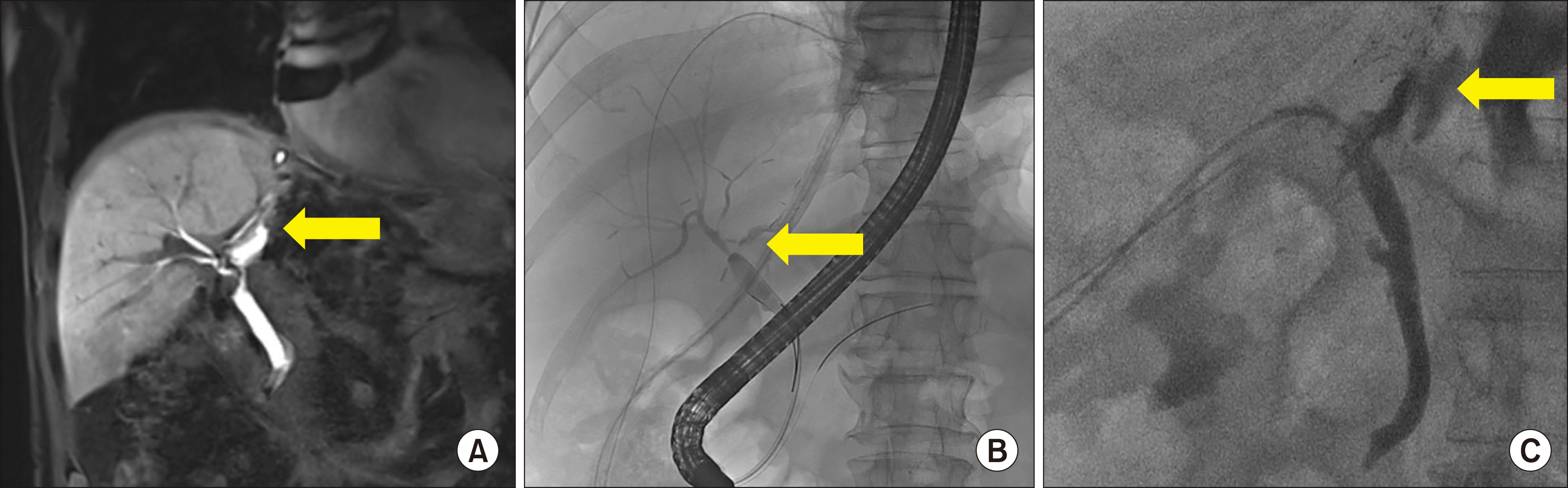
Forty patients had undergone MR Cholangiography with Gd-EOB-DTPA due to suspected biliary complications. Biloma was observed during routine follow-up CT in 10 cases (23.3%), and 11 cases (25.6%) in whom the drainage color had changed to greenish and 14 cases (32.5%) of biloma confirmed by CT at the emergency department due to abdominal pain. Anastomosis leakage was confirmed in 33 cases of the 37 suspected patients with leakage. Fifteen (34.8%) patients underwent endoscopic retrograde biliary drainage (ERBD) through ERCP, and six (13.9%) patients experienced ERCP failure and subsequently underwent percutaneous transhepatic biliary drainage (PTBD) (Fig. 1). Thirteen patients (30.2%) with large-sized biloma on CT taken with symptoms were visualized on MRI Cholangiography with Gd-EOB-DTPA after reducing the biloma cavity after percutaneous catheter drainage (PCD) insertion (Fig. 2). In the 20 min delay image, anastomosis can be well evaluated, and in the 40 min delay image, the contrast agent can confirmed the site of leakage.
Among 38 patients with confirmed leakage or stricture, 37 were found to have leakage or stricture on MRI and confirmed positive results. The sensitivity for bile-duct complication detection was 97.3% (37/38). The number of patients without biliary complication was two and no biliary complication was identified from GD-EOG-MRI in all of these patients. The specificity of the test was 100% (2/2) (Table 2). The connection between biloma and CBD was confirmed in the PCD tubogram in one patient among three who showed negative findings on MRI.
Differential diagnosis of cut surface leakage
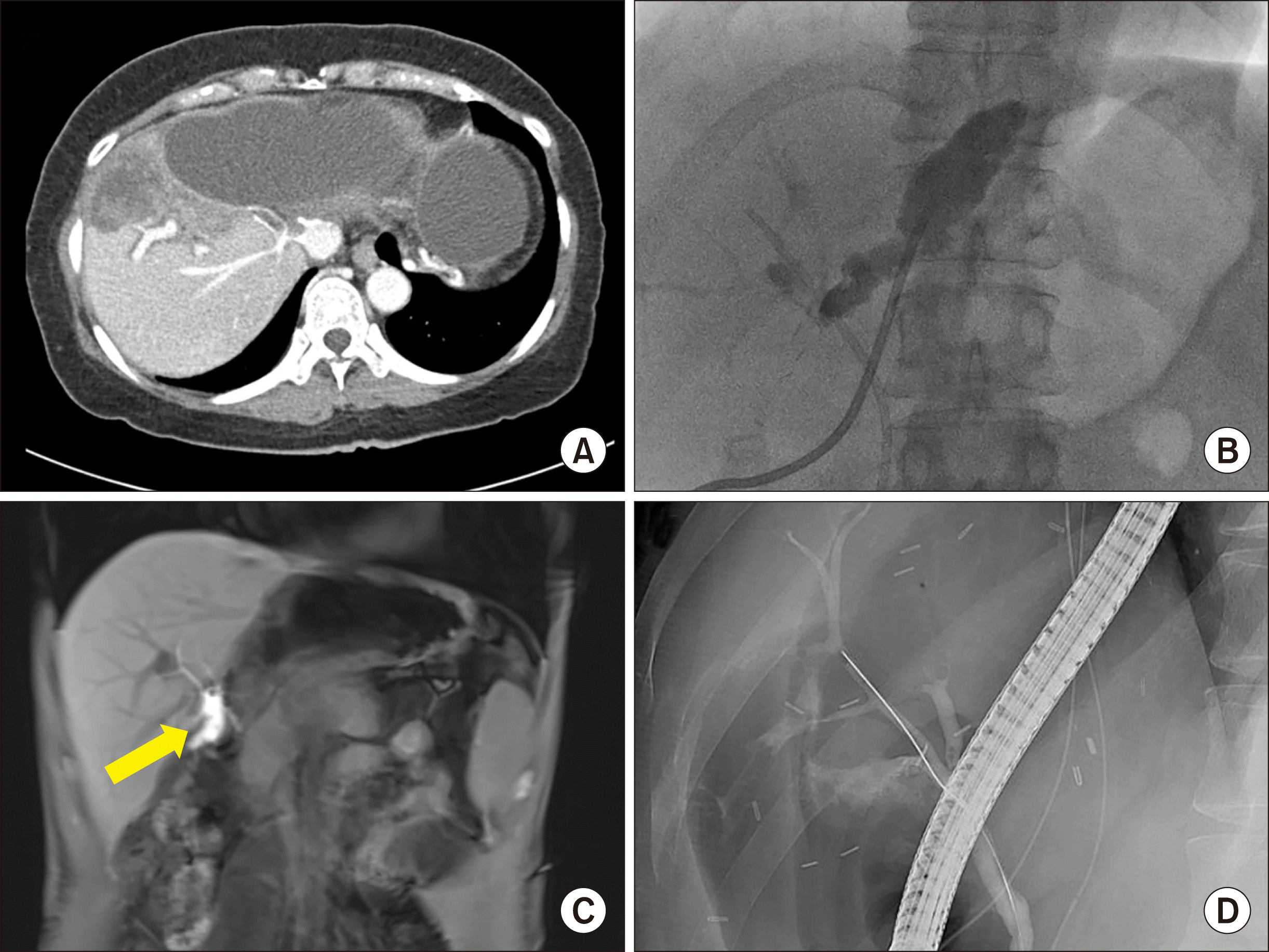
Among the patients suspected with biliary complication, two were diagnosed with the leakage of the cut surface, not of the anastomosis site. The PCD was placed into the biloma cavity in the two cases and the amount of drainage was decreased. The two patients were discharged after conservative management. The case was a 62-year-old man who had a Jackson-Pratt drain color change after LDLT. A biloma was identified around the liver cut surface on the postoperative 7 days abdominal CT scan (Fig. 3). The PCD was placed into the biloma cavity thereafter MR Cholangiography with Gd-EOB-DTPA with a 40 min delay image was performed for the differential diagnosis of biliary complication. From the MR Cholangiography, no direct connection from the anastomosis to biloma was noted and the distance between biloma and the anastomosis site was a little more. This case was diagnosed as a cut surface leakage, percutaneous drainage without ERCP or PTBD was performed. The patient’s condition improved after drainage.
Evaluation of liver dysfunction
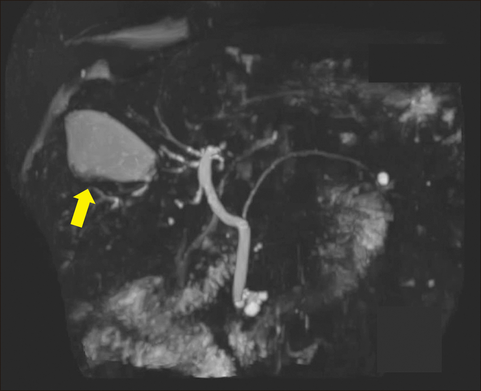
MR Cholangiography with Gd-EOB-DTPA was performed to evaluate the liver excretion in 3 cases with recurrent cholangitis due to bile duct complication or suspected liver dysfunction due to chronic rejection. Three cases had liver parenchymal damage due to chronic rejection and recurrent cholangitis, and one of the patient was admitted for acute onset hyperbilirubinemia on the liver function test in the outpatient clinic. Abdominal CT was performed to evaluate the structural defect of the bile duct, but the bile duct was not dilated. The result of the liver biopsy performed for the differential diagnosis of hyperbilirubinemia revealed no acute cellular rejection but confirmed the chronic rejection. MR Cholangiography with Gd-EOB-DTPA was performed to confirm hyperbilirubinemia caused by dysfunction of bile secretion due to chronic change. In a normal case, bile duct is usually observed in the 20 min delay image. However, the bile duct was not observed from the MR Cholangiography with Gd-EOB-DTPA even after a 40 min delay (Fig. 4). It was related to the dysfunction of the secretion and bile production. The bile transporter could be damaged due to chronic liver damage caused by rejection and recurrent cholangitis.
DISCUSSION
Gd-EOB-DTPA-enhanced MRI is an alternative technique to evaluate the biliary tree according to its biliary excretion.10,12,15 It has been utilized for the preoperative workup of potential living liver donors in the field of LT. Except in some cases with organic anion transporting polypeptide expression disorder in the donor, it demonstrated good or excellent agreement for visualizing the biliary tree even in a case with a complex anatomical variation.
Meanwhile, several attempts to noninvasively confirm biliary leakages, such as MRCP or hepatobiliary scan (DISIDA scan), have continued. Previous reports have revealed the usefulness of scans in the identification of leakage in hepatobiliary surgery, as well as research on images obtained using MRCP.
However, the two imaging tools were indeed difficult to accurately read because the image blurring was quite severe. DISIDA scans are functional studies of the gallbladder. Technetium-labeled analogs of iminodiacetic acid (IDA) or diisopropyl IDA-DISIDA are administered intravenously and are secreted by the hepatocytes into the bile, enabling the visualization of the liver and biliary tree.9,16 Although bile secretion can be confirmed, it is difficult to observe the surrounding structures accurately, and efforts were made to overcome this problem through fusion images with SPECT-CT.17 However, since it fuses two different image tools, it is inferior to MR cholangiography with GD-EOB-DTPB aspect of the accurate localization.
Magnetic resonance cholangiopancreatography (MRCP) is not a functional imaging modality, it is an imaging method to confirm the structure, and the structure also has blurring compared to CT, and therefore it is not a favorable tool to observe the functional images such as leakage. MR cholangiography with Gd-EOB-DTPA can be considered to have the advantages of both the methods. By using a contrast agent, it is possible to obtain a slightly clearer, sequential, and functional image, which has the greater advantage of enabling the simultaneous evaluation of the structure and function with one tool. Moreover, as the leakage point is the cut surface rather than the anastomosis, differentiation is easy as shown in Fig. 2, reducing unnecessary procedures.
If the bile duct was not observed in the Gd-EOB DTPA MRI 40-minute delay image, the main cause of the hyperbilirubinemia was liver dysfunction. When liver dysfunction is identified, it helps to avoid insignificant bile duct intervention and reconfirm long-term strategies. ERCP or PTBD procedures can cause problems such as bleeding and pancreatitis. In the immediate postoperative period after LT, coagulopathy sometimes occurs and can cause major complications. Additionally, in the case of cut surface leakage, diagnosis with ERCP alone is difficult, and leakage sites may not be identified without accurate cannulation technique; therefore, it will be helpful for the interventionist to proceed with an intervention if the leakage point can be predicted and guided using the MRI.
The limitation of this study is that most of the patients diagnosed with complications finally underwent biliary intervention. Therefore, one may consider if the procedure is justified. Additionally, because the cost of this test does not allow its frequently use, there can be a debate about the procedure when the anastomosis leakage is obvious.
However, if unnecessary ERCP or biopsy procedures can be reduced through the confirmation of cut surface leakage or liver dysfunction, there might be an advantage in considering the cost and risk aspects. Depending on the insurance coverage in each country and whether or not the inspection is set, application at all transplant centers may be difficult. However, we consider its relevance as sufficient in that the most accurate and clean images can be obtained as a tool for diagnosing noninvasive bile duct problems after the previously reported hepatobiliary surgery.
In conclusion, MR cholangiography with GD-EOB-DTPB 40-min delay image is a useful, safe, and non-invasive diagnostic tool for the evaluation of bile duct complication and liver dysfunction after LT. It can not only show a clear reconstruction image but also its functional status. Although it has a cost and insurance coverage problem, we consider it as favorable in terms of reducing unnecessary repetitive invasive procedures through accurate diagnosis without any complication related examination.
 XML Download