

 PDF
PDF Citation
 Print
Print


INTRODUCTION
Familiarity with the anatomy is paramount for conduct of safe surgery for a surgeon. The availability of cross- sectional imaging has widely helped surgeons review the anatomy and avoid an intra-operative surprise or disaster. They say that there is nothing called as a normal anatomy when it comes to the hepato-biliary region with reported anomalies exceeding 40%.1 While anomalies with the biliary and arterial system are common, portal vein (PV) anomalies are least common.2 A preduodenal PV (PDPV) has been reported in about 100 cases but a combination of PDPV with preduodenal common bile duct (PDCBD) is extremely rare with only 9 cases being reported.3,4 Further, the embryological basis for the peculiar anatomy observed in our cases has never been reported earlier. We report this case that presented with jaundice and briefly review the relevant embryology and the reported cases in literature.
Go to : 
CASE
A 50-year-old lady presented with jaundice and pain in right hypochondrium of 2 months’ duration. She was icteric and on abdominal examination the gall bladder was not palpable. No other abnormality was noticed. Laboratory investigations revealed normal hemogram and deranged liver functions (total bilirubin 12 mg/dl; direct 9.7 g/dl, alkaline phosphatase 452 U/L and normal transaminases). An ultrasound of abdomen reported dilated intrahepatic and common bile duct (CBD) with a cut off at its distal end. A contrast-enhanced computed tomography (CECT) of the abdomen with a magnetic resonance cholangiopancreatography (MRCP) was reported as cholelithiasis with choledocholithiasis. A laparoscopic cholecystectomy with CBD exploration (CBDE) was planned. During the surgery, there were dense adhesions around the gallbladder/CBD and after some meticulous dissection, the CBD was identified. However, there were two tubular structures and the anatomy was unclear. So a decision was taken to convert to an open surgery. On laparotomy, we noticed that the two tubular structures were actually a PDPV and PDCBD (Fig. 1). What was more interesting was that the PDPV was on the right lateral aspect of the PDCBD, crossing it in it lower part, possibly causing its compression and leading to development of choledocholithiasis. A cholecystectomy, CBDE with stone clearance was done and a roux- en-y choledochojejunostomy was added keeping the obstruction by PDPV in mind. Post surgery, we reviewed the imaging and it corroborated our intra-operative findings (Fig. 2).
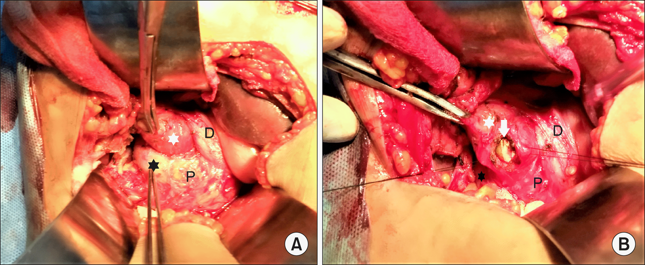 | Fig. 1Intraoperative picture showing preduodenal portal vein (PD PV) and preduodenal CBD. PD PV (black star) to the right of PDCBD (white star). The duodenum (D) is coursing behind and pancreas (P) is in front of these structures.
|
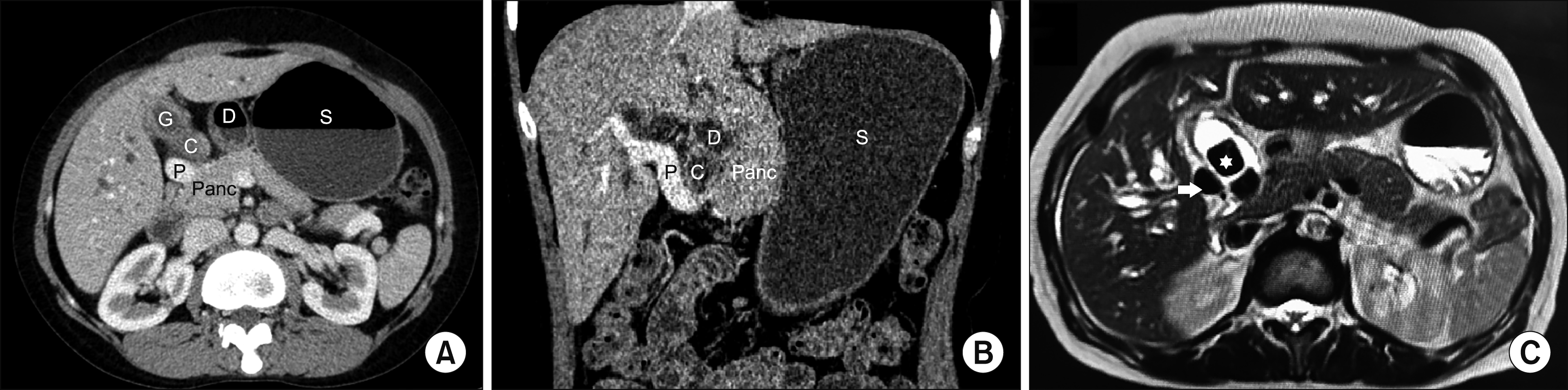 | Fig. 2Pre-operative imaging of the patient. (A) Axial CT scan image showing portal vein (P) and CBD (C) in front of duodenum (D) and pancreas (Panc). (B) Coronal reformatted CT scan image showing portal vein (P) crossing the CBD (C) and causing compression. Both these structures are in front of duodenum (D) and pancreas (Panc). (C) MRI T2 weighted axial image showing choledocholithiasis (white star) and portal vein (white arrow) to right of CBD. S, stomach; G, Gallbladder.
|
Go to :
DISCUSSION
A PDPV was first described by Knight5 in 1921 and the first surgery in a case of PDPV was done by Schnitzler6 in 1926 for duodenal obstruction. A PDPV can be associated with various anomalies but an associated PDCBD is the least common. First described by Stengel7 in 1934, only 9 cases have been reported so far.3,4
Embryologically, the PV is formed by paired vitelline veins having three communications; the cranial intra-hepatic, the middle retroduodenal and the caudal preduodenal. The cranial and caudal communications along with the proximal right and distal left vitelline veins disappear to form the ‘S’ shaped retroduodenal PV.8 Development of the pancreas and the biliary system occurs simultaneously with the formation of the PV, as the ventral pancreatic bud rotates clockwise (seen from cephalic end) around the duodenum to meet the dorsal bud and fuses with it. This rotation places the PV and CBD posterior to both the pancreas and the duodenum (Fig. 3B).
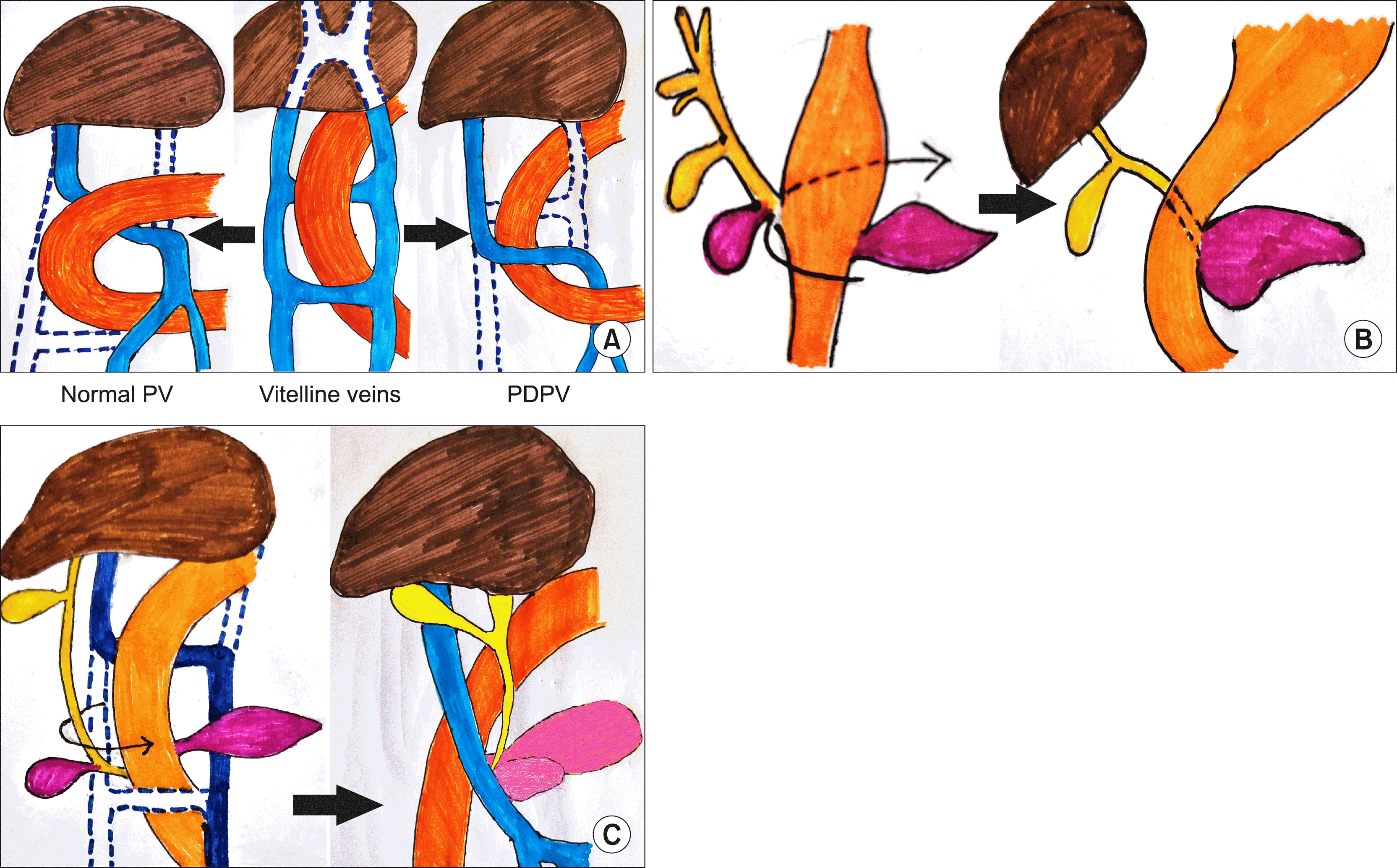 | Fig. 3Illustration showing the embryological development. (A) Development of a normal (PV) and preduodenal portal vein (PDPV) from the paired vitelline veins. (B) Development of CBD and pancreas with normal counterclockwise rotation of the duodenum. (C) Reverse rotation during development leading to a preduodenal portal vein and CBD in our case (blue–portal vein; yellow–CBD; pink–pancreas; brown–liver, and orange–stomach/duodenum).
|
An isolated PDPV occurs when the middle communication disappears and the caudal communication persists (Fig. 3A). The embryology behind a PDCBD is unclear. It could arise due to counter-clockwise rotation of the duodenum.9 In our case it seems the cause of both PDPV and PDCBD is likely to be due to a common occurrence - the counter- clockwise rotation (Fig. 3C). This hypothesis probably explains the finding of the PDPV on the right lateral aspect of the PDCBD and the absence of intestinal malrotation. To the best of our knowledge, this unique anatomy and the embryological basis has never been reported in literature. In cases reported in literature, where specified, the PDPV was found to the left of PDCBD which can arise due to primary abnormal formation of PV and reverse rotation of duodenum or due to situs inversus and reversed rotation of duodenum.
A summary of cases reported in literature is placed at Table 1.3,4,7,9-13 Ours is only the 10th case, albeit with a unique altered anatomy and the embryological basis for the same. The age at diagnosis is equally split between adults and children/neonates with a slight female preponderance. Most cases have been associated with some degree of intestinal malrotation. In addition to these cases there has been one case reported of an isolated PDCBD without a PDPV.14 This was a neonate who was operated for duodenal atresia and while performing a duodenoduodenostomy, the PDCBD was accidentally transected which was repaired over a stent.
Table 1
Summary of all cases of preduodenal portal vein associated with preduodenal CBD reported in literature
| Sl | Year | Author | Age/sex | Diagnosis | Other associated anomalies | Treatment |
|---|---|---|---|---|---|---|
| 1 | 1934 | Stengel7 | 9 m/F | At autopsy | Malrotation, ventro-dorsal pancreas, abnormal ramification of celiac axis and superior mesenteric artery | - |
| 2 | 1962 | Debray et al.10 | 65 y/M | At autopsy | Malrotation, ventral pancreas, partial annular pancreas, no tail of pancreas, inversion of major vessels | - |
| 3 | 1971 | Braun and Cuendet11 | 8 d/M | Duodenal atresia | Malrotation, duodenal atresia, common mesentery | Duodenojejunostomy |
| 4 | 1971 | Braun and Cuendet11 | 4 d/M | Duodenal web | Duodenal web, mobile right colon | Excision of web, duodenoplasty |
| 5 | 1976 | Uchiyama et al.12 | 3 m/M | Biliary atresia | Malrotation, congenital biliary atresia | R-Y portoenterostomy |
| 6 | 1985 | Patti et al.13 | 1 d/F | Esophageal and duodenal atresia | Esophageal and duodenal atresia | Duodenojejunostomy |
| 7 | 1996 | Leung et al.9 | 32 y/F | Choledocholithiasis | Short small bowel mesentery | Choledochoduodenostomy |
| 8 | 2004 | Yi et al.3 | 75 y/M | At autopsy | Situs inversus totalis, malrotation, short pancreas, bilobed/accessory spleen, and abnormal ramification of the celiac axis, superior mesenteric artery and renal arteries | - |
| 9 | 2009 | Shah et al.4 | 75 y/F | Hepatolithiasis | Polysplenia, missing tail of pancreas | Left lateral Sectionectomy |
| 10 | 2020 | Present case | 50/F | Choledocholithiasis | None | Choledochojejunostomy |
![]()
What could be the clinical significance of this anomaly? Three cases have been diagnosed at autopsy meaning that such cases could remain asymptomatic throughout their life. The significance of this anomaly would be on two accounts. First, the anomalous relationship could lead to difficulties in hepato-biliary-pancreatic surgery where a failure to recognize the altered arrangement of the portal vein and CBD early in dissection could lead to accidents and disastrous consequences like injury to the PV. Second, the abnormal arrangement may cause obstruction to the CBD leading to choledocholithiasis, as in our case, and/or hepatolithiasis.4,9
The hepatobilary region will continue to surprise surgeons with its anatomical variations. It is important to study the imaging meticulously to pick up anomalies, if any, to prevent intraoperative surprises or disasters especially in laparoscopic surgery because of the absence of tactile feedback and the total reliance on visual cues. In our case, the radiologists could not pick up the anomaly because of being unaware of this entity that is so rare. We too were wiser only after discovering the anomaly on conversion and when we looked up literature. This particular case had dense adhesions which took us about an hour to identify what we thought was the CBD and were lucky that we did not enter into the PV thinking it is the CBD. Though rare, surgeons must be aware of a PDPV and if associated with jaundice/choledocholithiasis, there probably is an associated PDCBD too.
Go to :
 XML Download