서론
수부 각성 수술
1. 적응증
2. 마취
Table 1.
(1) 가능하면 가는 주사 바늘을 사용한다(27 gauge, 0.41 mm 이하) [17] (Fig. 1).
(2) 주사기를 잡을 때 양손을 모두 사용하여 바늘이 흔들리지 않도록 한다.
(3) 바늘이 피부를 관통할 때 피부에 수직으로 삽입하여 접촉되는 피부 신경을 최소화한다[18].
(4) 바늘로 피부를 찌르지 말고, 피부를 밀어 올려 피부가 바늘로 들어가게 하면 통증을 줄일 수 있다. 주변 피부의 작은 진동이나 압력 등은 바늘이 피부를 관통할 때 초기 통증 감소에 도움이 된다.
(5) 2–10 mL의 마취액을 피하에 주사할 때 바늘이 움직이지 않도록 주의해야 한다.
(6) 넓은 범위를 마취할 때는 이전 주사로 피부 색이 하얗게 변한, 감각이 둔해진 부위 1 cm 이내에 다음 바늘을 삽입하여 통증을 줄여야 한다[19].
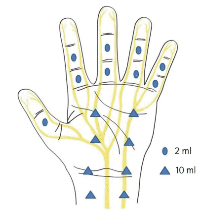
(7) 마취액은 충분한 양을 주사해야 한다. 단 손가락 수장부와 수배부는 각 수지(phalanx)에 2 mL면 충분하다(Fig. 2).
(8) 마취 범위는 절개 부위나 골절 부위에서 최소 2 cm 이상의 폭으로 충분히 마취액이 들어가야 한다.
(9) 마취 중 환자와 원활한 의사소통으로 통증 여부를 확인하여, 최소한의 통증으로 마취가 되도록 꾸준히 연습(training)하여야 한다.
(10) 전완부 건 이전술(tendon transfer) 등 광범위한 마취가 필요한 경우는 Blunt tipped filler cannula를 사용하여 주사하는 것이 도움이 된다[8].
3. 수술
수부 각성 수술의 적응증
1. 굴곡건의 치료
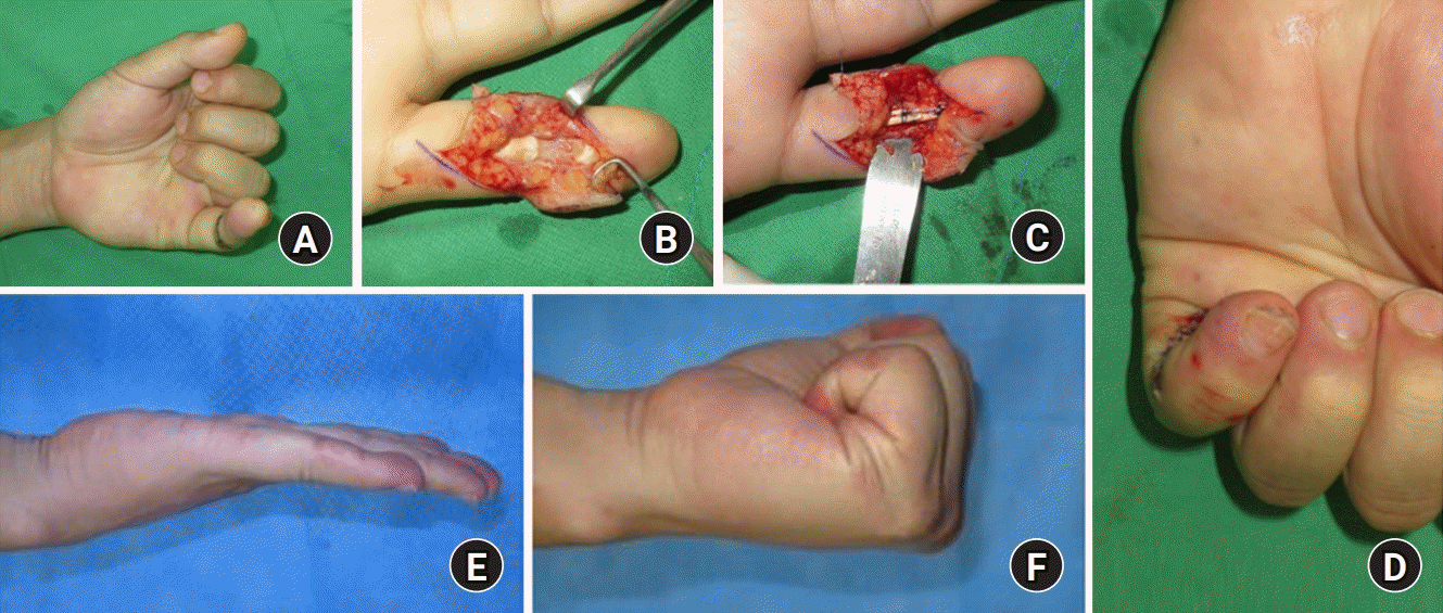 | Fig. 4.(A, B) Wide-awake local anesthesia for a case with a complete zone II flexor tendon profundus cut in the small finger. Inject 2 mL of 1% lidocaine with 1:100,000 epinephrine (buffered at a ratio of 10 mL of lidocaine/epinephrine to 1 mL of 8.4% sodium bicarbonate) in subcutaneous fat. (C, D) Venting the A4 pulley enhances the motion of the flexor digitorum profundus tendon. (E, F)The range of motion of the small finger 8 months later. Written informed consent was obtained for publication of these images. |
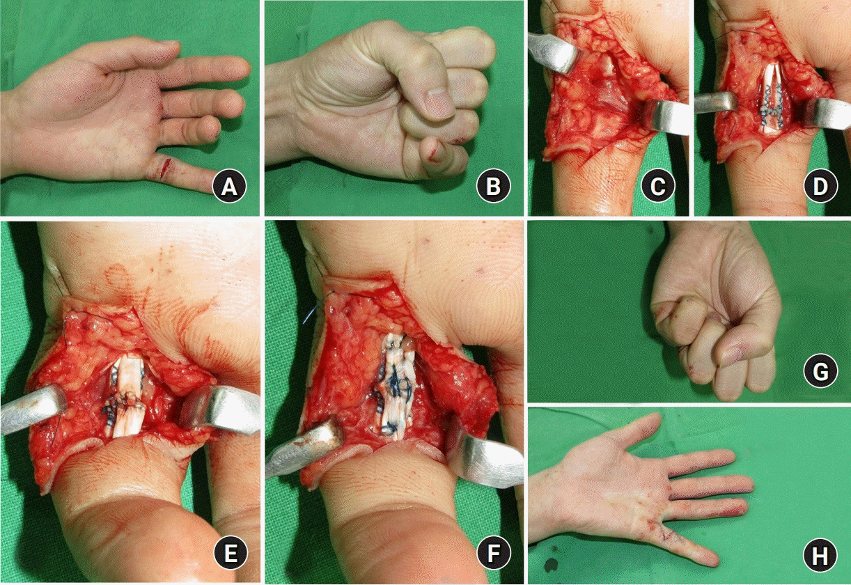 | Fig. 5.(A, B) Preoperative view of rupture of the flexor tendons to the left small finger in a 28-year-old man. (C) Ruptured the sharp margin of the fifth flexor digitorum sublimis (FDS) and flexor digitorum profundus (FDP) tendon was shown at zone II. Simultaneous suturing of the ruptured FDS and FDP tendon at zone II increased the size of the two tendons. (D–F) Full release of A4 pulley and partial venting of A2 pulley will prevent the motion limitation of finger flexion. (G, H) Intraoperative views show full flexion and extension of small fingers after tendon repair. Written informed consent was obtained for publication of these images. |
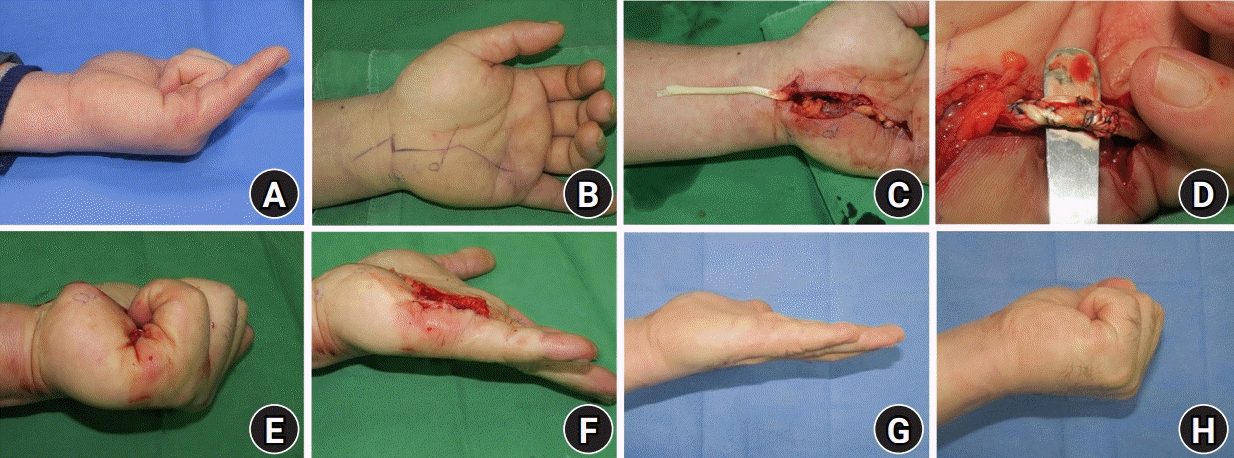 | Fig. 6.(A, B) A 46-year-old man, a professional golf player, had been fractured hook of hamate and attritional rupture of both fifth flexor digitorum tendon at zone III. (C) With wide-awake anesthesia, the small profundus tendon was reconstructed with the ring finger superficialis tendon transfer. (D) Interweaving sutures are made fourth flexor digitorum sublimis to the fifth flexor digitorum profundus with adjusting tension. (E, F) Intraoperative assessment of the motion after tendon transfer. (G, H) The range of motion of the small finger 6 months later. Written informed consent was obtained for publication of these images. |
2. 신전건의 치료
 | Fig. 7.A 33-year-old man had extensor tendon subluxation by sagittal band rupture. (A) At the dorsum of metacarpophalangeal (MCP) joint, inject 10 mL of 1% lidocaine with 1:100,000 epinephrine (buffered at a ratio of 10 mL of lidocaine/epinephrine to 1 mL of 8.4% sodium bicarbonate) in subcutaneous fat. (B) Extensor tendon subluxation during MCP joint flexion. (C, D) The patient can regain the stability of the extensor tendon. Written informed consent was obtained for publication of these images. |
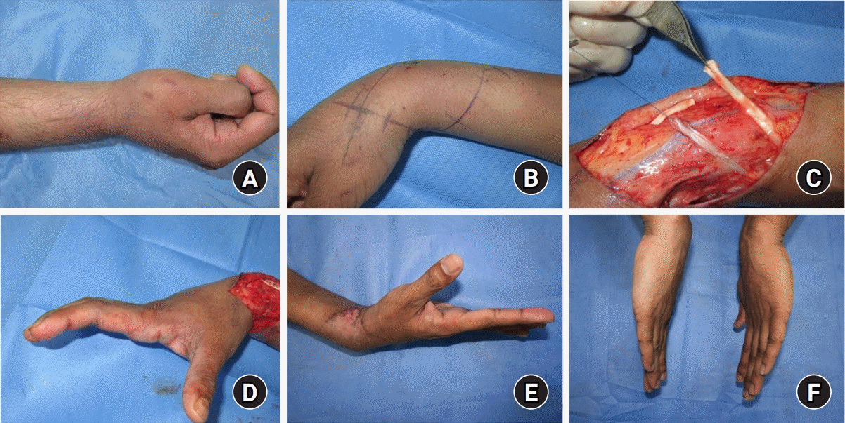 | Fig. 8.(A) A 29-year-old man had lower radial nerve palsy of the right arm for 2 years. (B) Around one incision over the radio-dorsal wrist, 50 mL of 0.5% lidocaine with 1:200,000 epinephrine was used to decrease the total amount of injected epinephrine. (C) Skin incision and dissection of both the palmaris longus tendon and extensor pollicis longus tendon, and flexor carpi radialis and extensor digitorum communis are dissected under tourniquet. (D) Intraoperative assessment of the motion after tendon transfer. (E, F) Finger and thumb extension 6 months later. Written informed consent was obtained for publication of these images. |
3. 건 박리
 | Fig. 9.(A) A 27-year-old man had previous fifth metacarpal fracture and limitation of metacarpophalangeal (MCP) joint flexion. (B) Severe adhesion of extensor tendon and was seen. (C) After tenolysis of extensor tenon, but no improvement of range of motion by MCP joint dorsal capsule thickening. (D) Immediate postoperative view. After dorsal capsulectomy, full flexion of the MP joint was possible. Written informed consent was obtained for publication of these images. |
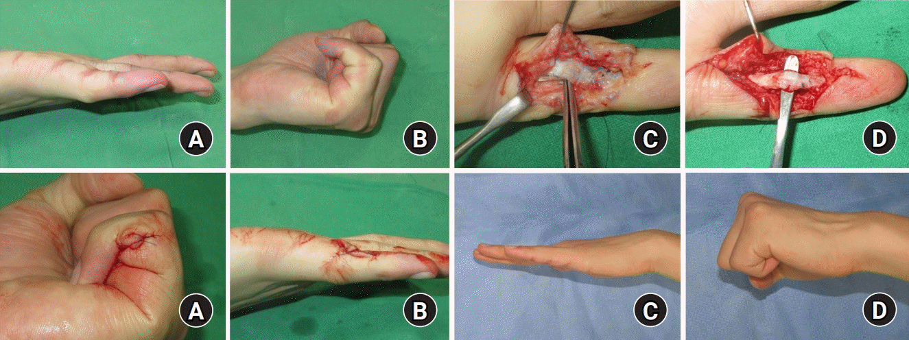 | Fig. 10.(A, B) Flexion and extension contracture of the interphalangeal joints of the small finger after flexor tendon repair. (C) After opening the tendon sheath, the authors found severe adhesions between the flexor digitorum sublimis and flexor digitorum profundus (FDP) tendons as well as between flexor tendons and pulleys. (D) Tenolysis with FDP tendon. (E, F) Test of full digital extension and flexion during surgery. (H, I) Postoperative view 6 months later. Full flexion and extension were possible. Written informed consent was obtained for publication of these images. |
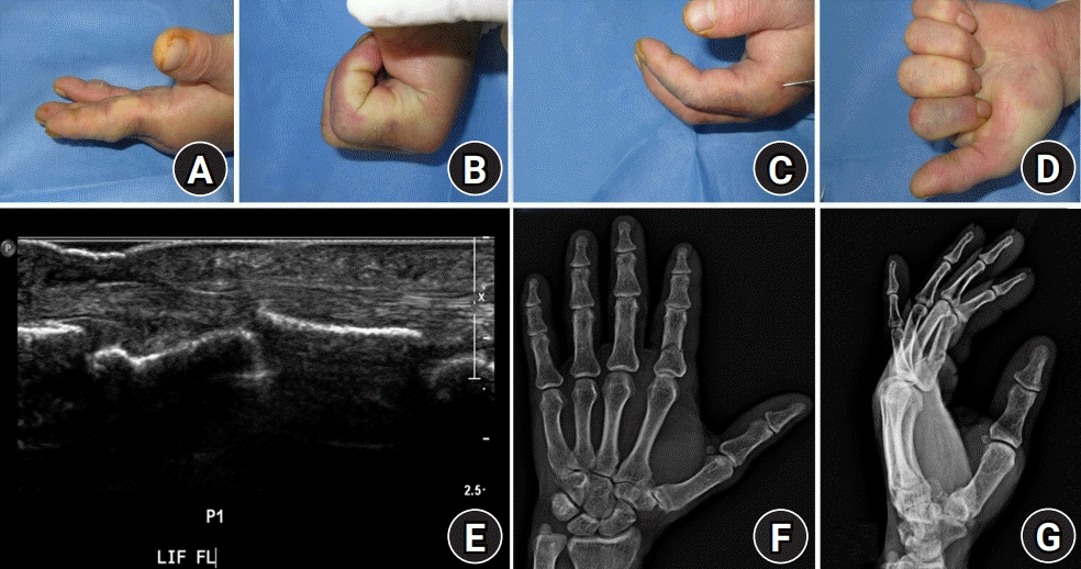 | Fig. 11.A 55-year-old man underwent K-wire fixation for a closed proximal phalanx fracture of the index finger. (A–D) Intraoperative view of the index finger motion before and after fixation of K-wire. (E) Ultrasonogram showed catching of the flexor digitorum profundus tendon. (F, G) Preoperative radiographic findings. Written informed consent was obtained for publication of these images. |


 PDF
PDF Citation
 Print
Print



 XML Download