Abstract
Purpose
Plantar wounds represent a frequent practice of the plastic and reconstructive surgeon. The uniqueness and high complexity of the microarchitecture and biomechanics of the plantar region explain the complex challenge of its reconstruction. With the advances in microsurgery, including the plantar subunit principle, both applied in the restoration of plantar defects through the use of sensorineural free flaps have allowed an optimal reconstruction.
Methods
A descriptive, retrospective study was carried out in a period of time established between January 2016 and January 2019, obtaining a total of 18 patients with plantar defects, reconstructed using sensory free flaps. Tissue stability, recovery of protective sensitivity, early ambulation, and correct use of footwear were evaluated.
Results
The most frequent etiology was secondary to oncological resections due to melanoma (n=12, 66.7%), followed by gunshot wounds (n=4, 22.2%). Subunit 3 was the most frequently involved in 38.9% (n=7). In the 88.8% of the cases, was used an anterolateral thigh flap (n=16) and the lateral antebrachial flap in 22.2% (n=2). The free flap survival rate was 100%. An average of seven points was obtained at 6 months based on the Semmes-Weinstein test. The mean for the return to their daily activities was 2.5 months. The patients of 94.4% (n=17) recovered ambulation and could footwear.
Go to : 
Pelvic limb injuries, specifically plantar defect, constitute a frequent and complex practice of the plastic and reconstructive surgeon. The most frequent causes include vehicular trauma, oncological resections, motor vehicular trauma, burns, or secondary to complications of chronic degenerative diseases [1-3].
The uniqueness and high complexity of the microarchitecture and biomechanics of the foot explain the complex challenge of its reconstruction. First, the epidermal thickness of the glabrous skin can be 10 to 13 mm, giving it a unique capacity for resistance and protection. Second, the strong dermal adherence to the deep plantar fascia through fibrous septa in honeycomb arrangement, allow adequate absorption and redistribution of loads. Third, the subcutaneous plantar tissues (well-organized lobes of adipose tissue) act together to form specialized structures to withstand compressive and shear forces, due to their high deformability. Fourth, the highly specialized sensory-motor integration of the foot together with the central nervous system play a fundamental role in the standing, ambulation, and proprioceptive abilities. Last but not least, the foot constitutes a key piece in the aesthetics of the pelvic limb, so the contour and compactness in its subunits are important psychological and cultural considerations, as is the use of footwear [4-7].
Throughout history, a wide variety of therapeutic possibilities have been developed for plantar reconstruction. However, with the advent of microsurgery in the 1960s and with the continuous development of new techniques and principles of reconstruction, the use of sensatel free flaps has become highly relevant in this anatomical region [8-10].
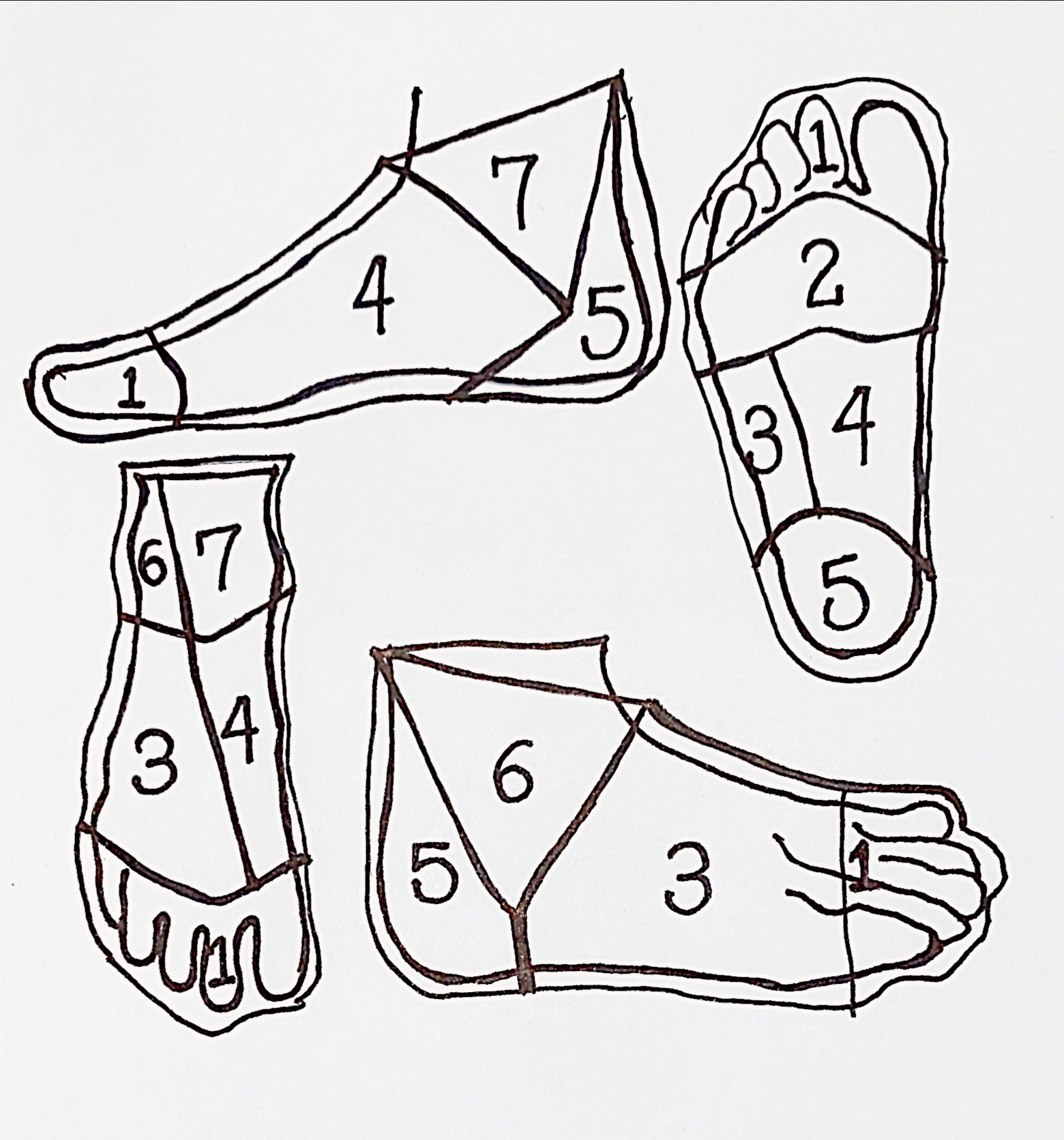
Hollenbeck’s plantar subunit principle [11] (Table 1, Fig. 1) is a watershed in the treatment of plantar lesions. This principle analyzes the specific characteristics of the plantar skin and also recognizes that each subunit has different biophysical, functional, and aesthetic demands. Hollenbeck et al. [11] divide the ankle and plantar region into seven subunits, each of which requires specific anatomical, biomechanical, and aesthetic characteristics for optimal restitution. Thus, if units 3, 4, 6, and 7 (low load units) are well reconstructed with thin and flexible flaps, subunits 2 and 5 require more shear resistant tissues since they are high load sites and finally the subunit 1 could even depending on the case not be rebuilt. Therefore, at present, the reconstructive requirements demand a planning based on the anatomical subunits, in order to achieve maximum sensitive recovery, with restitution of anatomical harmony [7,8,12].
This publication aims to show the experience of 3 years in a specialized center in Mexico City, in reconstruction of plantar defects with free sensate flaps.
Go to : 
This study was exempt from ethics committees because it is a retrospective study that does not publish patient identities or location data, as well as being observational and retrospective.
A descriptive, retrospective study was carried out in a period of time established between January 2016 and January 2019 obtaining a total of 18 patients with plantar defects; all of them operated by the same micro-surgeon. The reconstructive requirement was evaluated based on the characteristics of the defect, such as size, skin coverage, bone involvement, and integrity of the deep plantar fascia; and classified according to Hollenbeck et al. [11].
The flaps used were the lateral antebrachial and anterolateral flaps of the subfascial thigh, including the lateral brachial sensory nerve and the lateral descending femoral nerve, respectively. An insite was performed for the restoration of the anatomical-functional subunit, arterial anastomosis, two venous anastomoses, and end-to-end nerve coaptation, as well as adherence of the deep fascia to the plantar fascia and splint in anatomical position. The resulting donor area was managed with split-thickness skin graft vs. primary closure.
Demographic data such as age, sex, diagnosis, size of the defect, affected plantar subunit, flap performed, recipient vessels and nerves, time of reincorporation to daily activities, and sensory-protective evaluation were collected 6 months and 1 year of the postoperative with the Semmes-Weinstein test. Those cases that did not have the variables to collect and those that did not comply with the minimum follow-up were discarded.
Go to : 
Eighteen patients were obtained for the reconstruction of defects with transfer of free sensate tissue during the designated period of time (Table 2). Fifty percent were females and 50% were males. The mean age was 53.6 years (range, 45 to 62.5 years). The most frequent etiology was secondary to oncological resections due to melanoma (n=12, 66.7%), followed by gunshot wounds (n=4, 22.2%) and finally motor vehicle trauma (n=2, 22.2%).
Demographic data
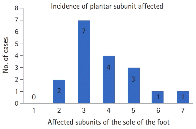
In our study, subunit 3 was the most frequently involved in 38.9% (n=7); subunit 4 in 22.2% (n=4), subunit 5 in 16.7% (n=3), subunit 2 in 11.1% (n=2), and finally subunits 6 and 7 with 5.5%, respectively (Fig. 2). For vascular assessment and preoperative evaluation, Doppler ultrasonography was performed in 77.8% (n=14) and contrast computed tomography in 22.2% (n=4).
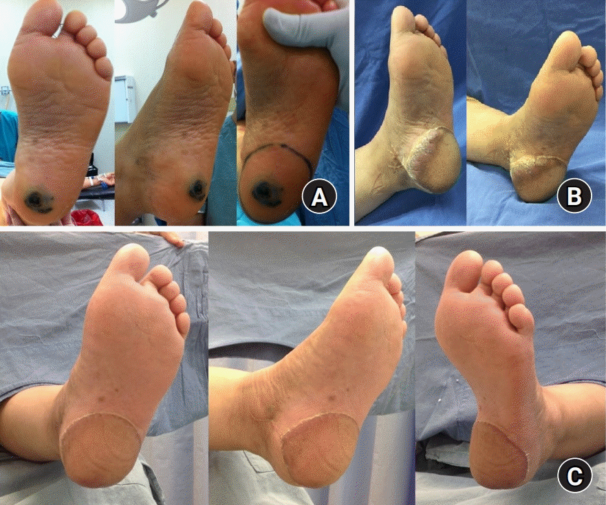
Two main donor sites for free sensate flaps, the anterolateral thigh (ALT) flap (Fig. 3) in 88.9% (n=16), and the lateral forearm flap (Fig. 4) in 11.1% (n=2) were used. The most frequent recipient vessel was the pedis artery, followed by the posterior and anterior tibial arteries. For nerve coaptation, the deep peroneal nerve was used in most cases as the receptor nerve, followed by the medial plantar nerve.
The survival rate of free flaps was 100% at 1-year follow-up (n=18). One case (5.6%) suffered a complication that required reoperation in a patient undergoing ALT due to a wound from gunshot wounds, with bleeding (organized hematoma), without compromising the vascular pedicle and without a flap loss. The mean follow-up was 1.8 years with at least six serial postoperative follow-up visits.
For the assessment of sensitivity and sensory-protective capacity, all patients underwent the Semmes-Weinstein test using the eight plantar points, at 6- and 12-month follow-up. Observing an average of seven points at 6 months and 7.3 at 12 months, which translates into a recovery of protective sensitivity. The mean for reincorporation to their daily activities was 2.5 months (range, 2 to 4 months). In 94.4% of the cases (n=17) were able to walk without assistance, in an average time of 3.8 months. Finally, 94.4% (n=17) were able to wear footwear in less than a year (Table 3).
Postoperative evaluations
Go to : 
The skin covering of the plantar region differs from any other part of the body, due to its complex microarchitecture and unique biomechanics to which it is tested on a daily basis. For this reason, it makes its reconstruction an unprecedented challenge, especially when the surgical principle of “Replace like with like” is taken as the cornerstone [1,13-15].
Throughout history, multiple reconstructive strategies have been proposed for plantar defects; skin graft, regional and distal flaps. Today, it is widely known that the exclusive use of skin grafts should not be considered as the first therapeutic option, due to the low weight-bearing capacity, associating with a high rate of chronic ulceration [16-18]. As a result, the indiscriminate use of regional flaps became a frequent practice, with the advance flaps in V–Y [19-21], rotational and transposition standing out [22]. However, these have been linked with restrictions in the arches of movement, comorbidities in the donor area, joint stiffness, late limb amputations, and border hyperkeratosis; therefore, their use has been limited for the reconstruction of suprafascial defects with small size [8,16,17].
Nowadays, advances in microsurgery with the use of free sensate flaps can provide a completely functional and aesthetic reconstruction, where the therapeutic objectives should be emphasized toward the return to walking without assistance, restoration of protective sensitivity (deep pressure, vibration, temperature, and two-point discrimination), restoration of the deep plantar fascia, and tissue capability for redistribution of loads (differs for each subunit), all without interfering with the proper fit of the footwear [11,17,24].
In general, the evidence showed in the literature, as well as in our study, has shown that fasciocutaneous flaps represent the ideal tissue for the reconstruction of plantar defects secondary to oncological resections and high-energy trauma [25]. Its main indication lies in plantar defects with a subfascial compromise, secondary reconstructions, or exposure of deep structures (bone, nerve, or vascular tissue) [8,11,16].
As reported in the literature as in our study, the ALT and the forearm flap have been the most frequently used free flaps [8]. The versatility of the ALT flap allows it to be modified according to the needs of the defect when it is referring to its shape and volume, since it can be divided into its individual perforators to improve its contour and thin up to 4–7 mm without presenting increased risk of necrosis, respectively [26,27]. Likewise, it provides a cutaneous island of up to 370 cm2, which is why use in wide and complex defects has advantages over other free flaps [28-30].
On the other hand, the forearm flap stands out for its great adaptability, wide cutaneous island, and the volume necessary to restore defects in plantar regions, being an ideal flap for the use of footwear [31,32]. Both the forearm flap and the ALT allow harvesting as sensorineural flaps, through antebrachial cutaneous nerves and lateral femoral cutaneous, respectively [8,33]. Other flaps described include the medial plantar, gracilis, scapular, dorsal pedis, among others. However, these do not offer superiority over those used in our study [5,16,34-36].
The sensory-protective capacity of free flaps continues to be a matter of debate today. Although fasciocutaneous flaps per se have been shown to present a certain degree of sensory recovery, various studies have shown that nerve coaptation to the receptor site significantly improves early sensitivity and two-point discrimination ability [16,33,37]. In our series, nerve coaptation was performed outside the compromised subunit, in other to avoid aberrant sensitivity, presenting very encouraging results for our patients. In this context, it can be associated with our low rate of pressure ulcers, as reported by Oh et al. [5] and Sönmez et al. [38].
Chronic ulceration and border hyperkeratosis represent the most feared and frequent late complications reported in the literature, especially seen in reconstructed subunits with high load demand (subunit 2–5) [8,11]. In our series, we have observed a significant decrease in it (n=0); this explained by the type of flaps used and also by the complete restoration of the subunit involved since the border sites (anatomical interval between the flap and native plantar tissue) constitute the most frequent place of appearance due to differences in mobility and load redistribution in this interface [11]. The reduction of these complications is reflected, in an important decrease of secondary surgeries, where flap thinning surgeries can become obsolete.
In our study, long-term follow-up revealed good results, regardless of the flap used, with short periods of restart of ambulation being observed as well as early return to daily activities. As reported by Heidekrueger et al. [12], long-term aesthetic and functional results have an important influence on quality of life, and these in turn are a reflection of early and late complications during treatment.
It was observed that the choice of the flap type did not affect the rate of late major surgical complications (late instability and late amputation) as reported in the study by Cho et al. [25]. Agreeing with Jen and Wei [39], where the best reconstructive technique is not measured by the method, but by the skill and experience of the surgeon to determine the correct therapeutic method.
Finally, the authors acknowledge the limitations that a retrospective study presents and that an important limitation was that the two-point discrimination is not evaluated in each of the flaps, and that this could be evaluated in future studies. Although the majority of plantar defects developed secondary to oncological resections and high-energy trauma, it is thought that the presence of vascular insufficiency related to atherosclerosis due to chronic degenerative diseases can change the outlook and prognosis; for this reason, the evaluation of each plantar defect must be done individually.
Go to : 
Reconstruction of plantar defects represents an unprecedented challenge, due to the complex microarchitecture and biomechanics that each subunit is being tested for. For the authors of this article, each reconstruction should be approached in a systematic way, taking the subunit principle as a central point, seeking as an objective the anatomical, functional and aesthetic restoration; these determined by an adequate sensory-protective recovery, adequate skin coverage, walking capacity, and the ability to wear footwear.
We can conclude that both variants of free sensate flaps (ALT and antebrachial) constitute an adequate option for the integral reconstruction of plantar defects.
Go to : 
REFERENCES
1. Soltanian H, Garcia RM, Hollenbeck ST. Current concepts in lower extremity reconstruction. Plast Reconstr Surg. 2015; 136:815e–829e.

2. Godina M. Early microsurgical reconstruction of complex trauma of the extremities. Plast Reconstr Surg. 1986; 78:285–92.


3. Hinchliffe RJ, Forsythe RO, Apelqvist J, et al. Guidelines on diagnosis, prognosis, and management of peripheral artery disease in patients with foot ulcers and diabetes (IWGDF 2019 update). Diabetes Metab Res Rev. 2020; 36 Suppl 1:e3276.

4. Scaglioni MF, Rittirsch D, Giovanoli P. Reconstruction of the heel, middle foot sole, and plantar forefoot with the medial plantar artery perforator flap: clinical experience with 28 cases. Plast Reconstr Surg. 2018; 141:200–8.

5. Oh SJ, Moon M, Cha J, Koh SH, Chung CH. Weight-bearing plantar reconstruction using versatile medial plantar sensate flap. J Plast Reconstr Aesthet Surg. 2011; 64:248–54.


6. Hayashida K, Yamakawa S, Saijo H, Fujioka M. Foot reconstruction with the superficial circumflex iliac artery perforator flap under local anesthesia: two case reports. Medicine (Baltimore). 2019; 98:e13888.
7. Suh HS, Oh TS, Hong JP. Innovations in diabetic foot reconstruction using supermicrosurgery. Diabetes Metab Res Rev. 2016; 32 Suppl 1:275–80.


8. Crowe CS, Cho DY, Kneib CJ, Morrison SD, Friedrich JB, Keys KA. Strategies for reconstruction of the plantar surface of the foot: a systematic review of the literature. Plast Reconstr Surg. 2019; 143:1223–44.

9. Tamai S. History of microsurgery: from the beginning until the end of the 1970s. Microsurgery. 1993; 14:6–13.

10. Tamai S. The history of microsurgery. Plast Reconstr Surg. 2009; 124(6 Suppl):e282–94.
11. Hollenbeck ST, Woo S, Komatsu I, Erdmann D, Zenn MR, Levin LS. Longitudinal outcomes and application of the subunit principle to 165 foot and ankle free tissue transfers. Plast Reconstr Surg. 2010; 125:924–34.


12. Heidekrueger PI, Ehrl D, Prantl L, et al. Microsurgical reconstruction of the plantar foot: long-term functional outcomes and quality of life. J Reconstr Microsurg. 2019; 35:379–88.

13. D’Arpa S, Moschella F. Introduction to the current concepts in lower extremity reconstruction by the Italian Society for Microsurgery. J Reconstr Microsurg. 2017; 33(S 01):S01–2.

14. Löfstrand JG, Lin CH. Reconstruction of defects in the weight-bearing plantar area using the innervated free medial plantar (instep) flap. Ann Plast Surg. 2018; 80:245–51.


15. Hosseinian MA, Gharibi Loron A, Nemati Honar B. Reconstruction of the plantar toe with a distal reverse instep sensory island flap. Microsurgery. 2018; 38:667–73.


16. Lykoudis EG, Seretis K, Lykissas MG. Free sensate medial plantar flap for contralateral plantar forefoot reconstruction with flap reinnervation using end-to-side neurorrhaphy: a case report and literature review. Microsurgery. 2013; 33:227–31.


17. May JW Jr, Halls MJ, Simon SR. Free microvascular muscle flaps with skin graft reconstruction of extensive defects of the foot: a clinical and gait analysis study. Plast Reconstr Surg. 1985; 75:627–41.


18. Oliver-Allen H, Piper M, Vaughn C, Sbitany H. Immediate reconstruction for plantar melanoma: a paradigm shift. Ann Plast Surg. 2017; 78(5 Suppl 4):S194–8.
19. Scaglioni MF, Franchi A, Uyulmaz S, Giovanoli P. The bipedicled medial plantar flap: vascular enhancement of a reverse flow Y-V medial plantar flap by the inclusion of a metatarsal artery perforator for the reconstruction of a forefoot defect: a case report. Microsurgery. 2018; 38:698–701.

20. Eroğlu L, Güneren E, Keskin M, Uysal OA, Tomak Y. The extended V-Y flap for coverage of a mid-planatar defect. Br J Plast Surg. 2000; 53:708–10.

21. Giraldo F, De Haro F, Ferrer A. Opposed transverse extended V-Y plantar flaps for reconstruction of neuropathic metatarsal head ulcers. Plast Reconstr Surg. 2001; 108:1019–24.


22. Gahalaut P, Pinto J, Pai GS, Kamath J, Joshua TV. A novel treatment for plantar ulcers in leprosy: local superficial flaps. Lepr Rev. 2005; 76:220–31.


23. Macedo JL, Rosa SC, Neto AV, Silva AA, Amorim AC. Reconstruction of soft-tissue lesions of the foot with the use of the medial plantar flap. Rev Bras Ortop. 2017; 52:699–704.


24. Wang CL, Shau YW, Hsu TC, Chen HC, Chien SH. Mechanical properties of heel pads reconstructed with flaps. J Bone Joint Surg Br. 1999; 81:207–11.


25. Cho EH, Shammas RL, Carney MJ, et al. Muscle versus fasciocutaneous free flaps in lower extremity traumatic reconstruction: a multicenter outcomes analysis. Plast Reconstr Surg. 2018; 141:191–9.

26. Olivan MV, Busnardo FF, Faria JC, Coltro PS, Grillo VA, Gemperli R. Chimerical anterolateral thigh flap for plantar reconstruction. Microsurgery. 2015; 35:546–52.


27. Hong JP, Kim EK. Sole reconstruction using anterolateral thigh perforator free flaps. Plast Reconstr Surg. 2007; 119:186–93.


28. Sekido M, Yamamoto Y, Furukawa H, Sugihara T. Change of weight-bearing pattern before and after plantar reconstruction with free anterolateral thigh flap. Microsurgery. 2004; 24:289–92.


29. Chang NJ, Waughlock N, Kao D, Lin CH, Lin CH, Hsu CC. Efficient design of split anterolateral thigh flap in extremity reconstruction. Plast Reconstr Surg. 2011; 128:1242–9.


30. Yildirim S, Avci G, Aköz T. Soft-tissue reconstruction using a free anterolateral thigh flap: experience with 28 patients. Ann Plast Surg. 2003; 51:37–44.


31. Kadam D. Microsurgical reconstruction of plantar ulcers of the insensate foot. J Reconstr Microsurg. 2016; 32:402–10.


32. Zhu YL, Wang Y, He XQ, Zhu M, Li FB, Xu YQ. Foot and ankle reconstruction: an experience on the use of 14 different flaps in 226 cases. Microsurgery. 2013; 33:600–4.


33. Kuran I, Turgut G, Bas L, Ozkan T, Bayri O, Gulgonen A. Comparison between sensitive and nonsensitive free flaps in reconstruction of the heel and plantar area. Plast Reconstr Surg. 2000; 105:574–80.


34. Heymans O, Verhelle N, Lahaye T. Covering small defects on the weight bearing surfaces of the foot: the free temporal fasciocutaneous flap. Br J Plast Surg. 2005; 58:460–5.


35. Karşıdağ S, Akçal A, Turgut G, Uğurlu K, Baş L. Lower extremity soft tissue reconstruction with free flap based on subscapular artery. Acta Orthop Traumatol Turc. 2011; 45:100–8.


36. Emsen IM. Reconstruction with distally based dorsalis pedis fasciocutaneous flap for the coverage of distal toe-plantar defects. Can J Plast Surg. 2012; 20:e25–7.

37. Santanelli F, Tenna S, Pace A, Scuderi N. Free flap reconstruction of the sole of the foot with or without sensory nerve coaptation. Plast Reconstr Surg. 2002; 109:2314–24.


Go to : 


 PDF
PDF Citation
 Print
Print


 XML Download