Journal List > J Korean Med Sci > v.36(4) > 1146411

Lee, Kwon, Na, Choe, Seol, Kim, Kim, Park, Kim, Ryu, Hwang, and Shim: Clinical Practice Guidelines for Prenatal Aneuploidy Screening and Diagnostic Testing from Korean Society of Maternal-Fetal Medicine: (2) Invasive Diagnostic Testing for Fetal Chromosomal Abnormalities

Abstract

The Korean Society of Maternal Fetal Medicine proposed the first Korean guideline on prenatal aneuploidy screening and diagnostic testing, in April 2019. The clinical practice guideline (CPG) was developed for Korean women using an adaptation process based on good-quality practice guidelines, previously developed in other countries, on prenatal screening and invasive diagnostic testing for fetal chromosome abnormalities. We reviewed current guidelines and developed a Korean CPG on invasive diagnostic testing for fetal chromosome abnormalities according to the adaptation process. Recommendations for selected 11 key questions are: 1) Considering the increased risk of fetal loss in invasive prenatal diagnostic testing for fetal genetic disorders, it is not recommended for all pregnant women aged over 35 years. 2) Because early amniocentesis performed before 14 weeks of pregnancy increases the risk of fetal loss and malformation, chorionic villus sampling (CVS) is recommended for pregnant women who will undergo invasive prenatal diagnostic testing for fetal genetic disorders in the first trimester of pregnancy. However, CVS before 9 weeks of pregnancy also increases the risk of fetal loss and deformity. Thus, CVS is recommended after 9 weeks of pregnancy. 3) Amniocentesis is recommended to distinguish true fetal mosaicism from confined placental mosaicism. 4) Anti-immunoglobulin should be administered within 72 hours after the invasive diagnostic testing. 5) Since there is a high risk of vertical transmission, an invasive prenatal diagnostic testing is recommended according to the clinician's discretion with consideration of the condition of the pregnant woman. 6) The use of antibiotics is not recommended before or after an invasive diagnostic testing. 7) The chromosomal microarray test as an alternative to the conventional cytogenetic test is not recommended for all pregnant women who will undergo an invasive diagnostic testing. 8) Amniocentesis before 14 weeks of gestation is not recommended because it increases the risk of fetal loss and malformation. 9) CVS before 9 weeks of gestation is not recommended because it increases the risk of fetal loss and malformation. 10) Although the risk of fetal loss associated with invasive prenatal diagnostic testing (amniocentesis and CVS) may vary based on the proficiency of the operator, the risk of fetal loss due to invasive prenatal diagnostic testing is higher in twin pregnancies than in singleton pregnancies. 11) When a monochorionic twin is identified in early pregnancy and the growth and structure of both fetuses are consistent, an invasive prenatal diagnostic testing can be performed on one fetus alone. However, an invasive prenatal diagnostic testing is recommended for each fetus in cases of pregnancy conceived via in vitro fertilization, or in cases in which the growth of both fetuses differs, or in those in which at least one fetus has a structural abnormality. The guidelines were established and approved by the Korean Academy of Medical Sciences. This guideline is revised and presented every 5 years.

Graphical Abstract

jkms-36-e26-abf001.jpg

INTRODUCTION

Prenatal diagnosis is an important aspect of obstetrics as it can identify structural or functional abnormalities in a developing and growing fetus. Information on prenatal diagnosis is used for fetal treatment and monitoring and determination of delivery method, which can be divided into two (prenatal screening testing and invasive diagnostic testing). Prenatal screening testing include maternal serum screening and ultrasound examinations. Recently, as a prenatal screening test, the fetal DNA screening test via DNA analysis of a pregnant women's blood sample has been developed and used worldwide. Moreover, it has been utilized in clinical trials conducted not only in other countries but also in Korea. Prenatal screening testing can be safely performed on pregnant women and fetuses. However, this test can be used only for estimating the risk of fetal chromosomal abnormalities, and cannot be used as a confirmatory test.1
Chorionic villus sampling (CVS), amniocentesis, and cordocentesis are invasive diagnostic testing used for diagnosing genetic or congenital diseases. Invasive diagnostic testing can directly examine the chromosomes of a fetus by invasively collecting fetal villi (placenta), amniotic fluid, and umbilical cord blood samples. Moreover, they can detect the presence of chromosomal abnormalities in a fetus, with an accuracy rate of nearly 100%.12
Since the specimen must be collected via the uterus, the testing must be performed by a highly skilled expert. Its disadvantage is that it is expensive. Moreover, serious complications, such as miscarriage, bleeding, and infection, may occur. The fetal abortion rate within 2 weeks after the procedure is about 3%.34 Therefore, invasive diagnostic testing cannot be performed on all pregnant women. Clinical examination guidelines on testing and methods used are necessary. Although there are several existing clinical treatment guidelines already known in other countries, they have not yet been properly developed. By establishing domestic medical guidelines on prenatal diagnosis tests, we can reduce unnecessary invasive tests in the future and can prevent false artificial abortion. Moreover, these guidelines can facilitate health promotion.
In relation to this reason, clinical practice guidelines (CPG) are required to determine which prenatal diagnosis method should be used. Although there are several CPGs already known in other countries, those in Korea have not yet been properly developed. Hence, the establishment of domestic CPGs on prenatal diagnosis tests can promote national health by reducing unnecessary invasive tests and parental anxiety due to incorrect information. In addition, this CPG can be used as a basis for establishing an effective prenatal management system at the government level and for overcoming low birth rate. Furthermore, it can be utilized as basic research data for the development of a new prenatal diagnosis method for effective prenatal care.
After reviewing the guidelines developed in other countries that have already been universalized previously, the establishment of a new clinical question is considered insignificant. Rather, the development of medical guidelines that combine recommendations and contents based on high-quality CPGs in other countries will be advantageous. Therefore, a CPG for invasive diagnostic testing was developed using the adaptation process.

METHODS

Process of guideline development

After reviewing the existing guidelines in other countries, the Korean CPG on invasive diagnostic testing (https://www.guideline.or.kr/evaluation/sub2.php) was developed using the adaptation process.

Establishment of a CPG committee

The working committee was organized by the Korean Society for Maternal Fetal Medicine (KSMFM). To define key questions for CPG, the committee comprised 12 maternal fetal medicine specialists and 11 methodology experts from medical schools and hospitals. Moreover, they conducted planning, guideline search, selection of key clinical questions, guideline assessment, selection, and adaptation to develop the guidelines.

Process of CPG development

1) Guideline search

PubMed, Embase (Elsevier version), and Cochrane Library were explored to collect all types of published CPG. Previous studies on invasive prenatal testing for fetal chromosome abnormalities were searched using the following key words: ‘practice guidelines,’ ‘guidelines,’ ‘recommendation,’ ‘level of evidence,’ and ‘evidence grade.’ CPGs without recommendations, those that are outdated, those with unrelated topic, or those with missing data were excluded. After further elimination of duplicates, 41 guidelines were finally selected for review.

2) Selection of key clinical questions

Initially, 12 key questions were tentatively selected based on the needs and public opinion of clinicians. The importance and urgency of the key questions were assessed using a 5-point Likert scale. Those rated as ≥ 4 by > 90% of the respondents were included as key questions according to the modified Delphi method, which was described in previous studies.5 Multiple repeat surveys have reduced the key question from 12 to 11. The final question was edited and revised to ensure consistency and clarity of grammar.

3) Guideline assessment, selection and adaptation

To evaluate the quality of the selected guidelines, we used the Korean Appraisal of Guidelines for Research & Evaluation II (K-AGREE II), which was approved by the AGREE Research Trust.6 Fifteen existing guidelines have been identified as potentially suitable for adaptation. Based on the results of the K-AGREE II evaluation, a recommendation matrix with 15 final guidelines was prepared. The recommendations for 11 major questions were compiled based on the guidelines selected by three authors.

4) Additional search of evidence

The references were reviewed based on the selected guidelines to assess the level of evidence.

5) Writing guidelines and determining the recommendation level

The guidelines were assessed and evidence was summarized. The level of evidence (Table 1) and recommendations (Table 2) were determined based on the Scottish Intercollgeate Guideline Network translated into Korean by the National Evidence-Based Healthcare Collaborating Agency (NECA, https://www.guideline.or.kr/evaluation/file/koreaguide.pdf).7
Table 1

Levels of evidence from the ‘SIGN Grading System 1999–2012’

jkms-36-e26-i001
Levels Definition
1++ High quality meta-analyses, systematic reviews of RCTs, or RCTs with a very low-risk of bias
1+ Well-conducted meta-analyses, systematic reviews, or RCTs with a low-risk of bias
1− Meta-analyses, systematic reviews, or RCTs with a high-risk of bias
2++ High quality systematic reviews of case control or cohort or studies
High quality case control or cohort studies with a very low-risk of confounding or bias and a high probability that the relationship is causal
2+ Well-conducted case control or cohort studies with a low-risk of confounding or bias and a moderate probability that the relationship is causal
2− Case control or cohort studies with a high-risk of confounding or bias and a significant risk that the relationship is not causal
3 Non-analytic studies, e.g., case reports, case series
4 Expert opinion
SIGN = Scottish Intercollegiate Guidelines Network, RCT = randomized controlled trial.
Table 2

Grades of recommendations from the ‘SIGN Grading System 1999–2012’

jkms-36-e26-i002
Grades Definition
A At least one meta-analysis, systematic review, or RCT rated as 1++, and directly applicable to the target population; or A body of evidence consisting principally of studies rated as 1+, directly applicable to the target population, and demonstrating overall consistency of results
B A body of evidence including studies rated as 2++, directly applicable to the target population, and demonstrating overall consistency of results; or Extrapolated evidence from studies rated as 1++ or 1+
C A body of evidence including studies rated as 2+, directly applicable to the target population and demonstrating overall consistency of results; or
Extrapolated evidence from studies rated as 2++
D Evidence level 3 or 4; or Extrapolated evidence from studies rated as 2+
SIGN = Scottish Intercollegiate Guidelines Network, RCT = randomized controlled trial.

6) Consensus methodology for guideline adoption

The members of the KSMFM subcommittee were instructed to provide a score for each draft recommendation using a 9-point Likert scale. A score of 1 indicates strong disagreement and 9 strong consent. The average of each voting score was calculated, and it was used to assign items to one of three groups: 1–3 (disagreement), 4–6 (neutrality), and 7–9 (agreement). Based on these scores, the degree of consensus was evaluated.

7) Assessment and review conducted by external experts

There was a public hearing on the background, contents, and evidence of the guidelines during the 24th KSMFM Conference held in Seoul in June 2018.

8) Approval

After several revisions by the working committee, the guidelines were finally approved by the Korean Academy of Medical Sciences in March 2019.

Patients for whom the guidelines are applicable

The main target population is pregnant women in Korea. The major topic is invasive diagnostic testing for fetal chromosome abnormalities. The query for literature search was made with consideration of the target population and potential audience.

Professionals for whom the guideline is recommended

The primary users of this guideline are physicians who care for pregnant women. These guidelines can be used by medical students for learning purposes or as a reference for pregnant women to obtain the latest medical knowledge.

Principles and methods of guideline updates

The developed guidelines are listed on the website of the Korean Medical Guidelines Information Center (‘Clinical Practice Guidelines for Prenatal Aneuploidy Screening & Diagnostic Tests,’ https://www.guideline.or.kr/guide/view.php?number=1085&cate=A). Feasibility within the national insurance system will be reviewed and re-evaluated. The guidelines are revised every 5 years after publication. The KSMFM Committee is responsible for this update.

RESULTS

Key questions and recommendations

  • • KQ1. Should an invasive prenatal diagnostic testing for fetal genetic disorders be recommended for all pregnant women aged over 35 years?

Recommendation: Considering the increased risk of fetal loss in invasive prenatal diagnostic testinging for fetal genetic disorders, it is not recommended for all pregnant women aged over 35 years (Level of evidence: 2++, recommendation grade: B).
Summary of evidence: In older pregnant women, there is a high risk of complications, such as infertility, fetal malformations, gestational diabetes, and hypertension.8910 In the past, pregnant women aged 35 years or older (32 years or older in the case of twin pregnancies) belonged to the high-risk group. Hence, invasive prenatal diagnostic testing for fetal genetic disorders is recommended.1112 However, several studies have shown that age itself (over 35 years) is not a risk factor for chromosomal abnormalities, such as submicroscopic partial imbalances.13 Considering the risk of fetal loss based on an invasive diagnostic test, invasive prenatal diagnostic testing for fetal genetic disorders is not recommended for all pregnant women aged over 35 years.114
  • • KQ2. Is CVS better than amniocentesis among pregnant women who will undergo invasive prenatal diagnostic testing for fetal genetic disorders in the first trimester of pregnancy?

Recommendation: Because early amniocentesis performed before 14 weeks of pregnancy increases the risk of fetal loss and malformation, CVS is recommended for pregnant women who will undergo invasive prenatal diagnostic testing for fetal genetic disorders in the first trimester of pregnancy. However, CVS before 9 weeks of pregnancy also increases the risk of fetal loss and deformity. Thus, CVS is recommended after 9 weeks of pregnancy (Level 1+, Grade A).
Summary of evidence: The World Health Organization (WHO) analyzed their CVS data. Results showed that when CVS is performed after 10 weeks of pregnancy, the procedure itself does not increase the fetal loss rate or cause fetal deformities, except in cases of fetal death or malformation due to abnormalities that the fetus initially had. The WHO stated that CVS is the only method used for the prenatal diagnosis of genetic and chromosomal diseases in early pregnancy, and it can be safely performed by a highly skilled individual.15 In addition, in the study of Kuliev et al.16, which performed CVS between 9 and 12 weeks of pregnancy, the risk of fetal limb reduction defects did not increase in the general population. The incidence of limb reduction defects after CVS was 5.2–5.7 per 10,000 people, and the incidence rate in the general population was 4.80–5.97 per 10,000 people.
By contrast, in a study conducted on 1,916 patients who had early amniocentesis (before 13 weeks of gestation) and 1,775 individuals who had mid-trimester genetic amniocentesis (after 15 weeks of pregnancy), the rate of fetal loss in women who had early amniocentesis was significantly higher than that in women who had mid-trimester amniocentesis (7.6% vs. 5.9%, P = 0.012).17 The incidence of equinovarus in the early amniocentesis group was also significantly higher than that in the mid-trimester amniocentesis group (1% ± 3% vs. 0% ± 1%, P < 0.001). The incidence of membrane rupture associated with the procedure was significantly higher in the early and mid-trimester amniocentesis groups (3.5% vs. 1.7%, P < 0.001).17 In other studies, early amniocentesis conducted before 13 weeks of gestation was associated with a significantly higher fetal loss rate and incidence of equinovarus compared with mid-trimester amniocentesis group.1819 Sundberg et al.20 conducted a study comparing early amniotic puncture (11–13 weeks of pregnancy) and CVS (10–12 weeks of pregnancy). Results showed that the incidence of fetal equinovarus in the early amniocentesis group significantly increased. Moreover, there was a significantly higher number of amniotic fluid leakage cases after the procedure. The fetal loss rates were 4.8% (n = 27) in the CVS group and 5.4% (n = 30) in the early amniocentesis group (P = 0.66).
Meanwhile, Brambati et al.21 showed that the fetal loss rate within the first 4 weeks after CVS performed at 6–7 weeks of gestation was significantly higher than after CVS performed at later gestational weeks (7.2% vs. 2.5%). Moreover, they reported that when CVS was performed in early pregnancy, the blood vessels of the chorionic plate could be damaged. This leads to hypoxic damage in the embryo since the boundary of the placenta cannot be clearly observed on ultrasonography and the path of the needle is difficult to control. Therefore, CVS is advantageous compared with early amniocentesis conducted in the first trimester of pregnancy. However, CVS should be prevented in early pregnancy.
  • • KQ3. What should we do when the CVS results indicate mosaicism?

Recommendation: Amniocentesis is recommended to distinguish true fetal mosaicism from confined placental mosaicism (Level 2++, Grade C).
Summary of evidence: Mosaicism occur when maternal cells are contaminated with fetal specimens after an invasive prenatal diagnosis, which may achieve false-positive results. To prevent this phenomenon, amniocentesis must be conducted away from the placenta if possible. The first 1–2 mL of amniotic fluid should be discarded with the syringe, and the remaining sample should be collected with a new syringe. When the CVS results indicate mosaicism, amniocentesis is recommended to determine whether it is localized in the placenta. In a retrospective study of 11,200 patients who underwent CVS, the incidence rate of mosaicism was 1.3%, and it was confined to the cytotrophoblast of the placenta. However, approximately 90% of patients had normal amniocentesis results. 22 Mosaicism confined to the placenta causes pregnancy complications, such as fetal growth restriction and fetal death due to placental dysfunction. However, there is no chromosomal abnormality observed in the fetus itself.11 In a retrospective study of 115 patients who underwent CVS and 230 controls, the incidence rates of stillbirth, preterm birth, pre-eclampsia, and gestational diabetes were similar. However, patients who underwent CVS had a three-fold higher fetal growth restriction.23 Localized placental mosaicism can be associated with trisomy or uniparental disomy (UPD), which can cause fetal abnormalities if an imprinted gene is present on a particular chromosome. Chromosomes 6, 7, 11, 14, and 15 are monophilic chromosomes that can cause phenotypic abnormalities. Hence, if there is mosaicism with trisomy in these chromosomes, the UPD test using a single nucleotide polymorphism (SNP) array should be considered when performing amniocentesis. In particular, Prader-Willi syndrome and Angelman syndrome are UPD-related diseases. Therefore, when the CVS result indicates chromosomal mosaicism, genetic counseling and amniocentesis may be recommended.11114 Parents should be informed that assessments are important after childbirth because the fetus can have either a normal or abnormal development.
  • • KQ4. In the case of pregnancy between Rh-negative women and Rh-positive men, should immunoglobulins be administered after an invasive prenatal diagnostic testing?

Recommendation: Anti-immunoglobulin should be administered within 72 hours after the invasive diagnostic testing (Level 2++, Grade B).
Summary of evidence: In a prospective study, of 655 Rh-negative pregnant women who underwent amniocentesis, 361 gave birth to Rh-positive newborns.24 Five (1.4%) patients who did not receive anti-D immunoglobulin vaccine developed antibodies. However, none of the newborns presented with abnormalities. Of 115 Rh-negative pregnant women underwent amniocentesis, those who gave birth to Rh-positive newborns were included in the analysis.25 Approximately 3.4% of women who underwent amniocentesis were sensitized. This rate was higher than that of women who did not undergo amniocentesis and were sensitized. In addition, sensitization was commonly performed before 28 weeks of pregnancy. Of four sensitized newborns, two had exchange transfusions; however, during follow-up, the children did not present with specific developmental disorders at the age of 2 years. The clinical guidelines of RCOG in 2011 showed that the volume of maternal fetal blood loss was significantly associated with the production of anti-D antibody.26 In the UK, Rh-negative pregnant women have been receiving anti-D immunoglobulins after delivery since 1969. This clinical guideline shows that before 1969, 46 per 100,000 newborns died from RhD alloimmune disease. However, the rate decreased to 1.6 per 100,000 newborns in 1990. However, even if these clinical guidelines are followed, alloimmune diseases cannot be completely prevented. About 18%–27% of women who receive anti-D immunoglobulin after 28 weeks during their first pregnancy develop alloimmune diseases. Therefore, vaccines must be provided before this time. For successful immunological prevention, anti-D immunoglobulins should be administered within 72 hours. Even if anti-D immunoglobulin was not administered within 72 hours, it must be provided within 10 days to protect the fetus to some extent.26
  • • KQ5. If a pregnant woman has hepatitis B, C or AIDS, does an invasive diagnostic testing increase the incidence of vertical transmission?

Recommendation: Since there is a high risk of vertical transmission, an invasive prenatal diagnostic testing is recommended according to the clinician's discretion with consideration of the condition of the pregnant woman (Level 2++, Grade B).
Summary of evidence:
  • 1) Hepatitis B

  • The vertical transmission rate was approximately 15% in women who tested positive for hepatitis B antigen but did not receive immunological prophylaxis. In pregnant women with hepatitis B e-antigen (HBeAg) and hepatitis B surface antigen (HBsAg), the vertical transmission rate was ≥ 90%. Therefore, immunological prophylaxis is important because it can reduce the vertical transmission rate to 1.5% in women who tested positive for hepatitis B antigen and up to 10% in those who tested positive for HBeAg.27 In a cohort study of pregnant women who tested positive for hepatitis B antigen, the risk of vertical transmission was about three times higher in those who experienced amniotic fluid puncture.28 When the virus level was low, there was no significant difference in the vertical transmission rate. However, when the virus level was 7 log10 copies/mL or higher, the rate was as high as 50% in women who underwent amniocentesis. In other studies, the vertical transmission rate was not high in pregnant women who tested negative for HBeAg compared with those who tested positive for HBeAg.2930 In 2014, the Society of Obstetricians and Gynaecologists of Canada (SOGC) stated that amniocentesis, which is performed without the needle passing through the placenta, may reduce vertical transmission in pregnant women who tested positive for HBeAg. However, there is no sufficient clinical evidence that can support this notion.31 In the case of pregnant women with HBeAg, it is important for her to undergo HBV copy number test before invasive prenatal testing. When virus copy level is high, administration of an antiviral drug should be considered before the invasive prenatal testing, or the test should be re-determined.

  • 2) Hepatitis C

  • According to a report by the American College of Obstetricians and Gynecologists (ACOG) in 2007, there was no significant difference in terms of the vertical transmission rate between pregnant women with hepatitis C who underwent amniocentesis and those who did not.32 Delamare et al.33 conducted a study on 22 pregnant women with hepatitis C. Of these patients, 16 who underwent amniocentesis in the second trimester of pregnancy were assessed. Hepatitis C virus RNA was found in the amniotic fluid in one patient. However, none of the newborns, including one whose amniotic fluid samples after birth tested positive, had the virus. Therefore, even if the data analyzed were limited, the current study presented a low vertical transmission rate.

  • 3) AIDS

  • Amniocentesis is an extremely significant risk factor for vertical transmission even in patients who received antiviral drugs. A retrospective study assessed 553 pregnant women with AIDS, and results showed that the risk of vertical transmission was four times higher in those who underwent amniocentesis than in those who did not.34 However, with multiple highly active antiviral therapies, the rate of vertical transmission has decreased. Maiques et al.35 conducted a comparative study before and after 1997 when the use of antiviral treatment began. The vertical transmission rates were 30% among pregnant women who underwent amniocentesis before 1997 and 16.2% among those who did not. However, the rate decreased to 0% after 1997. Subsequently, Mandelbrot et al.36 compared highly activated antiviral therapy with zidovudine monotherapy, with vertical transmission rates of 0% and 6.1%, respectively. Hence, the highly activated antiviral therapy was found to have an excellent effect. By contrast, if no treatment was provided, the vertical transmission rate was 25%. Somigliana et al.37 and Shapiro et al.38 have reported that if pregnant women with AIDS have a low viral load or if they have received highly activated antiviral therapy before pregnancy or have started this therapy at least 2 weeks prior to amniocentesis even though they have a high viral load, the vertical transmission rate may not be high. Studies about the effect of CVS on vertical transmission among pregnant women with AIDS are limited. In the case of a pregnant woman with hepatitis B, C or AIDS, invasive diagnostic testing are recommended with caution because they may increase the incidence of vertical transmission.114313940

  • • KQ6. Should antibiotics be used before or after an invasive diagnostic testing?

Recommendation: The use of antibiotics is not recommended before or after an invasive diagnostic testing (Level 2++, Grade B).
Summary of evidence: Giorlandino et al.41 conducted a prospective randomized control trial at the largest center in Italy in 2009. Unlike previous studies, this study showed that the use of antibiotics reduced the incidence of miscarriage or premature rupture of membranes among women who underwent amniocentesis. This result is in accordance with that of previous studies.424344 In 2007, a retrospective study revealed that there was no difference in terms of the incidence of complications between the group who received antibiotics and the group who did not.45 In 2012, a systematic review of studies did not confirm the efficacy of antibiotics.46 Therefore, the International Society of Ultrasound in Obstetrics & Gynecology (ISUOG) and the SOGC do not recommend the use of antibiotics when performing an invasive prenatal diagnostic testing.47 No randomized controlled trial has assessed the efficacy of prophylactic antibiotics in women who underwent cordocentesis. Boulot et al.48 reported that antibiotics can be used to reduce the risk of fetal death when performing cordocentesis. However, other studies have reported that the complications were not caused by sterile manipulation alone without antibiotics.49505152 The ACOG does not recommend the use of antibiotics to prevent intrauterine infections before an invasive diagnostic testing.32
  • • KQ7. Can the chromosomal microarray test be used as an alternative to the conventional cytogenetic test for pregnant women who will undergo an invasive prenatal diagnostic testing?

Recommendation: The chromosomal microarray (CMA) test as an alternative to the conventional cytogenetic test is not recommended for all pregnant women who will undergo an invasive diagnostic testing (Level 2++, Grade B).
Summary of evidence: In 2012, Wapner et al.53 conducted a multicenter study comparing the conventional cytogenetic test and CMA with the help of the National Institute of Child Health and Human Development in the United States. The CMA included all conventional cytogenetic test results, and it was more advantageous than cytogenetic testing among fetuses who presented with one or more major malformations on ultrasonography. In particular, based on the CMA results, 6.0% of fetuses with normal cytogenetic test results but with structural abnormalities on prenatal sonography presented with clinically significant deletions or duplications. In cases where the NT thickness was > 3.5 mm, the CMA results indicated a high incidence rate of pathogenic copy number variation (CNV) even though the cytogenetic test results were normal.5455 By contrast, in pregnant women who underwent cytogenetic testing due to maternal old age or an abnormal maternal serum screening test result, if there were no structural fetal abnormalities on ultrasonography and no abnormal cytogenetic test results, only 1.7% presented with significantly abnormal CMA results. In a systematic review of previous studies, CMA abnormalities were found in approximately 6.5% of fetuses with normal cytogenetic test results but with structural abnormalities, and CMA abnormalities were confirmed in 1.0%–1.1% of fetuses with normal cytogenetic and ultrasonographic results.56 CMA is superior to cytogenetic testing in terms of technology. In particular, the time from sample collection to reporting can be decreased because the cells do not need to be cultured. In addition, the test report rate of CMA was higher in stillborn fetuses, and a higher number of genetic test results could be confirmed compared with that of cytogenetic test results.57 However, there are several limitations. That is, it is difficult to clinically and accurately explain whether a specific CMA result has never been reported before or if gene duplication mutations with various clinical results will appear. CMA often report test results as benign, pathologic, and uncertainty of variation (VUS). Approximately 1.4% of cases involved VUS. CMA tests cannot be used to diagnose chromosomal balance translocations or inversions, and less than 20% of mosaicism is difficult to detect using CMA. Based on several guidelines, microarray tests should be performed when a fetus present with structural abnormalities or when invasive diagnostic genetic tests are performed on a stillborn child.145859
  • • KQ8. Can amniocentesis be performed before 14 weeks of gestation?

Recommendation: Amniocentesis before 14 weeks of gestation is not recommended because it increases the risk of fetal loss and malformation (Level 1++, Grade A).
Summary of evidence: A previous study has compared 1,916 patients who had early amniocentesis (before 14 weeks of pregnancy) and 1,775 patients who underwent mid-trimester amniocentesis (after 15 weeks of pregnancy). Results showed that the rate of fetal loss was significantly higher in those who had early amniocentesis than in those who had mid-trimester amniocentesis (7.6% vs. 5.9%, P = 0.012).17 The incidence rate of equinovarus was significantly higher in the early amniocentesis group than in the mid-trimester amniocentesis group (1% ± 3% vs. 0% ± 1%, P < 0.001). Membrane rupture due to the procedure was significantly higher in the early amniocentesis group than in the mid-trimester amniocentesis group (3.5% vs. 1.7%, P < 0.001).17 In a previous study, the risk of club foot in patients who had early amniocentesis was 10 times higher than that of the normal population. The incidence rate of bilateral club foot was high in the early amniocentesis group.19 In a study by Sundberg et al.,20 the incidence rate of equinovarus was significantly higher, and amniotic fluid leakage was more commonly observed in the early amniocentesis group than in the mid-trimester amniocentesis group. Winsor et al.18 has shown that early amniocentesis was associated with an increased rate of fetal loss.
  • • KQ9. Can CVS be performed before 9 weeks of gestation?

Recommendation: CVS before 9 weeks of gestation is not recommended because it increases the risk of fetal loss and malformation (Level 2++, Grade B).
Summary of evidence: CVS is the only method used for the prenatal diagnosis of genetic and chromosomal diseases in early pregnancy, and it can be safely performed by an expert.15 The WHO analyzed their CVS data. Results showed that if CVS was performed after 10 weeks of pregnancy, the procedure itself did not increase the fetal loss rate and did not cause birth defects, except in the case of fetal death due to fatal malformations inherent to the fetus.1560 In a previous study, when CVS was performed between 9 and 12 weeks of gestation, the risk of fetal limb reduction defects was not higher than that of the general population. The incidence rate of limb loss defects after CVS was 5.2–5.7 per 10,000 people, and the incidence rate in the general population was 4.8–6.0 per 10,000 people.16 However, CVS performed before 10 weeks of gestation is associated with a higher risk of fetal loss, fetal limb defects, and facial malformations compared with CVS conducted after 10 weeks of pregnancy. Botto et al.61 reported that the transverse digital deficiencies (TDDs) of the fetus were more commonly observed when CVS was performed before 11 weeks of pregnancy. Moreover, they showed that early CVS is correlated with an increased risk of fetal TDD. Moreover, other studies showed a higher incidence of fetal limb loss when early CVS was performed.6263 Brambati et al.21 showed that the incidence rates of fetal loss were significantly higher within the first 4 weeks after the test in women who underwent early CVS (at 6–7 weeks of gestation) than in those who underwent late CVS (7.2% vs. 2.5%). When CVS is performed in early pregnancy, the borders of the placenta cannot be observed clearly on ultrasonography, and it is difficult to control the path of the needle, which can damage the blood vessels of the chorionic plate and can subsequently lead to hypoxic injury in the embryo. Kuliev et al.16 showed that unnecessary procedures can be prevented among embryos with prenatal death-related diseases that can be diagnosed on ultrasonography after 9 weeks of pregnancy. Hence, CVS should be performed after 9 weeks of gestation.
  • • KQ10. Is the risk of fetal loss due to an invasive prenatal diagnostic testing higher in twin pregnancy than in singleton pregnancy?

Recommendation: Although the risk of fetal loss associated with invasive prenatal diagnostic testing (amniocentesis and CVS) may vary based on the proficiency of the operator, the risk of fetal loss due to invasive prenatal diagnostic testing is higher in twin pregnancies than in singleton pregnancies (Level 2++, Grade C).
Summary of evidence: Recent studies have revealed the risk of fetal loss after amniocentesis. In 2006, the case-control study of Millaire et al.64 reported that the risk of fetal loss after amniocentesis was 3.0%. Meanwhile, in 2013, Lenis-Cordoba et al.65 showed that the risks of fetal loss in women who underwent amniocentesis and those who did not were 2.7% and 0.8%, respectively. In another study, the risk of fetal loss after amniocentesis conducted during the mid-trimester in a twin pregnancy was 3.2%.66 A meta-analysis showed that the overall risk of fetal loss due to amniocentesis was 3.07%, and the risk of fetal loss at 24 weeks before pregnancy was 2.54%. Moreover, the fetal loss rate was 2.59% in case-control studies.67 Studies on the risk of fetal loss after CVS are still limited. However, a meta-analysis showed that the risk of fetal loss after CVS was 3.84%.67 CVS can be performed using several methods, which were as follows: testing via the abdomen or the cervix, use of one or two needles, and testing once or twice via the uterus. The risk of fetal loss between these methods did not significantly differ.67 In a study comparing the risk of fetal loss due to amniocentesis and the risk of fetal loss due to CVS, the fetal loss rates were 2.9% and 3.2%,68 4.0%, and 3.85% (P= 0.95, log rank test),69 respectively. There were no significant differences between the two methods. When planning CVS or amniocentesis, the risk of fetal loss should be considered. Only few studies have compared the risk of fetal loss between singleton and twin pregnancies. However, the risk of fetal loss was higher in twin pregnancies than in singleton pregnancies.666769
  • • KQ11. When performing an invasive prenatal diagnostic testing in a monochorionic twin pregnancy, is it sufficient to test one fetus alone?

Recommendation: When a monochorionic twin is identified in early pregnancy and the growth and structure of both fetuses are consistent, an invasive prenatal diagnostic testing can be performed on one fetus alone. However, an invasive prenatal diagnostic testing is recommended for each fetus in cases of pregnancy conceived via in vitro fertilization (IVF), or in cases in which the growth of both fetuses differs, or in those in which at least one fetus has a structural abnormality (Level 2++, Grade B).
Summary of evidence: When performing amniocentesis in dichorionic twin pregnancies, both fetuses should be tested using different needles. In the case of amniocentesis using this method, the risk of duplicating tests on one fetus is about 1.8%.707172 Moreover, indigo carmine should be injected into the amniotic cavity prior to the removal of the needle during amniocentesis of the first fetus to prevent duplicate testing. Methylene blue is not recommended due to the risk of fetal malformation.7374 When amniocentesis is performed in a monochorionic twin pregnancy, the examination of one fetus alone may be sufficient. This method can be used when a monochorionic pregnancy is confirmed on ultrasonography before 14 weeks of pregnancy, and the growth and structure of the two fetuses must be similar. Otherwise, amniocentesis is recommended for both fetuses. In addition, in cases of IVF pregnancy, individual amniocentesis is recommended for the two fetuses,69707576 because, intertwin crown-rump length discordance above 10% in the 1st trimester period is generally known to be associated with adverse perinatal outcome and chromosomal anomaly.77 When there is more than 10% intertwin discordance of CRL, the risk of fetal chromosomal anomalies is 9.2%, the risk of structural anomalies is 14.0 to 27.3%, and the risk of spontaneous fetal loss less than 24 weeks is 15.0% to 25.0%.777879 When there is more than 15% intertwin discordance, the risk of fetal chromosomal anomalies is 17.0%, the risk of structural anomalies is about 29.0%, and the risk of spontaneous fetal loss less than 24 weeks is 17.0% to 37.1%.7778 In dichorionic twin pregnancy, the placenta of both fetuses should be assessed using different needles. Sample errors due to cross-contamination have been reported in approximately 1% of cases.76778081 To prevent this risk, samples must be collected close to the area where the umbilical cord is attached if possible. This prevents collecting samples around the membrane, which separates the fetuses. When performing CVS in a monochorionic twin pregnancy, the test can be performed only once. In cases in which the growth or structure of the fetuses differs or in cases of IVF pregnancies, CVS should be avoided, and amniocentesis must be performed twice for each fetus.7082 However, whether an invasive prenatal diagnostic testing should be performed once in a monochorionic twin pregnancy or it must be conducted independently for each fetus remains controversial. Since there are cases in which fetuses have different chromosomes in monochorionic twin pregnancy,8384 the number of tests should be determined based on several factors such as intertwin growth, structural anomaly, and types of fertilization.

SUMMARY

Invasive prenatal diagnostic testing has been recommended for women with advanced maternal age, particularly those with a previous history of pregnancy with a baby with chromosomal abnormalities, parents with a chromosomal abnormality, those with a significant medical history, those with a family history of hereditary diseases, or those at high risk of maternal serum screening. However, fetal loss or injuries may occur after the tests. Moreover, pregnant women should cover the high costs because invasive diagnostic testing in Korea are not covered by the national insurance. This guideline was developed to reduce unnecessary invasive prenatal diagnostic testing for fetal chromosome abnormalities by validating the target of the tests. Moreover, it aimed to present appropriate methods that can be used when performing diagnostic tests. Based on these guidelines, more effective prenatal management systems can be developed.

ACKNOWLEDGMENTS

The authors are thankful to Yu-Kyung Lee of Soonchunhyang University and Eui-Soo Shin of the Asan Medical Library.

Notes

Funding: This research was supported by a grant of the Korea Health Technology R&D Project through the Korea Health Industry Development Institute (KHIDI), funded by the Ministry of Health & Welfare, Republic of Korea (grant No. HC15C1336).

Disclosure: The authors have no potential conflicts of interest to disclose.

Author Contributions:

  • Conceptualization: Hwang HS, Shim JY.

  • Data curation: Lee JY, Kwon JY, Na S, Choe SA, Seol HJ, Kim M, Kim MA, Park CW, Kim K, Ryu HM, Hwang HS, Shim JY.

  • Formal analysis: Lee JY, Hwang HS.

  • Funding acquisition: Ryu HM.

  • Investigation: Lee JY, Ryu HM, Shim JY.

  • Methodology: Lee JY, Ryu HM, Hwang HS, Shim JY.

  • Supervision: Hwang HS.

  • Writing - original draft: Lee JY, Hwang HS.

  • Writing - review & editing: Lee JY, Hwang HS.

References

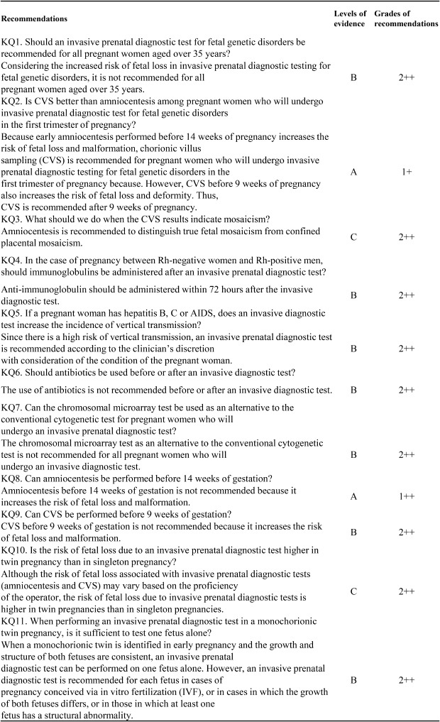
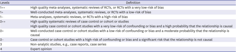
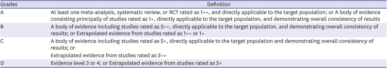
1. American College of Obstetricians and Gynecologists' Committee on Practice Bulletins–Obstetrics, Committee on Genetics. Society for Maternal-Fetal Medicine. Practice bulletin No. 162: prenatal diagnostic testing for genetic disorders. Obstet Gynecol. 2016; 127(5):e108–22.
2. Tabor A, Alfirevic Z. Update on procedure-related risks for prenatal diagnosis techniques. Fetal Diagn Ther. 2010; 27(1):1–7. PMID: 20051662.
crossref pmid
3. Alfirevic Z, Navaratnam K, Mujezinovic F. Amniocentesis and chorionic villus sampling for prenatal diagnosis. Cochrane Database Syst Rev. 2017; 9:CD003252. PMID: 28869276.
crossref
4. Centers for Disease Control and Prevention. Chorionic villus sampling and amniocentesis: recommendations for prenatal counseling. MMWR Recomm Rep. 1995; 44(RR-9):1–12.
5. Redman CW, Dollery E, Jordan JA. Development of the European colposcopy core curriculum: use of the Delphi technique. J Obstet Gynaecol. 2004; 24(7):780–784. PMID: 15763789.
crossref pmid
6. Ministry of Health & Welfare, Korean Academy of Medical Science. Korean Appraisal of Guidelines for Research & Evaluation II. Seoul: Korean Academy of Medical Science;2009.
7. SIGN, Network SIG. SIGN 50: a guideline developer's handbook. Updated 2020. Accessed May 6, 2020. https://www.sign.ac.uk/assets/sign_grading_system_1999_2012.pdf.
8. Ogawa K, Urayama KY, Tanigaki S, Sago H, Sato S, Saito S, et al. Association between very advanced maternal age and adverse pregnancy outcomes: a cross sectional Japanese study. BMC Pregnancy Childbirth. 2017; 17(1):349. PMID: 29017467.
crossref pmid pmc
9. Alldred SK, Takwoingi Y, Guo B, Pennant M, Deeks JJ, Neilson JP, et al. First trimester ultrasound tests alone or in combination with first trimester serum tests for Down's syndrome screening. Cochrane Database Syst Rev. 2017; 3:CD012600. PMID: 28295158.
crossref
10. Sheen JJ, Wright JD, Goffman D, Kern-Goldberger AR, Booker W, Siddiq Z, et al. Maternal age and risk for adverse outcomes. Am J Obstet Gynecol. 2018; 219(4):390.e1–390.15. PMID: 30153431.
crossref
11. Wilson RD, Davies G, Gagnon A, Desilets V, Reid GJ, Summers A, et al. Amended Canadian guideline for prenatal diagnosis (2005) change to 2005-techniques for prenatal diagnosis. J Obstet Gynaecol Can. 2005; 27(11):1048–1062. PMID: 16529673.
pmid
12. Audibert F, Gagnon A. No. 262-prenatal screening for and diagnosis of aneuploidy in twin pregnancies. J Obstet Gynaecol Can. 2017; 39(9):e347–61. PMID: 28859779.
crossref
13. Grati FR, Molina Gomes D, Ferreira JC, Dupont C, Alesi V, Gouas L, et al. Prevalence of recurrent pathogenic microdeletions and microduplications in over 9500 pregnancies. Prenat Diagn. 2015; 35(8):801–809. PMID: 25962607.
crossref pmid
14. Ghi T, Sotiriadis A, Calda P, Da Silva Costa F, Raine-Fenning N, Alfirevic Z, et al. ISUOG Practice Guidelines: invasive procedures for prenatal diagnosis. Ultrasound Obstet Gynecol. 2016; 48(2):256–268. PMID: 27485589.
crossref pmid
15. WHO/PAHO consultation on CVS, evaluation of chorionic villus sampling safety. Prenat Diagn. 1999; 19(2):97–99. PMID: 10215062.
pmid
16. Kuliev A, Jackson L, Froster U, Brambati B, Simpson JL, Verlinsky Y, et al. Chorionic villus sampling safety. Report of World Health Organization/EURO meeting in association with the Seventh International Conference on Early Prenatal Diagnosis of Genetic Diseases, Tel-Aviv, Israel, May 21, 1994. Am J Obstet Gynecol. 1996; 174(3):807–811. PMID: 8633647.
crossref pmid
17. The Canadian Early and Mid-trimester Amniocentesis Trial (CEMAT) Group. Randomised trial to assess safety and fetal outcome of early and midtrimester amniocentesis. Lancet. 1998; 351(9098):242–247. PMID: 9457093.
18. Winsor EJ, Tomkins DJ, Kalousek D, Farrell S, Wyatt P, Fan YS, et al. Cytogenetic aspects of the Canadian early and mid-trimester amniotic fluid trial (CEMAT). Prenat Diagn. 1999; 19(7):620–627. PMID: 10419609.
crossref pmid
19. Farrell SA, Summers AM, Dallaire L, Singer J, Johnson JA, Wilson RD. Club foot, an adverse outcome of early amniocentesis: disruption or deformation? CEMAT. Canadian Early and Mid-Trimester Amniocentesis Trial. J Med Genet. 1999; 36(11):843–846. PMID: 10544229.
pmid pmc
20. Sundberg K, Bang J, Smidt-Jensen S, Brocks V, Lundsteen C, Parner J, et al. Randomised study of risk of fetal loss related to early amniocentesis versus chorionic villus sampling. Lancet. 1997; 350(9079):697–703. PMID: 9291904.
crossref
21. Brambati B, Simoni G, Travi M, Danesino C, Tului L, Privitera O, et al. Genetic diagnosis by chorionic villus sampling before 8 gestational weeks: efficiency, reliability, and risks on 317 completed pregnancies. Prenat Diagn. 1992; 12(10):789–799. PMID: 1475247.
crossref pmid
22. Goldberg JD, Wohlferd MM. Incidence and outcome of chromosomal mosaicism found at the time of chorionic villus sampling. Am J Obstet Gynecol. 1997; 176(6):1349–1352. PMID: 9215195.
crossref pmid
23. Baffero GM, Somigliana E, Crovetto F, Paffoni A, Persico N, Guerneri S, et al. Confined placental mosaicism at chorionic villous sampling: risk factors and pregnancy outcome. Prenat Diagn. 2012; 32(11):1102–1108. PMID: 22961322.
crossref pmid
24. Tabor A, Jerne D, Bock JE. Incidence of rhesus immunisation after genetic amniocentesis. Br Med J (Clin Res Ed). 1986; 293(6546):533–536.
crossref
25. Murray JC., Karp LE, Williamson RA, Cheng EY, Luthy DA.Rh isoimmunization related to amniocentesis. Am J Med Genet. 1983; 16(4):527–534. PMID: 6419606.
crossref pmid
26. Royal College of Obstetricians and Gynaecologists. The Use of Anti-D Immunoglobulin for Rhesus D Prophylaxis. Green-top Guideline No. 22. London: RCOG;2011.
27. Centers for Disease Control. Hepatitis B virus: a comprehensive strategy for eliminating transmission in the United States through universal vaccination: recommendations of the Advisory Committee on Immunization Practices (ACIP). Part 1: immunization of infants, children, and adolescents. MMWR Morb Mortal Wkly Rep. 1991; 40(RR-13):1–19. PMID: 1898620.
pmid
28. Yi W, Pan CQ, Hao J, Hu Y, Liu M, Li L, et al. Risk of vertical transmission of hepatitis B after amniocentesis in HBs antigen-positive mothers. J Hepatol. 2014; 60(3):523–529. PMID: 24269471.
crossref pmid
29. Towers CV, Asrat T, Rumney P. The presence of hepatitis B surface antigen and deoxyribonucleic acid in amniotic fluid and cord blood. Am J Obstet Gynecol. 2001; 184(7):1514–1518. PMID: 11408875.
crossref pmid
30. Grosheide PM, Quartero HW, Schalm SW, Heijtink RA, Christiaens GC. Early invasive prenatal diagnosis in HBsAg-positive women. Prenat Diagn. 1994; 14(7):553–558. PMID: 7971756.
crossref pmid
31. Gagnon A, Davies G, Wilson RD, Audibert F, Brock JA, Campagnoto C, et al. Prenatal invasive procedures in women with hepatitis B, hepatitis C, and/or human immunodeficiency virus infections. J Obstet Gynaecol Can. 2014; 36(7):648–653. PMID: 25184985.
crossref pmid
32. American College of Obstetricians and Gynecologists. ACOG Practice Bulletin No. 88, December 2007. Invasive prenatal testing for aneuploidy. Obstet Gynecol. 2007; 110(6):1459–1467. PMID: 18055749.
pmid
33. Delamare C, Carbonne B, Heim N, Berkane N, Petit JC, Uzan S, et al. Detection of hepatitis C virus RNA (HCV RNA) in amniotic fluid: a prospective study. J Hepatol. 1999; 31(3):416–420. PMID: 10488698.
crossref pmid
34. Tess BH, Rodrigues LC, Newell ML, Dunn DT, Lago TD. Breastfeeding, genetic, obstetric and other risk factors associated with mother-to-child transmission of HIV-1 in Sao Paulo State, Brazil. Sao Paulo Collaborative Study for Vertical Transmission of HIV-1. AIDS. 1998; 12(5):513–520. PMID: 9543450.
pmid
35. Maiques V, García-Tejedor A, Perales A, Córdoba J, Esteban RJ. HIV detection in amniotic fluid samples. Amniocentesis can be performed in HIV pregnant women? Eur J Obstet Gynecol Reprod Biol. 2003; 108(2):137–141. PMID: 12781400.
pmid
36. Mandelbrot L, Jasseron C, Ekoukou D, Batallan A, Bongain A, Pannier E, et al. ANRS French Perinatal Cohort (EPF). Amniocentesis and mother-to-child human immunodeficiency virus transmission in the Agence Nationale de Recherches sur le SIDA et les H'epatites Virales French Perinatal Cohort. Am J Obstet Gynecol. 2009; 200:160.e1–160.e9. PMID: 18986640.
37. Somigliana E, Bucceri AM, Tibaldi C, Alberico S, Ravizza M, Savasi V, et al. Early invasive diagnostic techniques in pregnant women who are infected with the HIV: a multicenter case series. Am J Obstet Gynecol. 2005; 193(2):437–442. PMID: 16098867.
crossref pmid
38. Shapiro DE, Sperling RS, Mandelbrot L, Britto P, Cunningham BE. Risk factors for perinatal human immunodeficiency virus transmission in patients receiving zidovudine prophylaxis. Pediatric AIDS Clinical Trials Group protocol 076 Study Group. Obstet Gynecol. 1999; 94(6):897–908. PMID: 10576173.
pmid
39. Society for Maternal-Fetal Medicine (SMFM). Berry SM, Stone J, Norton ME, Johnson D, Berghella V. Fetal blood sampling. Am J Obstet Gynecol. 2013; 209(3):170–180. PMID: 23978246.
crossref pmid
40. Royal College of Obstetricians and Gynaecologists. Amniocentesis and chorionic villus sampling: green-top guideline No. 8. Updated 2013. Accessed May 6, 2020. https://www.rcog.org.uk/globalassets/documents/guidelines/gtg_8.pdf.
41. Giorlandino C, Cignini P, Cini M, Brizzi C, Carcioppolo O, Milite V, et al. Antibiotic prophylaxis before second-trimester genetic amniocentesis (APGA): a single-centre open randomised controlled trial. Prenat Diagn. 2009; 29(6):606–612. PMID: 19294678.
crossref pmid
42. Alfirevic Z, Pilu G. Antibiotic prophylaxis for amniocentesis. Prenat Diagn. 2009; 29(11):1094. PMID: 19859907.
crossref pmid
43. Ferrazzi E. Antibiotic prophylaxis before second-trimester genetic amniocentesis. Prenat Diagn. 2010; 30(2):188. PMID: 20101625.
crossref pmid
44. Hobbins JC, Pilu G, Abuhumad A, Alfirevic Z, Bahado-Singh RO, Benacerraf BR, et al. Antibiotic prophylaxis before amniocentesis. Prenat Diagn. 2011; 31(12):1213–1214. PMID: 22120439.
crossref pmid
45. Gramellini D, Fieni S, Casilla G, Raboni S, Nardelli GB. Mid-trimester amniocentesis and antibiotic prophylaxis. Prenat Diagn. 2007; 27(10):956–959. PMID: 17607664.
crossref pmid
46. Mujezinovic F, Alfirevic Z. Technique modifications for reducing the risks from amniocentesis or chorionic villus sampling. Cochrane Database Syst Rev. 2012; 8(8):CD008678.
crossref
47. Wilson RD, Gagnon A, Audibert F, Campagnolo C, Carroll J, Wilson RD, et al. Prenatal diagnosis procedures and techniques to obtain a diagnostic fetal specimen or tissue: maternal and fetal risks and benefits. J Obstet Gynaecol Can. 2015; 37(7):656–668. PMID: 26366824.
crossref pmid
48. Boulot P, Deschamps F, Lefort G, Sarda P, Mares P, Hedon B, et al. Pure fetal blood samples obtained by cordocentesis: technical aspects of 322 cases. Prenat Diagn. 1990; 10(2):93–100. PMID: 2343027.
crossref
49. Johnstone-Ayliffe C, Prior T, Ong C, Regan F, Kumar S. Early procedure-related complications of fetal blood sampling and intrauterine transfusion for fetal anemia. Acta Obstet Gynecol Scand. 2012; 91(4):458–462. PMID: 22356474.
pmid
50. Tangshewinsirikul C, Wanapirak C, Piyamongkol W, Sirichotiyakul S, Tongsong T. Effect of cord puncture site in cordocentesis at mid-pregnancy on pregnancy outcomes. Prenat Diagn. 2011; 31(9):861–864. PMID: 21706506.
crossref pmid
51. Tongsong T, Wanapirak C, Kunavikatikul C, Sirirchotiyakul S, Piyamongkol W, Chanprapaph P. Cordocentesis at 16-24 weeks of gestation: experience of 1,320 cases. Prenat Diagn. 2000; 20(3):224–228. PMID: 10719326.
crossref pmid
52. Aina-Mumuney AJ, Holcroft CJ, Blakemore KJ, Bienstock JL, Hueppchen NA, Milio LA, et al. Intrahepatic vein for fetal blood sampling: one center's experience. Am J Obstet Gynecol. 2008; 198(4):387.e1–387.e6. PMID: 18191806.
crossref
53. Wapner RJ, Martin CL, Levy B, Ballif BC, Eng CM, Zachary JM, et al. Chromosomal microarray versus karyotyping for prenatal diagnosis. N Engl J Med. 2012; 367(23):2175–2184. PMID: 23215555.
crossref
54. Jansen FA, Blumenfeld YJ, Fisher A, Cobben JM, Odibo AO, Borrell A, et al. Array comparative genomic hybridization and fetal congenital heart defects: a systematic review and meta-analysis. Ultrasound Obstet Gynecol. 2015; 45(1):27–35. PMID: 25319878.
crossref pmid
55. Grande M, Jansen FA, Blumenfeld YJ, Fisher A, Odibo AO, Haak MC, et al. Genomic microarray in fetuses with increased nuchal translucency and normal karyotype: a systematic review and meta-analysis. Ultrasound Obstet Gynecol. 2015; 46(6):650–658. PMID: 25900824.
crossref pmid
56. Callaway JL, Shaffer LG, Chitty LS, Rosenfeld JA, Crolla JA. The clinical utility of microarray technologies applied to prenatal cytogenetics in the presence of a normal conventional karyotype: a review of the literature. Prenat Diagn. 2013; 33(12):1119–1123. PMID: 23983223.
crossref pmid pmc
57. Reddy UM, Page GP, Saade GR, Silver RM, Thorsten VR, Parker CB, et al. Karyotype versus microarray testing for genetic abnormalities after stillbirth. N Engl J Med. 2012; 367(23):2185–2193. PMID: 23215556.
crossref
58. Society for Maternal-Fetal Medicine (SMFM). Dugoff L, Norton ME, Kuller JA. The use of chromosomal microarray for prenatal diagnosis. Am J Obstet Gynecol. 2016; 215(4):B2–9.
59. Committee on Genetics and the Society for Maternal-Fetal Medicine. Committee opinion No.682: microarrays and next-generation sequencing technology: the use of advanced genetic diagnostic tools in obstetrics and gynecology. Obstet Gynecol. 2016; 128(6):e262–8. PMID: 27875474.
60. Borrell A, Costa D, Delgado RD, Fuster JJ, Soler A, Cararach J, et al. Transcervical chorionic villus sampling beyond 12 weeks of gestation. Ultrasound Obstet Gynecol. 1996; 7(6):416–420. PMID: 8807757.
crossref pmid
61. Botto LD, Olney RS, Mastroiacovo P, Khoury MJ, Moore CA, Alo CJ, et al. Chorionic villus sampling and transverse digital deficiencies: evidence for anatomic and gestational-age specificity of the digital deficiencies in two studies. Am J Med Genet. 1996; 62(2):173–178. PMID: 8882399.
crossref pmid
62. Calzolari E, Manservigi D, Garani GP, Cocchi G, Magnani C, Milan M. Limb reduction defects in Emilia Romagna, Italy: epidemiological and genetic study in 173,109 consecutive births. J Med Genet. 1990; 27(6):353–357. PMID: 2359096.
crossref pmid pmc
63. Olney RS, Khoury MJ, Alo CJ, Costa P, Edmonds LD, Flood TJ, et al. Increased risk for transverse digital deficiency after chorionic villus sampling: results of the United States Multistate Case-Control Study, 1988–1992. Teratology. 1995; 51(1):20–29. PMID: 7597654.
crossref pmid
64. Millaire M, Bujold E, Morency AM, Gauthier RJ. Mid-trimester genetic amniocentesis in twin pregnancy and the risk of fetal loss. J Obstet Gynaecol Can. 2006; 28(6):512–518. PMID: 16857119.
crossref pmid
65. Lenis-Cordoba N, Sánchez MA, Bello-Muñoz JC, Sagalá-Martinez J, Campos N, Carreras-Moratonas E, et al. Amniocentesis and the risk of second trimester fetal loss in twin pregnancies: results from a prospective observational study. J Matern Fetal Neonatal Med. 2013; 26(15):1537–1541. PMID: 23544929.
crossref pmid
66. Cahill AG, Macones GA, Stamilio DM, Dicke JM, Crane JP, Odibo AO. Pregnancy loss rate after midtrimester amniocentesis in twin pregnancies. Am J Obstet Gynecol. 2009; 200(3):257.e1–257.e6. PMID: 19136086.
crossref
67. Agarwal K, Alfirevic Z. Pregnancy loss after chorionic villus sampling and genetic amniocentesis in twin pregnancies: a systematic review. Ultrasound Obstet Gynecol. 2012; 40(2):128–134. PMID: 22125091.
crossref pmid
68. Wapner RJ, Johnson A, Davis G, Urban A, Morgan P, Jackson L. Prenatal diagnosis in twin gestations: a comparison between second-trimester amniocentesis and first-trimester chorionic villus sampling. Obstet Gynecol. 1993; 82(1):49–56. PMID: 8515925.
crossref pmid
69. Simonazzi G, Curti A, Farina A, Pilu G, Bovicelli L, Rizzo N. Amniocentesis and chorionic villus sampling in twin gestations: which is the best sampling technique? Am J Obstet Gynecol. 2010; 202(4):365.e1–365.e5. PMID: 20060095.
crossref
70. Audibert F, Gagnon A, Douglas Wilson R, Audibert F, Blight C, Brock JA, et al. Prenatal screening for and diagnosis of aneuploidy in twin pregnancies. J Obstet Gynaecol Can. 2011; 33(7):754–767. PMID: 21749753.
pmid
71. Evans MI, Goldberg JD, Horenstein J, Wapner RJ, Ayoub MA, Stone J, et al. Selective termination for structural, chromosomal, and mendelian anomalies: international experience. Am J Obstet Gynecol. 1999; 181(4):893–897. PMID: 10521749.
crossref pmid
72. Casals G, Borrell A, Martínez JM, Soler A, Cararach V, Fortuny A. Transcervical chorionic villus sampling in multiple pregnancies using a biopsy forceps. Prenat Diagn. 2002; 22(3):260–265. PMID: 11920906.
crossref pmid
73. Kidd SA, Lancaster PA, Anderson JC, Boogert A, Fisher CC, Robertson R, et al. A cohort study of pregnancy outcome after amniocentesis in twin pregnancy. Paediatr Perinat Epidemiol. 1997; 11(2):200–213. PMID: 9131711.
crossref pmid
74. McFadyen I. The dangers of intra-amniotic methylene blue. Br J Obstet Gynaecol. 1992; 99(2):89–90. PMID: 1554680.
crossref pmid
75. Morin L, Lim K, Morin L, Lim K, Bly S, Butt K, et al. Ultrasound in twin pregnancies. J Obstet Gynaecol Can. 2011; 33(6):643–656. PMID: 21846456.
crossref pmid
76. Weisz B, Rodeck CH. Invasive diagnostic procedures in twin pregnancies. Prenat Diagn. 2005; 25(9):751–758. PMID: 16170858.
crossref pmid
77. Grande M, Goncé A, Stergiotou I, Bennasar M, Borrell A. Intertwin crown-rump length discordance in the prediction of fetal anomalies, fetal loss and adverse perinatal outcome. J Matern Fetal Neonatal Med. 2016; 29(17):2883–2888. PMID: 26466907.
crossref pmid
78. Litwinska E, Syngelaki A, Cimpoca B, Sapantzoglou I, Nicolaides KH. Intertwin discordance in fetal size at 11–13 weeks' gestation and pregnancy outcome. Ultrasound Obstet Gynecol. 2020; 55(2):189–197. PMID: 31710737.
crossref pmid
79. Harper LM, Roehl KA, Odibo AO, Cahill AG. First-trimester growth discordance and adverse pregnancy outcome in dichorionic twins. Ultrasound Obstet Gynecol. 2013; 41(6):627–631. PMID: 22744892.
crossref pmid
80. Eddleman KA, Stone JL, Lynch L, Berkowitz RL. Chorionic villus sampling before multifetal pregnancy reduction. Am J Obstet Gynecol. 2000; 183(5):1078–1081. PMID: 11084544.
crossref pmid
81. Brambati B, Tului L, Guercilena S, Alberti E. Outcome of first-trimester chorionic villus sampling for genetic investigation in multiple pregnancy. Ultrasound Obstet Gynecol. 2001; 17(3):209–216. PMID: 11309169.
crossref pmid
82. van den Berg C, Braat AP, Van Opstal D, Halley DJ, Kleijer WJ, den Hollander NS, et al. Amniocentesis or chorionic villus sampling in multiple gestations? Experience with 500 cases. Prenat Diagn. 1999; 19(3):234–244. PMID: 10210122.
crossref pmid
83. Schmid O, Trautmann U, Ashour H, Ulmer R, Pfeiffer RA, Beinder E. Prenatal diagnosis of heterokaryotypic mosaic twins discordant for fetal sex. Prenat Diagn. 2000; 20(12):999–1003. PMID: 11113914.
crossref pmid
84. Shalev SA, Shalev E, Pras E, Shneor Y, Gazit E, Yaron Y, et al. Evidence for blood chimerism in dizygotic spontaneous twin pregnancy discordant for Down syndrome. Prenat Diagn. 2006; 26(9):782–784. PMID: 16927328.
crossref pmid
TOOLS
Similar articles