

 PDF
PDF Citation
 Print
Print


Dear Editor,
Jumping translocations (JTs) are rare but recurrent cytogenetic abnormalities in hematological malignancies and solid tumors [1]. Of the JTs reported in hematological malignancies, those involving chromosome 3q are the second most frequently observed donor segment [2-4]. However, to date, the impact of JTs involving 3q13.3 on the pathogenesis of leukemia has not been elucidated [4]. We describe the first RNA-sequencing (RNA-seq) data of a de novo acute myeloid leukemia (AML) patient with a JT involving 3q13.3 and trisomy 8 to investigate the structural variation and altered gene expression associated with JTs involving 3q13.3. The Institutional Review Board (IRB) of Samsung Medical Center, Seoul, Korea, approved this study (IRB No. SMC 2020-02-145-001). Informed consent for genetic studies were obtained from the patient before his death.
The patient was a 72-year-old man who was referred for a bone marrow (BM) study at Samsung Medical Center in April 2018 owing to the observation of 30% blasts on a peripheral blood smear. The BM study revealed 66% of blast cells with monocytoid differentiation. The patient was diagnosed as having AML, not otherwise specified, subtype acute monocytic leukemia. The patient was treated with decitabine for induction therapy due to his age; however, he died during the re-induction therapy owing to leukostasis and tumor lysis syndrome.
Conventional karyotyping was performed as previously described [5], and fluorescence in situ hybridization (FISH) analysis was performed on interphase nuclei using the LSI RPN1 SpectrumGreen/MECOM SpectrumOrange Probe (Vysis, Abbott Park, IL, USA), according to the manufacturer’s instructions. Copy number variation was analyzed using the CytoScan Dx Assay (Affymetrix Inc., Thermo Fisher Scientific, Santa Clara, CA, USA), according to the manufacturer’s instructions.
RNA was extracted from a BM aspirate using the RevertAid First Strand cDNA Synthesis Kit (Thermo Fisher Scientific, Waltham, MA, USA). Paired-end libraries were prepared using the TruSeq RNA Sample Prep Kit (V2; Illumina, San Diego, CA, USA) and sequenced using a HiSeq 2500 instrument (Illumina). The sequence reads were mapped using STAR (version 2.4.0; https:// github.com/alexdobin/STAR) and quantified using RSEM (version 1.3.1; https://github.com/deweylab/RSEM). RSEM was also applied to generate the normalized gene expression in terms of transcripts per million values and to quantify the isoform-level expression of LSAMP. RNA expression was analyzed using Student t-test for each gene, and P < 0.05 was considered signifi cant; the fusion transcript was detected using ChimeraScan (version 0.4.5) and deFuse (version 0.6.2). The fusion transcript detected by RNA-seq was confirmed by Sanger sequencing.
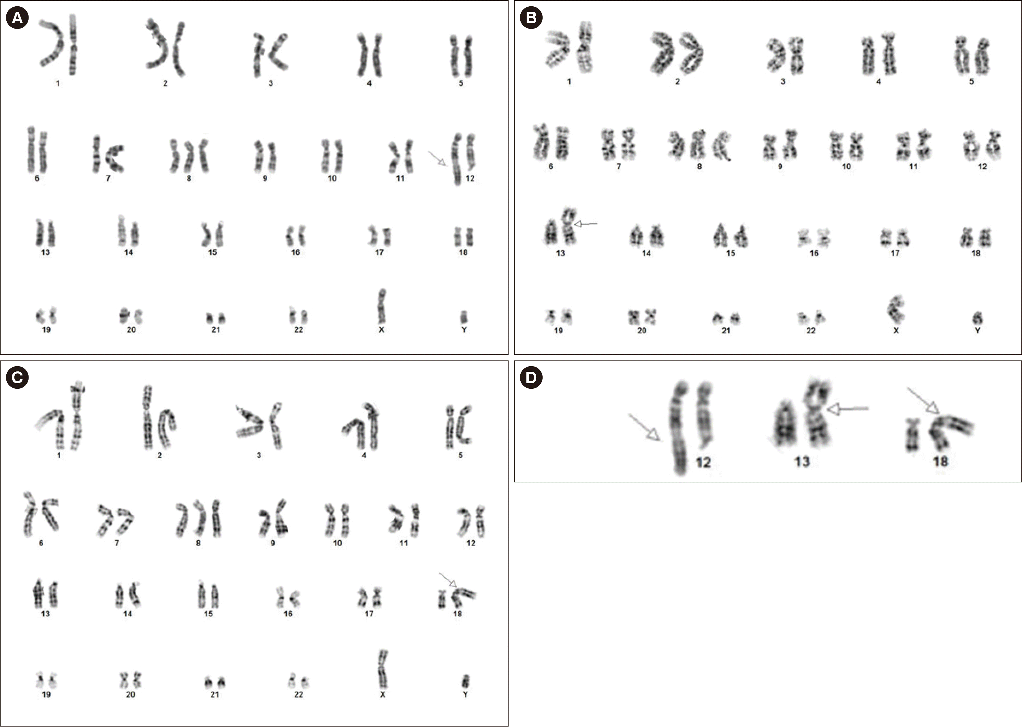
Conventional cytogenetic analysis of the BM aspirate demonstrated three related cytogenetically aberrant clones (Fig. 1). The jumping donor chromosome bands ranging from 3q13.3 to qter translocated to three recipient chromosome regions: 12q24.3, 13p13, and 18p11.3. The G-banding karyotypic results were: 47,XY,+8,der(18)t(3;18)(q13.3;p11.3)[10]/47,XY,+8,der(12) t(3;12)(q13.3;q24.3)[4]/47,XY,+8,der(13)t(3;13)(q13.3;p13) [4]/46,XY[2]. FISH analysis using RPN1/MECOM, a dual color dual fusion probe, detected 24.0% of cells (48/200) with three MECOM signals on 3q26.2.
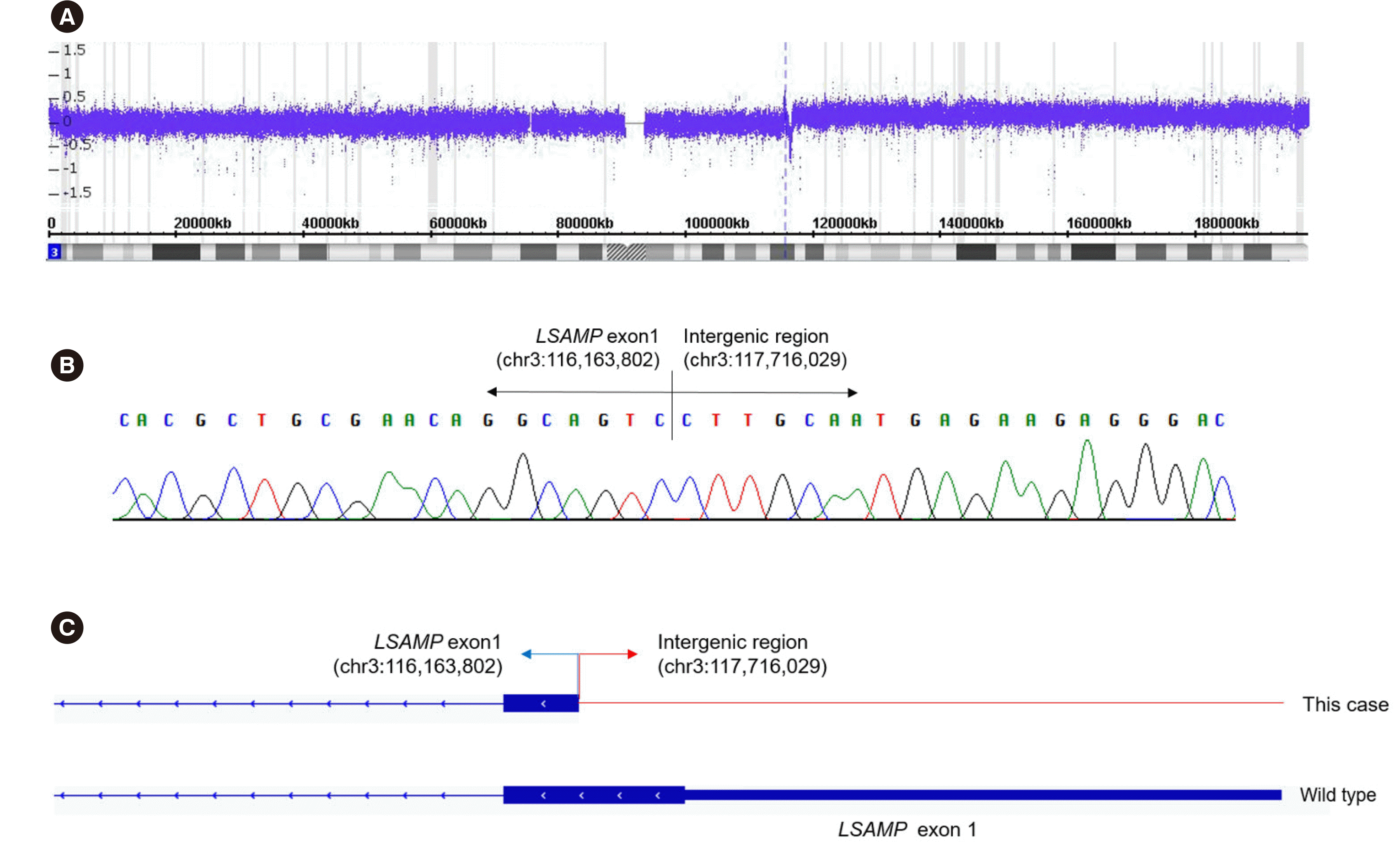
The chromosomal microarray showed partial trisomy of 3q13. 32-q29 (genomic position 117,967,114–197,851,986) and a single copy gain of 8p23.3-8q24.3 (genomic position 158,048– 146,295,771), which likely indicates trisomy 8. Moreover, a 385kb submicroscopic interstitial deletion at 3q13.31 (genomic position 116,234,782–116,620,270) was observed.
RNA-seq was performed to analyze the structural changes formed via the JT. A breakpoint resulting in a fusion between LSAMP exon 1 located on chromosome 3q13.31 and the intergenic region located at genomic position chr3:117,716,029 was observed (Fig. 2). The breakpoint was localized near the 3´ end of exon 1, spanning the 5´ untranslated region and the start codon of the LSAMP gene.
Gene expression was compared with the RNA expression data of 3q13.3 JT-negative AML patients. AML with a JT involving 3q13.3 showed significantly higher expression of LSAMP (P = 0.005). Quantification of the isoform-level gene expression of LSAMP revealed that isoform 1 transcripts were significantly increased (P = 0.003), whereas isoform 2 transcripts did not significantly differ. Of the genes located at the JT breakpoint, DL-GAP1, located on chromosome 18p11.31, showed overexpression. Overexpression of genes located on chromosome 3q (ACPP, NUDT16P, and EPHB3) or chromosome 8 (CHRNA6, ENPP2, MAPK15, and SDC2) was also observed.
JT-caused truncation or loss of the gene located near the donor chromosome breakpoint has been observed [6]. LSAMP is a putative tumor suppressor gene, previously reported to be inactivated by recurrent deletion and translocation in various cancer types [7-10]. Although LSAMP is mostly downregulated [7-10], Yen, et al. [9] reported that the correlation between LSAMP deletion and expression varied depending on the isoforms measured. In our study, LSAMP upregulation was detected, and its expression varied between its isoforms, suggesting alternative mRNA transcription or premature truncation. Our data showed the possibility that the rearrangement and aberrant LSAMP expression could be associated with the pathogenesis of AML with a JT involving 3q13.3.
 XML Download