Journal List > Lab Med Online > v.11(1) > 1146340

한국인 신경섬유종증 1형 환자에서 관찰된 NF1 유전자 변이 분포

초록

배경

신경섬유종증 1형(NF1)은 가장 높은 빈도로 발생하는 상염색체 우성 유전질환의 하나로, NF1 유전자에 다양한 질병연관변이가 발생하는 것으로 보고되었다. 본 연구는 한국인 NF1 환자에서 NF1 유전자 변이의 빈도와 특성을 확인하고자 수행되었다.

방법

본 연구에는 서울성모병원에 의뢰된 NF1 유전자 검사 462건이 포함되었다. NF1 유전자 변이는 messenger RNA (mRNA) 및 일부 genomic DNA 직접염기서열분석법과 multiplex ligation-dependent probe amplification (MLPA) 방법으로 분석하였다.

결과

전체 462명 중 303명(65.6%)의 환자에서 231종류의 변이를 검출하였다. 217종류의 변이를 질병연관변이로 보고하였고, 이 중106개(48.4%)의 변이는 이전 보고가 없는 새로운 변이였다. 가장 많은 환자들에게서 검출된 변이는 절단형 돌연변이로, 177명의 환자에서 135종류의 변이를 검출하였다. 짜깁기 변이는 42명의 환자에서 39종류, 과오변이는 44명의 환자에서 36종류가 검출되었다. MLPA에서만 확인되었던 엑손 단위 이상의 큰 결실은 25명의 환자에서 8종류가 관찰되었다. mRNA 분석에서 4건의 aberrant transcript를 발견하였다. NF1 유전자의 변이 검출 률은 NIH 진단기준 충족 수에 따라 유의하게 높았다.

결론

본 연구에서는 한국인 NF1 환자가 지닌 다양한 종류의 변이를 검출하였다. 이러한 다양한 스펙트럼의 변이를 검출하기 위해서 mRNA 및 gDNA에 대한 염기서열분석과 MLPA 검사를 통합적으로 수행하여야 하며 새로 검출한 변이를 규명하기 위해 추가적 평가가 필요할 것으로 생각된다.

Abstract

Background

Neurofibromatosis type 1 (NF1) is one of the most common autosomal dominant disorders in humans, and many different variants in the NF1 gene have been observed. The aim of this study was to investigate the genetic variant spectrum of NF1 patients in Korea.

Methods

A total of 462 cases were enrolled for NF1 analysis at Seoul St. Mary’s Hospital. NF1 was analyzed through Sanger sequencing of messenger RNA (mRNA) and genomic DNA (gDNA), and/or multiplex ligation-dependent probe amplification (MLPA) analysis.

Results

We identified 231 types of NF1 variants in 303 out of the 462 patients (65.6%). Of these, 217 variants were classified as pathogenic or likely pathogenic, and 48.4% (N=106) of these were novel changes. Truncating variants encompassing frameshift and nonsense variants were most commonly observed (135 types of variants in 177 patients), followed by splicing defect variants (39 types in 42 patients) and missense variants (36 types in 44 patients). There were 8 distinctive large deletions in 25 patients, detected via MLPA analysis. Interestingly, 4 cases showed aberrant transcripts that were identified through mRNA Sanger sequencing. The frequency of NF1 mutation detected was significantly higher according to the number of satisfying National Institutes of Health (NIH) diagnostic criteria.

Conclusions

This study revealed a wide spectrum of NF1 variants in Korean NF1 patients. A comprehensive analytical strategy that combines mRNA and gDNA analyses and MLPA is required to detect sequence variants. Additionally, it is important to define the effect of newly detected variants on the clinical course.

서 론

신경섬유종증 1형(neurofibromatosis type 1, NF1)은 2,500-3,000명당 1명 정도의 빈도로 발병하는 상염색체 우성 유전질환이다. 환자의 절반 정도는 가족성으로 발생하고 나머지는 가족력 없이 산발성으로 발생하며[1, 2], 17번 염색체 장완에 위치한 NF1 유전자의 변이가 주 원인으로 알려져 있다[2, 3]. NF1 유전자는 60개의 엑손으로 구성된 유전자로, 항상 mRNA로 전사되는 57개의 엑손과 그 외 3개의 엑손이 11 kb에서 13 kb 크기의 mRNA transcript를 형성한다. Genomic DNA (gDNA) 수준에서는 350 kb 정도를 차지하며, 염기의 유사성이 높아 검사 과정에 혼란을 야기할 수 있는 30여 종 이상의 위유전자(pseudogene)가 존재하기 때문에, 진단 오류를 방지하기 위해 mRNA를 이용한 염기서열분석이 일반적으로 수행되고 있다[4-6].
NF1 유전자는 종양억제유전자인 neurofibromin 1 단백질을 코딩하고 있으며, 그 기능을 소실할 경우 피부반점, 액와/서혜부 주근깨, 시신경교종, 특징적 골격계 이상 및 양성 신경섬유종에서 악성 신경초종(malignant peripheral nerve sheath tumor)에 이르는 종양이 발생할 수 있다[1, 2]. 지금까지 3,000여 종 이상의 NF1 변이가 Human Genome Mutation Database (HGMD)에 보고되었으며, 변이의 종류는 하나의 아미노산 과오변이(missense mutation)에서 전체 엑손 결실(deletion)까지 다양하며, 전형적인 인트론 위치(canonical splice acceptor and donor sites) 외에서도 짜깁기 변이가 발생하는 것으로 알려져 있다[7]. 따라서 NF1 환자에서의 유전학적 진단을 위해서는 엑손 및 deep intron을 포함한 여러 짜깁기 변이를 모두 검출할 수 있는 mRNA 분석을 시행하며, large gene rearrangement를 검출할 수 있는 Multiplex Ligation Dependent Probe Amplification (MLPA)을 적용할 필요가 있다[3, 4, 6, 8].
현재까지 국내에서 진행된 NF1 환자의 유전진단에 대한 연구는 대상 환자의 수가 적고 특정 검사법 만을 적용하는 등 전반적인 변이를 파악하는 데에는 다소 제한이 있다[9-13]. 본 연구에서는 gDNA와 mRNA 염기서열분석 및 MLPA를 이용하여 한국인 NF1 환자의 유전자 변이 분포를 파악하고자 하였다.

재료 및 방법

1. 대상 환자

2010년 2월부터 2019년 5월까지 서울성모병원에 NF1 유전자 검사가 의뢰된 488건 중 소아 혈액암 25건과 최종 판독 결과를 보고하지 않은 1건을 제외한 462건에 대해서 분석하였다. 의뢰된 검사 중 401건은 NF1 감별을 위해 의뢰되었으나 구체적인 임상정보를 얻을 수 없었다. 임상정보를 확인할 수 있는 61건의 경우, 환자의 진단 시 나이, 성별을 포함한 의무기록을 후향적으로 검토하여 표현형 데이터를 수집하였다. 이 중 25명의 환자가 NIH consensus development conference (1988)[2]에서 제시한 진단기준 중 2개 이상을 만족하여 NF1 확진을 받고 검사가 의뢰되었고, 진단기준 중 하나를 만족하여 NF1 감별을 위해 의뢰된 환자는 30명, 그 외 6명의 경우 위의 진단기준에 해당하는 항목이 없었으나 감별 목적으로 본 검사가 의뢰되었다. 연구에 포함된 환자 중 형제 관계일 것으로 추정되는 두 명의 환자(L075, L076) 이외에 명백한 혈연관계는 없었다. 본 연구는 가톨릭중앙의료원 임상연구심사위원회의 승인(KC19RESI0720)을 얻어 연구 윤리를 준수하여 시행하였다.

2. NF1 유전자 변이 검출 방법

1) mRNA 직접염기서열분석(Sanger sequencing)

환자의 말초혈액에서 High Pure RNA Isolation Kit (Roche, Mann-heim, Germany)를 이용하여 RNA를 분리하고 Transcriptor First Strand cDNA Synthesis Kit (Roche)로 cDNA를 합성하였다. 염기서열분석을 위한 시발체(primer)는 Primer 3 (http://bioinfo.ut.ee/primer3)를 이용하여 제작하였다. 중합효소연쇄반응(polymerase chain reaction, PCR)은 Thermal Cycler C1000 또는 S1000 (Bio-Rad, Foster City, CA, USA)에서 96°C 5분 반응시킨 후 95°C 30초, 58°C 30초, 72°C 60초 조건을 34회 반복하고 72°C에서 5분간 반응시켜 이후 4°C에서 유지하였다. PCR 산물을 전기영동으로 확인하고 3500xL Genetic Analyzer (Thermo Fisher Scientific, Foster City, CA, USA)으로 염기서열을 분석하였다. mRNA 분석 시 양호한 염기서열분석 결과를 얻기 어려운 부위인 엑손 19, 38, 39, 49, 52는 gDNA를 추가로 분석하였다. mRNA 염기서열분석에서 변이가 검출된 경우 해당 부위에 대한 gDNA 염기서열분석을 시행하여 확인하였다.
NF1 유전자는 NM_001042492.2를 기준으로 판독하여 기술하였다. 검출된 변이는 HGMD (Human Genome Mutation Database, Institute of Medical Genetics, Cardiff, Wales; http://www.hgmd.cf.ac.uk/ac/index.php), ClinVar (https://www.ncbi.nlm.nih.gov/clinvar/), dbSNP (https://www.ncbi.nlm.nih.gov/snp/) 데이터베이스에서 기존 보고 여부를 확인하고 ExAC, gnomAD, 1000 Genomes 데이터베이스에서 인구집단별 해당 변이의 빈도를 조사하였다. 네 가지 in-silico (Align GVHD, MutationTaster, PolyPhen, SIFT) 분석을 시행하였고, 이를 American College of Medical Genetics and Genomics (ACMG) 변이의 해석에 대한 종합적 권고지침[14]에 따라 pathogenic, likely pathogenic, variant of unknown significance (VUS), likely benign, benign으로 분류하였다. 질병연관 가능성이 높은 것으로 간주(PVS1)되는 무의미 변이, 틀이동 변이, 짜깁기 공여자/수여자 위치 변이, 시작 코돈 위치의 변이, 엑손 단위 이상의 큰 결실은 pathogenic 혹은 likely pathogenic으로 분류하였고, 선행된 연구에서 질병연관변이로 확인된 과오변이도 pathogenic으로 분류하였다. 기존에 보고되지 않았지만 해당 인구집단에서의 빈도가 낮은 과오변이, in-frame insertion 또는 in-frame deletion의 경우 동일한 위치에서의 아미노산 치환이 pathogenic으로 보고되었거나, 종간 보존성이 높은 위치에서의 변이로 다수의 in-silico 분석에서 질병과 관계가 높을 것으로 예측되는 변이는 likely pathogenic으로 분류하였다. 그 외의 변이는 미분류염기변이(variant of unknown significance, VUS)로 분류하였다.

2) Multiplex Ligation Dependent Probe Amplification (MLPA)

NF1 유전자의 엑손 단위 결실이나 중복은 SALSA MLPA Probemix P081-D1 NF1 mix 1 & P082-Cw NF1 mix2 (MRC-Holland, Amsterdam, The Netherlands)를 이용하여 분석하였다. DNA에 probe를 첨가하여 어닐링(annealing) 하고 PCR로 증폭한 후 3500xL Genetic Analyzer (Thermo Fisher Scientific)로 분석하고 GeneMarker version 1.91 (SoftGenetics, State College, PA, USA)로 판독하였다. 각각의 peak를 정상 대조군과 비교하여 비율이 0.7 미만은 결손, 1.3을 초과하면 중복으로 평가하였고[15], 결손의 비율이 0일 경우 동형접합(homozygous) 결손, 0.40-0.65인 경우 이형접합(he-terozygous) 결손으로 판독하였다.

3. 통계

NF1 유전자 변이 유무에 따라 진단 시 연령 및 표현형을 비교하였다. SPSS ver 25.0 (SPSS Inc., Chicago, IL, USA)을 이용하여 Student’s t-test, 카이제곱 검정(chi-square test) 또는 Fisher의 정확한 검정(Fisher’s exact test)을 시행하였다. P값 0.05 미만을 유의한 것으로 판단하였다.

결 과

1. NF1 유전자 변이 빈도 및 분포

분석된 462명 중 303명(65.6%)에서 NF1 유전자의 질병연관변이가 검출되었다. 그 중 182명에서 검출된 111개의 변이는 기존에 보고된 변이였고, 나머지 121명에서 120개의 새로운 변이가 발견되었다. 분석방법에 따라서는 mRNA 및 gDNA 염기서열분석을 통해 검출된 경우가 278명(91.3%)로 대부분을 차지하였고, MLPA에서만 검출 가능했던 엑손 단위 이상의 큰 결실은 25명(8.7%)의 환자에서 관찰되었다(Table 1).
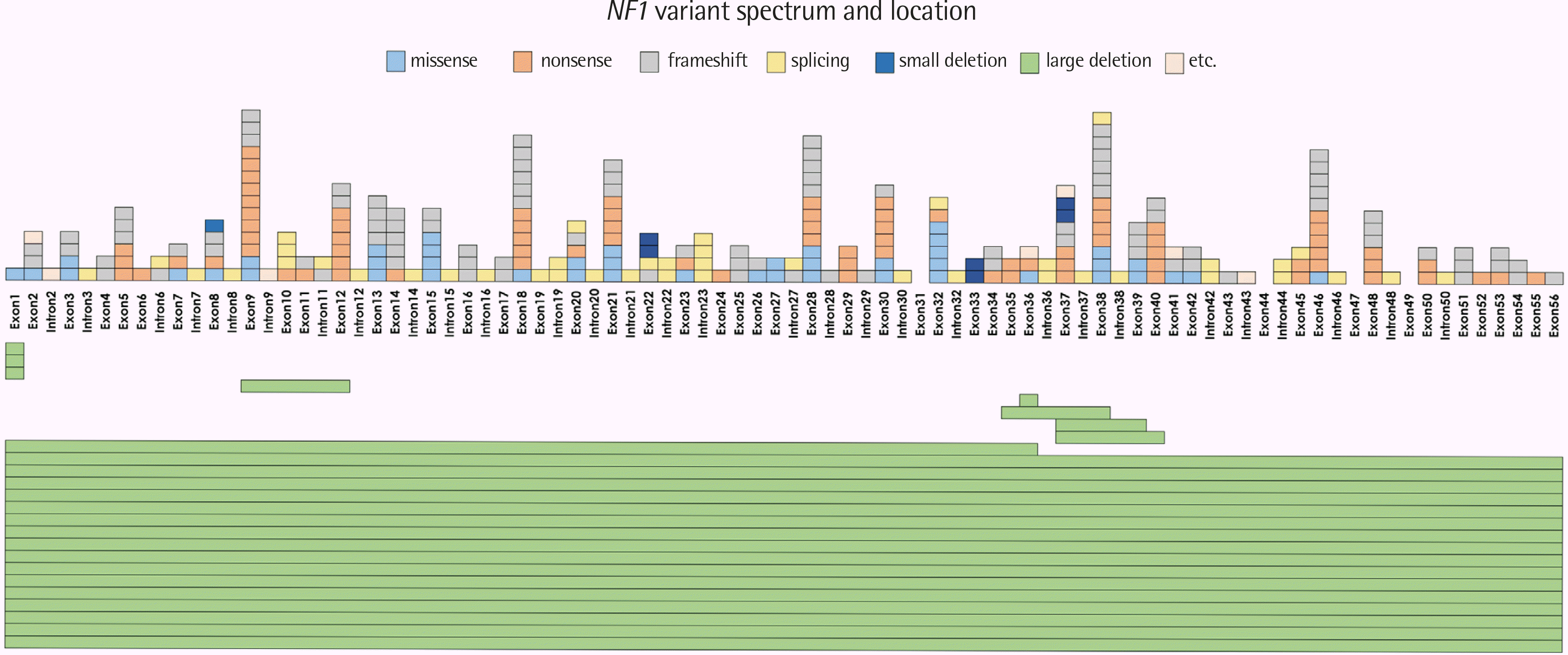
변이의 위치는 NF1 유전자 전체 엑손 결실이 총 16명의 환자에서 검출되었고 엑손 9에서의 변이가 15개(15/278, 5.4%), 다음으로 엑손 38 (14/278, 5.0%), 엑손 18과 28 (12/278, 4.3%), 엑손 46 (11/278, 4.0%) 순이었다. 그 외 부위에서 변이 빈도는 최대 8개(0-2.9%)까지 다양하게 나타났다(Fig. 1). 반복 검출된 변이는 전체 엑손 결실이 가장 많았고, c.910C>T (N=9), c.2041C>T (N=5), c.6852_6855delTTAC (N=4), c.6854dupA (N=4) 순이었다.
변이의 유형별로 보면, 무의미 변이(nonsense mutation) 및 틀이동 변이(frameshift mutation)를 포함한 절단형 돌연변이(truncating mutation)가 177명(58.4%)에서 관찰되었고, in-frame deletion은 7명(2.3%)의 환자에서 관찰되었다. 짜깁기(splicing)변이는 42명(13.9%)에서 관찰되었고, 이중 기존에 질병연관변이로 보고된 24개, 그리고 짜깁기 공여자 및 수여자부위에 발생한 15개의 변이가 있었다. mRNA 분석에서 aberrant transcript가 관찰된 경우, deep intronic mutation 가능성이 높을 것으로 예측되는 4예의 경우 likely pathogenic으로 보고하였다. 미량의 aberrant transcript가 검출된 2예의 경우, RNA 분리 지연에 의해 이러한 산물이 비정상적으로 발생하였을 가능성을 배제할 수 없어[3, 8] 재채혈 검체에서 확인하고자 하였으나 추적이 되지 않아 VUS로 분류하였다(Table 2). 44명(14.5%)의 환자에서 36개(15.6%)의 과오변이가 검출되었으며 여기에는 질병연관변이로 보고되었던 pathogenic 변이(21/36, 58.3%), likely pathogenic 변이(5/36, 13.9%), 그리고 VUS (10/36, 27.8%)가 포함되었다. 그 외에도 in-silico에서 짜깁기 과정에 영향을 줄 수 있다고 예측된 동의미(synonymous) 변이 1개(c.6033A>G)와 in-frame duplication 1개(c.5036_5041dup, p.Ile1679_Tyr1680dup)가 VUS로 분류되었다.
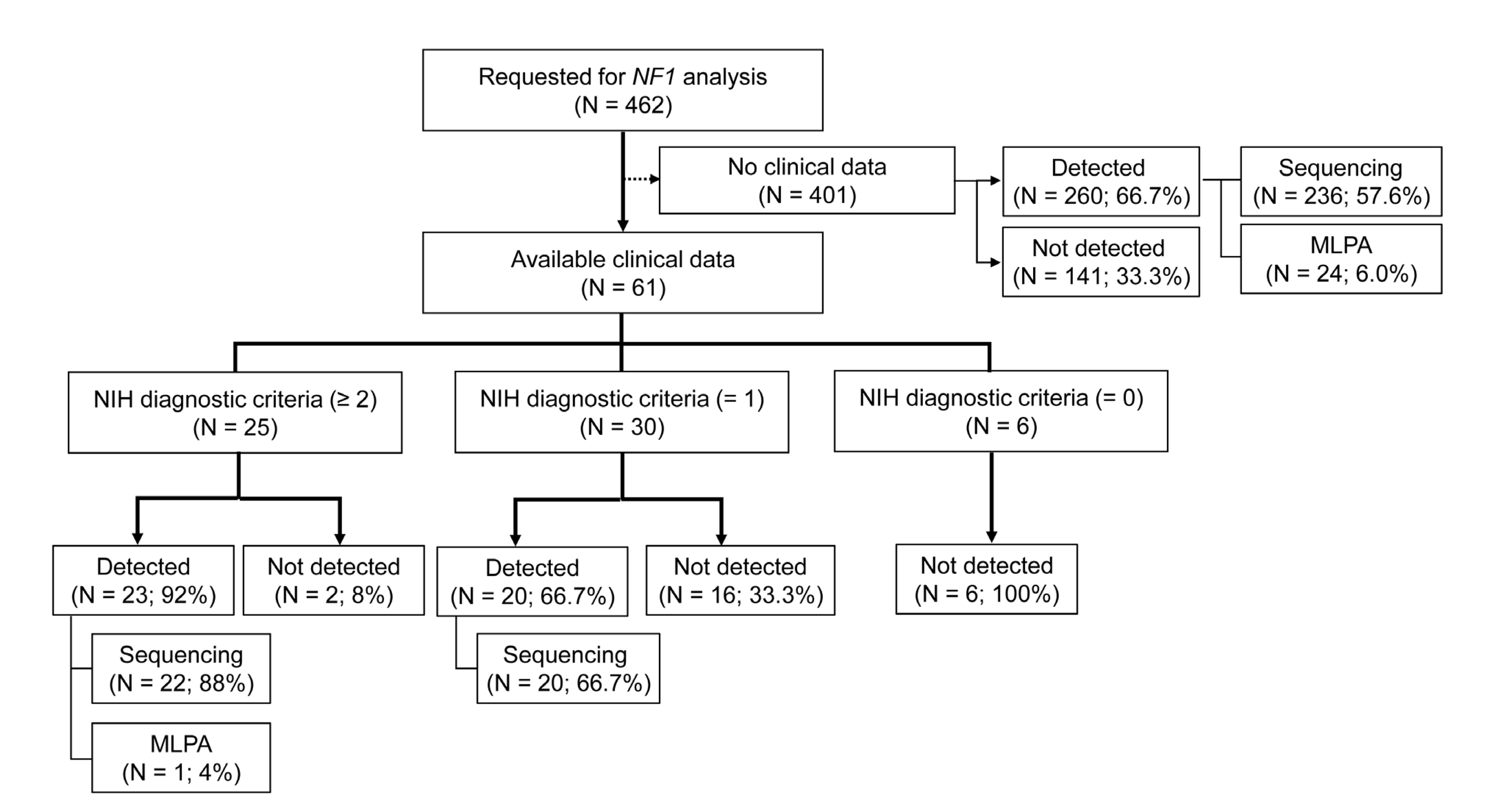
검사가 의뢰된 환자 중 임상정보를 수집할 수 있었던 61명 중 70.5% (42/61)에서 NF1 유전자 변이가 검출되었고, 이는 임상정보를 수집할 수 없었던 401건에서의 검출률 66.7% (260/401)와 유사한 수준이었다(Fig. 2). 비록 검사를 진행한 모든 환자에서 표현형 정보를 얻을 수 없었다는 제한이 있으나, 이들 61명에서의 표현형 분석을 통해 전체를 유추할 수 있을 것으로 생각된다. 표현형 정보를 얻을 수 있었던 61명의 환자에서 National Institutes of Health (NIH) Consensus conference [2]에서 제시한 NF1 임상진단 기준을 2개 이상 충족한 경우는 92.0% (23/25)에서 NF1 유전자의 변이가 발견되었으며, 하나의 진단 기준만 충족한 경우는 66.7% (20/30)에서 NF1 유전자의 변이가 확인되었다. NIH 임상적 진단 기준에 해당하는 항목이 없는 6명에서는 NF1 유전자 변이가 검출되지 않았다(Table 3, Fig. 2).
NF1 유전자 변이가 검출된 군과 검출되지 않은 두 군을 비교하면 변이가 검출된 환자(43명)가 그렇지 않은 환자(18명)에 비해 나이가 어렸고(P=0.014), 밀크커피반점(P=0.040), 신경섬유종(P=0.005), 뇌 과오종(brain hamartoma) (P=0.006)이 유의하게 높은 빈도로 관찰되었다. NF1 진단기준에 해당되지 않는 뇌 이외의 부위에서 발생한 mass lesion으로 검사를 의뢰한 경우 변이 검출률이 유의하게 낮았다(P<0.001) (Table 3).

고 찰

NF1은 질환의 표현형이 다양하게 나타날 수 있으며, 처음 검사한 시점에는 NIH에 제시된 기준을 충족시키지 않더라도 연령이 높아짐에 따라서 진단 기준에 합당한 임상 양상이 새롭게 나타날 수 있다. 질환을 조기에 진단하고 중대한 합병증을 예방할 수 있도록 질환이 의심되는 환자에서 검사를 시행하여 유전자 변이를 검출하고 빨리 치료를 시행하는 것이 중요하다[4]. 본 연구에서는 한국인 NF1 환자를 대상으로 단일 기관에서 NF1 변이를 검출한 결과를 후향적으로 검토하여 국내 환자들에서 나타나는 NF1 변이의 스펙트럼을 조사하였다. 전체 의뢰된 환자 중 65.6%에서 NF1 유전자 변이가 검출되었고, 이중에는 이전에 보고되지 않은 새로운 변이가 48.8%를 차지하여 기존 연구에서 보고된 30.8-66.2%와 유사하였다[13, 16-23]. 일부 영역에서 집중적으로 발생하는 것이 아니라, NF1 유전자 다양한 부위에서 검출되었다.
NF1 변이의 유형은 각 인종과 나라별로 다양하게 나타나지만, 대체로 절단형 돌연변이인 무의미 변이(9.0-37.5%)와 틀이동 변이(14.2-50.0%)의 비율이 높으며, 다음으로 짜깁기 변이(7.0-31.0%), 과오변이(7.7-30.0%), 엑손 단위 이상의 큰 결실(2.7-12.5%) 순으로 보고되었다. 각 연구마다 사용된 방법은 다양하였으나 검출된 변이의 양상은 유사하였다(Table 4). 본 연구에서는 절단형 돌연변이(135/231, 58.4%), 짜깁기 변이(39/231, 16.9%), 과오변이(36/231, 15.6%), 엑손 단위 이상의 큰 결실(8/231, 3.5%) 순이었고, 기존의 연구 보고와 비교했을 때 큰 차이는 없었다. NF1 유전자의 경우 deep intronic variant에 의한 짜깁기 변이가 드물지 않게 발생하며, 본 연구에서도 네 명의 환자에서 aberrant transcript가 검출되었다. 일반적으로 aberrant transcript가 관찰되는 경우 해석에 주의가 요구되며[3, 8], 특히 미량으로 검출되었다면 검체 처리 시간 지연 등에 의해 RNA가 손상되지는 않았는지 반드시 확인하고, 가능하다면 다시 채혈하여 확인하는 것이 필요하다. 또한 ACMG 권고에 따라 분류된 변이도 추가적인 연구를 통해 변경될 수 있기 때문에, 분류가 확실하지 않을 경우 부모 검사를 통한 신생(de novo) 변이 여부를 확인하고, 추가적인 기능적 검사(functional study), 분리 연구(segregation study) 등을 통해 변이의 위험도를 판단할 근거를 보다 확실하게 마련하는 것이 중요하다[13, 14, 21].
이전 연구[21]와 같이, 본 연구에서도 NIH consensus criteria (1988)에서 제시하는 임상진단 기준을 2개 이상 충족하는 환자군에서 NF1 의증 환자들에 비해 유의하게 NF1 유전자의 변이 검출률이 높게 나타났다. 그 이유로 우선 NF1과 유사한 임상증상을 보일 수 있는 다른 질환을 고려할 수 있으며, 면밀한 임상정보 확인 및 패널 유전자 검사가 도움이 될 수 있다. 최근 차세대염기서열분석(next-generation sequencing, NGS)의 지속적 향상 및 보급에 따라 NF1과 유사한 표현형을 보일 수 있는 유전자 패널을 함께 분석하여 질환의 원인 유전자를 찾고 진단하고 있다[20, 24]. 본 연구에 포함된 환자군에는 외부 병원에서 자세한 임상정보 제공 없이 검사가 의뢰된 경우가 다수 포함되어 있어 이러한 질환에 대한 평가와 감별에 제한이 있었다.
NIH 진단기준에 합당한 임상 양상 2개 이상을 충족할 경우 92%의 환자에서 변이가 검출되어 NF1 유전자의 변이가 NF1 환자의 80.9-92.4%에서 관찰된다는 기존의 보고와 일치하였다[21, 23]. 나아가 임상적으로 확인 가능한 증상이 한 가지에 국한되어 NIH 기준에 따른 NF1 진단에 미치지 못하는 경우에도 66.7%의 환자에서 NF1 유전자의 변이를 검출하였는데, 이는 증상이 제한적으로 발현된 질환의 초기에도 유전자 검사를 통하여 빠르고 정확한 진단을 할 수 있음을 보여주었다.
본 연구를 통하여 한국인 NF1 환자에서 다양한 NF1 유전자 변이를 검출하였다. 폭넓은 스펙트럼의 변이를 검출하기 위해서는 mRNA와 gDNA 직접염기서열분석, MLPA 등의 검사법을 종합적으로 수행할 필요가 있다. 향후 NGS 등을 활용하여 질병 원인 유전자 종류를 확대하고, 새롭게 검출된 유전자 변이의 임상적 의미를 규명하기 위한 노력이 필요할 것으로 생각되었다.

감사의 글

이 논문은 가톨릭대학교 서울성모병원의 연구비 지원을 받아 수행되었습니다.

Notes

이해관계

저자들은 본 연구와 관련하여 어떠한 이해관계도 없음을 밝힙니다.

REFERENCES

1. Reynolds RM, Browning GG, Nawroz I, Campbell IW. 2003; Von Recklinghausen's neurofibromatosis: neurofibromatosis type 1. Lancet. 361:1552–4. DOI: 10.1016/S0140-6736(03)13166-2.
pmid
2. Jett K, Friedman JM. 2010; Clinical and genetic aspects of neurofibromatosis 1. Genet Med. 12:1–11. DOI: 10.1097/GIM.0b013e3181bf15e3. PMID: 20027112.
pmid
3. Valero MC, Martín Y, Hernández-Imaz E, Marina Hernández A, Meleán G, Valero AM, et al. 2011; A highly sensitive genetic protocol to detect NF1 mutations. J Mol Diagn. 13:113–22. DOI: 10.1016/j.jmoldx.2010.09.002. PMID: 21354044. PMCID: PMC3128626.
pmid pmc
4. Messiaen L, Wimmer K, editors. 2008. NF1 mutational spectrum. Karger;Basel: p. 63–77. DOI: 10.1159/000126545.
5. Messiaen L, Wimmer K. 2012. Mutation analysis of the NF1 gene by cDNA-based sequencing of the coding region. Advances in neurofibromatosis research. Nova Science Publishers, Inc..
6. Evans DG, Bowers N, Burkitt-Wright E, Miles E, Garg S, Scott-Kitching V, et al. 2016; Comprehensive RNA analysis of the NF1 gene in classically affected NF1 affected individuals meeting NIH criteria has high sensitivity and mutation negative testing is reassuring in isolated cases with pigmentary features only. EBioMedicine. 7:212–20. DOI: 10.1016/j.ebiom.2016.04.005. PMID: 27322474. PMCID: PMC4909377.
7. Stenson PD, Mort M, Ball EV, Evans K, Hayden M, Heywood S, et al. 2017; The Human Gene Mutation Database: towards a comprehensive repository of inherited mutation data for medical research, genetic diagnosis and next-generation sequencing studies. Hum Genet. 136:665–77. DOI: 10.1007/s00439-017-1779-6. PMID: 28349240. PMCID: PMC5429360.
pmid pmc
8. Messiaen LM, Callens T, Mortier G, Beysen D, Vandenbroucke I, Van Roy N, et al. 2000; Exhaustive mutation analysis of the NF1 gene allows identification of 95% of mutations and reveals a high frequency of unusual splicing defects. Hum Mutat. 15:541–55. DOI: 10.1002/1098-1004(200006)15:6<541::AID-HUMU6>3.0.CO;2-N.
pmid
9. Jo YH, Kim HO, Song HR, Yoon KS. 2014; Identification of the NF1 gene mutation in Korean families with neurofibromatosis type 1. Genes & Genomics. 36:11–5. DOI: 10.1007/s13258-013-0132-2.
10. Jang MA, Kim YE, Kim SK, Lee MK, Kim JW, Ki CS. 2016; Identification and characterization of NF1 splicing mutations in Korean patients with neurofibromatosis type 1. J Hum Genet. 61:705–9. DOI: 10.1038/jhg.2016.33. PMID: 27074763.
pmid
11. Jeong SY, Park SJ, Kim HJ. 2006; The spectrum of NF1 mutations in Korean patients with neurofibromatosis type 1. J Korean Med Sci. 21:107–12. DOI: 10.3346/jkms.2006.21.1.107. PMID: 16479075. PMCID: PMC2733956.
pmid pmc
12. Kim MJ, Cheon CK. 2014; Neurofibromatosis type 1: a single center's experience in Korea. Korean J Pediatr. 57:410–5. DOI: 10.3345/kjp.2014.57.9.410. PMID: 25324867. PMCID: PMC4198956.
pmid pmc
13. Ko JM, Sohn YB, Jeong SY, Kim HJ, Messiaen LM. 2013; Mutation spectrum of NF1 and clinical characteristics in 78 Korean patients with neurofibromatosis type 1. Pediatr Neurol. 48:447–53. DOI: 10.1016/j.pediatrneurol.2013.02.004. PMID: 23668869.
pmid
14. Richards S, Aziz N, Bale S, Bick D, Das S, Gastier-Foster J, et al. 2015; Standards and guidelines for the interpretation of sequence variants: a joint consensus recommendation of the American College of Medical Genetics and Genomics and the Association for Molecular Pathology. Genet Med. 17:405–24. DOI: 10.1038/gim.2015.30. PMID: 25741868. PMCID: PMC4544753.
pmid pmc
15. De Luca A, Bottillo I, Dasdia MC, Morella A, Lanari V, Bernardini L, et al. 2007; Deletions of NF1 gene and exons detected by multiplex ligation-dependent probe amplification. J Med Genet. 44:800–8. DOI: 10.1136/jmg.2007.053785. PMID: 18055911. PMCID: PMC2652822.
pmid pmc
16. Ars E, Kruyer H, Morell M, Pros E, Serra E, Ravella A, et al. 2003; Recurrent mutations in the NF1 gene are common among neurofibromatosis type 1 patients. J Med Genet. 40:e82. DOI: 10.1136/jmg.40.6.e82. PMID: 12807981. PMCID: PMC1735494.
17. Lee MJ, Su YN, You HL, Chiou SC, Lin LC, Yang CC, et al. 2006; Identification of forty-five novel and twenty-three known NF1 mutations in Chinese patients with neurofibromatosis type 1. Hum Mutat. 27:832. DOI: 10.1002/humu.9446. PMID: 16835897.
pmid
18. Griffiths S, Thompson P, Frayling I, Upadhyaya M. 2007; Molecular diagnosis of neurofibromatosis type 1: 2 years experience. Fam Cancer. 6:21–34. DOI: 10.1007/s10689-006-9001-3. PMID: 16944272.
pmid
19. Sabbagh A, Pasmant E, Imbard A, Luscan A, Soares M, Blanché H, et al. 2013; NF1 molecular characterization and neurofibromatosis type I genotype-phenotype correlation: the French experience. Hum Mutat. 34:1510–8. DOI: 10.1002/humu.22392. PMID: 23913538.
pmid
20. Maruoka R, Takenouchi T, Torii C, Shimizu A, Misu K, Higasa K, et al. 2014; The use of next-generation sequencing in molecular diagnosis of neurofibromatosis type 1: a validation study. Genet Test Mol Biomarkers. 18:722–35. DOI: 10.1089/gtmb.2014.0109. PMID: 25325900. PMCID: PMC4216997.
pmid pmc
21. van Minkelen R, van Bever Y, Kromosoeto JN, Withagen-Hermans CJ, Nieuwlaat A, Halley DJ, et al. 2014; A clinical and genetic overview of 18 years neurofibromatosis type 1 molecular diagnostics in the Netherlands. Clin Genet. 85:318–27. DOI: 10.1111/cge.12187. PMID: 23656349.
pmid
22. Zhang J, Tong H, Fu X, Zhang Y, Liu J, Cheng R, et al. 2015; Molecular characterization of NF1 and neurofibromatosis Type 1 genotype-phenotype correlations in a Chinese population. Sci Rep. 5:11291. DOI: 10.1038/srep11291. PMID: 26056819. PMCID: PMC4460887.
pmid pmc
23. Calì F, Chiavetta V, Ruggeri G, Piccione M, Selicorni A, Palazzo D, et al. 2017; Mutation spectrum of NF1 gene in Italian patients with neurofibromatosis type 1 using Ion Torrent PGM™ platform. Eur J Med Genet. 60:93–9. DOI: 10.1016/j.ejmg.2016.11.001. PMID: 27838393.
pmid
24. Wu-Chou YH, Hung TC, Lin YT, Cheng HW, Lin JL, Lin CH, et al. 2018; Genetic diagnosis of neurofibromatosis type 1: targeted next-generation sequencing with Multiple Ligation-Dependent Probe Amplification analysis. J Biomed Sci. 25:72. DOI: 10.1186/s12929-018-0474-9. PMID: 30290804. PMCID: PMC6172719.
pmid pmc

Fig. 1
Identified NF1 variant spectrum and location. At the midline of the figure, the involved exons and/or introns of the NF1 gene are listed. Each block upward the midline represents one variation. Different kinds of variations are shown in different colors as indicated in the category above the chart. Large deletions are indicated by the green rectangle at the bottom of the midline. Here, each variant is displayed in the vertical direction. The different horizontal length of each rectangle indicates the deleted scope of each individual variant.
lmo-11-1-17-f1.tif
Fig. 2
Flow chart showing the NF1 gene analysis according to the clinical findings of enrolled patients. NIH, National Institutes of Health; Sequencing, Sanger sequencing of mRNA and gDNA; MLPA, multiplex ligation-dependent probe amplification.
lmo-11-1-17-f2.tif
Table 1
Detected variants of the NF1 gene
Pathogenic/LP VUS Total


Patient Variant Patient Variant Patient Variant
Truncating 177 (61.2%) 135 (62.2%) 0 (0%) 0 (0%) 177 (58.4%) 135 (58.4%)
Splicing 42 (14.5%) 39 (18.0%) 0 (0%) 0 (0%) 42 (13.9%) 39 (16.9%)
Missense 34 (11.8%) 26 (12.0%) 10 (71.4%) 10 (71.4%) 44 (14.5%) 36 (15.6%)
Large del 25 (8.7%) 8 (3.7%) 0 (0%) 0 (0%) 25 (8.3%) 8 (3.5%)
In-frame del 7 (2.4%) 5 (2.3%) 0 (0%) 0 (0%) 7 (2.3%) 5 (2.2%)
In-frame dup 0 (0%) 0 (0%) 1 (7.1%) 1 (7.1%) 1 (0.3%) 1 (0.4%)
Aberrant transcript 4 (1.4%) 4 (1.8%) 2 (14.3%) 2 (14.3%) 6 (2.0%) 6 (2.6%)
Synonymous 0 (0%) 0 (0%) 1 (7.1%) 1 (7.1%) 1 (0.3%) 1 (0.4%)
Total 289 217 14 14 303 231

Abbreviations: LP, likely pathogenic; VUS, variant of unknown significance; del, deletion; dup, duplication.

Table 2
NF1 variants causing aberrant transcripts
No ID Class variant AA change Location Test methods
1 L003 LP r.100_204del p.Val34_Met68del Exon 2 Sanger
2 L004 LP c.205-13T > G p.Arg69_Gly96del Intron 2 Sanger
3 L018 LP r.1063_1185del Splicing defect Intron 9 Sanger, MLPA
4 L067 LP r.4725_4835del p.His1576_Arg1612del Exon 36 Sanger, MLPA
5 V002 VUS r.1642_1845del204 Splicing defect Exon 15-16 Sanger, MLPA
6 V013 VUS c.6642+31T > G Splicing defect Intron 43 Sanger, MLPA

Abbreviations: AA, amino acid; LP, likely pathogenic; VUS, variant of unknown significance; Sanger, Sanger sequencing; MLPA, multiplex ligation-dependent probe amplification.

Table 3
Comparison of clinical characteristics of neurofibromatosis 1 patients with detected NF1 variants and those devoid of NF1 variants
Mutation Detected Not detected P OR (95% CI)
Number of patients 43 18
Age, yr* 13 (1-60) 22 (4-74) 0.014
Sex (N, %) 0.678 0.792 (0.263-2.385)
Male 24 (55.8%) 9 (50%)
Female 19 (44.2%) 9 (50%)
Number of satisfying NIH criteria (1988)
2 or more 23 (92.0%) 2 (8%) 0.002 9.200 (1.881-44.998)
One 20 (66.7%) 10 (33.3%) < 0.001 4.583 (2.779-7.559)
None 0 (0.0%) 6 (100%)
Clinical presentation (N, %)
1st degree family history 7 (16.3%) 1 (5.6%) 0.258 3.306 (0.376-29.045)
Café-au-lait spots 39 (90.7%) 9 (50.0%) < 0.001 9.750 (2.446-38.869)
Neurofibroma 18 (41.9%) 1 (5.6%) 0.005 12.240 (1.490-100.538)
Axillary/groin freckling 4 (9.3%) 1 (5.6%) 0.627 1.744 (0.181-16.778)
Lisch nodule 8 (18.6%) 2 (11.1%) 0.471 1.829 (0.348-9.603)
Optic glioma 2 (4.7%) 0 (0%) 0.352 1.049 (0.982-1.120)
Skeletal dysplasia 1 (2.3%) 0 (0%) 0.514 1.024 (0.978-1.072)
Brain hamartoma 14 (32.6%) 0 (0%) 0.006 1.483 (1.205-1.825)
Seizure, mental retardation 1 (2.3%) 0 (0%) 0.514 1.024 (0.978-1.072)
MPNST 1 (2.3%) 0 (0%) 0.514 1.024 (0.978-1.072)
Other mass lesion 0 (0%) 6 (33.3%) < 0.001 0.667 (0.481-0.924)

*Median (range).

Abbreviations: OR, odds ratio; CI, confidence interval; NIH, National Institutes of Health; MPNST, malignant peripheral nerve sheath tumors.

Table 4
Review of previous reports on sequence variants of the NF1 gene
Country Number of individuals Methods Detected numbers Types of variants Reference
Korea 78 individuals from 60 families DNA sequencing
mRNA sequencing
MLPA
FISH
52 variants in 78 individuals Missense 23.1%
Nonsense 34.6%
Splicing 21.2%
Indel 13.5%
Large del 7.7%
[13]
Spain 474 unrelated individuals DNA sequencing
mRNA sequencing
mRNA SSCP/HD
142 variants in 189 individuals Missense 8%
Nonsense 9%
Splicing 31%
Frameshift 50%
Others 2%
[16]
China 107 unrelated individuals DNA sequencing
DHPLC
microsatellite genotyping
Q-PCR
RT-PCR
68 individuals Missense 16.2%
Nonsense 29.4%
Splice site 13.2%
Frameshift 33.8%
Large del 7.4%
[17]
UK 169 unrelated individuals DNA sequencing
DHPLC MLPA
FISH
88 variants in 109 individuals Missense 15%
Nonsense 27%
Splicing 19%
Frameshift 27%
Large del 9%
Others 3%
[18]
France 565 unrelated individuals DNA sequencing
mRNA sequencing
microsatellite genotyping
MLPA
546 individuals Missense 7.7%
Nonsense 21.6%
Splicing 27.1%
Frameshift 30.0%
Large del 8.4%
Others 5.1%
[19]
Japan 86 unrelated individuals NGS
DNA sequencing
MLPA
67 variants in 80 individuals Missense 16.3%
Nonsense 37.5%
Splicing 10%
Frameshift 23.8%
Large del 12.5%
[20]
Netherlands 1985 unrelated individuals DNA sequencing
SSCP
MLPA
FISH
1178 individuals Missense 9.8%
Nonsense 29.4%
Splicing 19.7%
Frameshift 31.7%
Others 4.1%
[21]
China 109 individuals, 100 families DNA sequencing
mRNA sequencing
MLPA
89 variants in 100 families Missense 13.5%
Nonsense 23.6%
Splicing 16.9%
Frameshift 29.2%
Large del 12.4%
Others 4.5%
[22]
Italy 79 unrelated individuals NGS
MLPA
73 variants in 79 individuals Missense 30%
Nonsense 34%
Splicing 7%
Small del 24.7%
Insertion 1.4%
Large del 2.7%
[23]

Abbreviations: MLPA, multiplex ligation-dependent probe amplification; FISH, Fluorescence in situ hybridization; SSCP, single-strand conformation polymorphism; DHPLC, denaturing high performance liquid chromatography; RT-PCR, reverse transcriptase polymerase chain reaction; NGS, next-generation sequencing; Indel, insertion/deletion; del, deletion.

TOOLS
Similar articles