

 PDF
PDF Citation
 Print
Print


Introduction
Neck and shoulder pain is a frequent musculoskeletal problem with an estimated prevalence between 5.9% and 38.7% in a certain population1-3. Neck and shoulder pain syndrome is called “Katakori” in Japan and characterized by discomfort or spontaneous pain, mild pain on motion in the neck, upper scapula region, scapula region, or interscapular region with tense muscles at palpation as well as tender points or stiffness at those areas4. It might be correlated with prolonged static position of daily living, trauma, and degenerative disease in the cervical and shoulder region5,6. Unlike myofascial pain syndrome (MPS) or fibromyalgia (FM), there has been no previous research on effectiveness of extracorporeal shock wave therapy (ESWT) on neck and shoulder pain syndrome.
MPS is characterized by localized tenderness, taut band structure, convulsive response, and referred pain under compression and is defined as a localized pain syndrome caused by pain trigger points7. It can occur in the paracervical muscles, rotator cuff muscles, and lumbar muscles8,9; and trapezius is the most frequently involved muscle10. FM is a chronic condition that involves both central and peripheral sensitization and shares some of the features of MPS, such as hyperirritability, tender and trigger points11,12. Although MPS and FM are separate conditions, they may occur concomitantly.
ESWT is considered an effective treatment for MPS or FM13-17. However, there is no evidence that ESWT is effective on neck and shoulder pain syndrome which might be less severe form of disease than MPS and FM. There might be various disease entity which caused neck and shoulder pain syndrome and we should focus on underlying diseases to treat this disease entity. However, we hypothesized that symptomatic treatment for neck and shoulder pain syndrome might be effective and the ESWT could be an useful treatment modality for this. Therefore, the aim of current study is to verify the efficacy of ESWT on neck and shoulder pain syndrome.
Go to : 
Methods
1. Study design and ethics
This is a retrospective study with data collected from July 2019 to December 2019 and approved by Institutional Review Board of author’s hospital (IRB No. 2009-008-19338). The requirement for informed consent was waived due to nature of retrospective study design.
2. Subject
The patients who had treated with ESWT from July 2019 to December 2019 were accessed for eligibility. Eligible patients were adults over the age of 20 years and 23 patients who meet with inclusion criteria were enrolled. Inclusion criteria were patients who present with neck and one-sided shoulder pain and diagnosed as rotator cuff disease with neck strain treated with at least four times of consecutive ESWT treatment. Exclusion criteria were patients who presented with ipsilateral elbow pain and patients who were diagnosed with MPS or FM. Due to similar symptoms, we identified patients with MPS as presenting taut band and focal tenderness or referred pain on compression of taut band. Further, patients with additional systemic symptoms such as fainting, dizziness, fatigue, palpitation, etc. were diagnosed as FM. Patients without symptoms mentioned above regarding MPS and FM were all diagnosed with neck and shoulder pain syndrome.
3. Subgroups
Twenty-three patients were classified into group A (n=9) and group B (n=14). Group A was defined as the patients who presented with only pain and tenderness on neck (pain on upper scapular region with/without interscapular region) and group B was defined as the patients who presented with pain and tenderness on both neck and shoulder (pain on upper scapular with/without interscapular region and pain on scapular region). Upper scapular region refers to area around upper trapezius and levator scapulae. Scapular region refers to regions of infraspinatus, and interscapular region refers to rhomboid, and middle trapezius.
4. Demographics and assessment
Baseline demographic data including age, gender, duration of disease, and follow-up period was collected and described in Table 1. A total of 23 patients (male:female, 6:17; age, 56.61±16.28 years) were enrolled in this study. The evaluation of pain was conducted a total of five times using visual analogue scale (VAS) of pain which was at first visit, after each treatment and at final visit. Tenderness VAS was defined that severity of tenderness when examiner compressed the tender point of affected muscle. VAS is to estimate intensity on a scale of 0 to 10. A score of 10 indicated maximal pain or tenderness and 0 indicated no pain or tenderness18.
Table 1
Baseline demographics of the study subjects
| Variable | Total (n=23) | Group A (n=9) | Group B (n=14) | p-value |
|---|---|---|---|---|
| Age (yr) | 56.61±16.28 | 56.11±21.57 | 52.00±12.44 | 0.34 |
| Sex, male:female | 6 (26.1):17 (73.9) | 4 (17.4):5 (21.7) | 2 (8.7):12 (52.2) | 0.2 |
| Onset (mo) | 12.65±8.90 | 11.11±8.27 | 13.64±9.44 | 0.73 |
| Follow-up (wk) | 4.52±0.73 | 4.78±0.97 | 4.44±0.53 | 0.2 |
| At first visit | ||||
| Pain VAS | 5.48±2.37 | 5.11±3.37 | 5.71±1.54 | 0.4 |
| Tenderness VAS | 5.98±1.89 | 6.83±2.4 | 5.43±1.28 | 0.36 |
| NDI | 18.04±8.86 | 16.22±9.78 | 19.21±8.40 | 0.6 |
| Shoulder ROM | ||||
| Forward flexion (°) | 159.57±28.03 | 162.22±28.63 | 157.86±28.60 | 0.9 |
| External rotation (°) | 72.17±15.65 | 73.33±20.00 | 71.43±12.92 | 0.99 |
| Internal rotation* | 10.17±3.49 | 10.67±3.28 | 9.86±3.70 | 0.6 |
![]()
In addition, the evaluation of function was conducted two times which was performed at first visit and last visit. It was completed with evaluating neck disability index (NDI) and shoulder range of motion (ROM; forward flexion [FF], external rotation at neutral [ER], and internal rotation at back [IR]). NDI consists of 10 items and each item is scored from 0 to 5. A higher score indicates a more serious dysfunction in the cervical area. When the sum of measured scores is in the range 0 to 4, it indicates no disability. Scores within each range indicate as follows: 5 to 14, mild disability; 15 to 24, moderate disability; 25 to 34, severe disability; and 35 or higher, complete disability8. Regarding shoulder ROM, degrees of active FF and ER were measured by a goniometer. IR was measured by the vertebral spinous process that could be reached with the tip of the patient’s thumb and was converted into continuously numbered groups: T1–12 to 1–12, L1–5 to 13–17, and buttock to 18. Further, any other complications were noted during or after each radial ESWT treatment such as chest discomfort or pain, ecchymosis, and petechiae.
5. Application of ESWT
Radial or focused extracorporeal shock wave (Zeus Wave Integrated pain-treatment system; Wever Instruments, Uijeongbu, Korea) applied in the current study. The therapy was performed at a pulse rate of 1,500 to 2,000. Pulse frequency was 7 Hz and pulse intensity was 4 to 5 bar (0.23–0.45 mJ/mm2) depended on patient’s tolerability to pain during the procedure. It was performed by an orthopedic surgeon and the focused type ESWT was used rather than radial ESWT in cases of focused type pain and tenderness rather than diffuse type pain and tenderness. After applying ultrasound gel, treatment was performed with ESWT on tender area of affected muscles. Patients in the current study have received ESWT treatment one session per week for 4 weeks.
6. Other treatment
Patients in the current study received nonsteroidal anti- inflammatory medication (Naxosol, 2T #2; Hanmi Pharm. Corp., Ltd., Seoul, Korea) for 4 weeks. They were also educated to perform the active assisted stretching exercise of shoulder joint to each direction which was FF, ER, IR, and adduction as a home-based stretching exercise program.
7. Statistical analysis
The statistical analysis was processed with PASW Statistics version 18.0 software (IBM Corp., Armonk, NY, USA). The continuous data are expressed as the mean±standard deviation. Wilcoxon signed-ranks test was performed to determine the effect of therapy in each group at a certain period. The p-values of < 0.05 were considered significant.
Go to :
Results
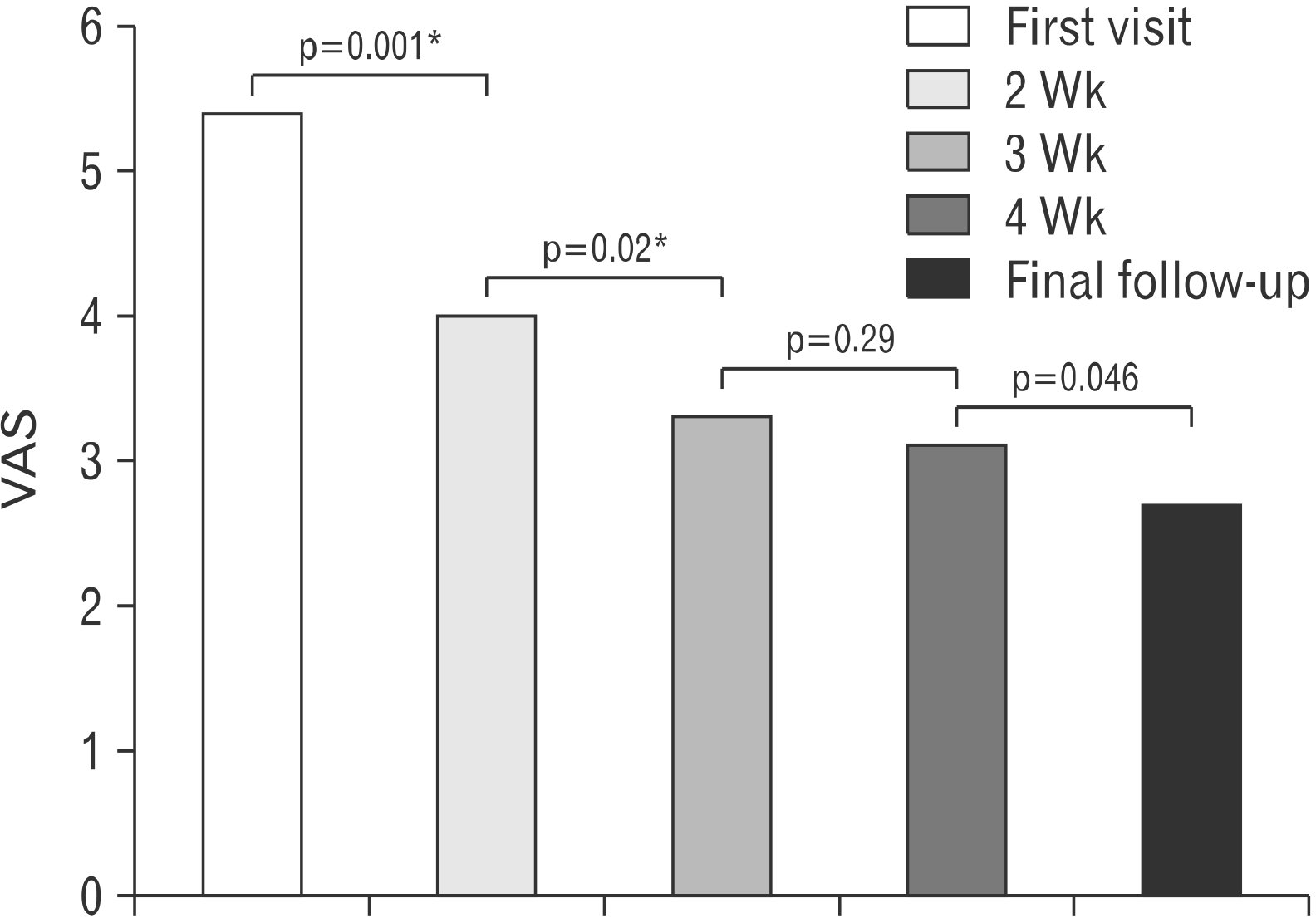
Regarding the pain VAS, the decrease of pain VAS was significant at each follow-up during first 2 weeks. Pain VAS was 5.5±2.4 at first visit, 4.0±1.8 (p=0.001) at first follow-up, and 3.3±2.1 (p=0.02) at second follow-up. The decreasing pattern of pain VAS was continued over third week to last follow-up; however, there was no statistical difference in comparison with previous follow-up at third week (Fig. 1).
In the subgroup analysis, group A revealed significant decrease of pain VAS from 5.11±3.37 at first visit to second week follow-up (3.44±1.67, p=0.04) and third week follow-up (2.56±1.81, p=0.02) in comparison with previous follow-up; however, there was no significant decrease at fourth week (2.67±2.55, p=0.87) and last follow-up (2.22±2.54, p=0.19) in comparison with previous follow-up. Group B revealed significant decrease of pain VAS 5.71±1.54 at first visit to second week follow-up (4.36±1.87, p=0.004); however, there was no significant decrease at third week (3.79±2.26, p=0.17), fourth week (3.36±1.91, p=0.23), and last follow-up (3.07±2.06, p=0.1) in comparison with previous follow-up (Table 2).
Table 2
VAS for pain and tenderness after consecutive ESWT
| VAS | At first visit (1 wk) | Follow-up | |||||||
|---|---|---|---|---|---|---|---|---|---|
|
|
|||||||||
| 2 Wk | 3 Wk | 4 Wk | Last | ||||||
|
|
|
|
|
|
|||||
| Mean±SD | Mean±SD | p-value | Mean±SD | p-value | Mean±SD | p-value | Mean±SD | p-value | |
| Total | |||||||||
| Pain | 5.48±2.37 | 4.0±1.8 | 0.001* | 3.3±2.14 | 0.02* | 3.09±2.15 | 0.29 | 2.74±2.24 | 0.046* |
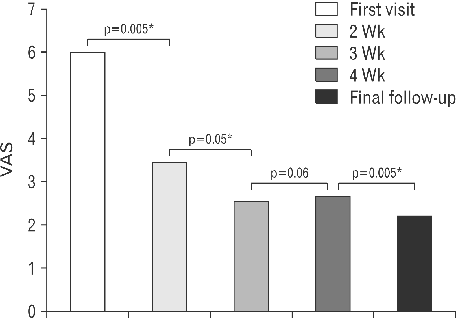
| Tenderness | 5.98±1.89 | 5.17±1.83 | 0.005* | 4.61±1.67 | 0.05* | 4.09±1.92 | 0.06 | 3.52±2.04 | 0.005* |
| Group A | |||||||||
| Pain | 5.11±3.37 | 3.44±1.67 | 0.04* | 2.56±1.81 | 0.02* | 2.67±2.55 | 0.87 | 2.22±2.54 | 0.19 |
| Tenderness | 6.83±2.40 | 5.89±1.83 | 0.04* | 4.56±2.07 | 0.03* | 4.22±2.59 | 0.67 | 3.33±2.55 | 0.07 |
| Group B | |||||||||
| Pain | 5.71±1.54 | 4.36±1.87 | 0.004* | 3.79±2.26 | 0.17 | 3.36±1.91 | 0.23 | 3.07±2.06 | 0.1 |
| Tenderness | 5.43±1.28 | 4.71±1.73 | 0.046* | 4.64±1.45 | 0.78 | 4.00±1.47 | 0.01* | 3.64±1.74 | 0.03* |
![]()
Regarding the tenderness VAS, the VAS at first visit was 5.98±1.89 and this decreased at first follow-up (5.17±1.83, p=0.005), second follow-up (4.61±1.67, p=0.05), and third follow-up (4.09±1.92, p=0.06) in comparison with previous follow-up (Fig. 2). In the subgroup analysis, group A revealed significant decrease of tenderness VAS 6.83±2.4 at first visit to second week follow-up (5.89±1.83, p=0.04) and third week follow-up (4.56±2.07, p=0.03) in comparison with previous follow-up; however, there was no significant decrease at fourth week (4.22±2.59, p=0.67) and last follow-up (3.33±2.55, p=0.07) in comparison with previous follow-up. Group B revealed significant decrease of tenderness VAS 5.43±1.28 at first visit to second week follow-up (4.71±1.73, p=0.046), and at fourth week (4.00±1.47, p=0.01) and last follow-up (3.64±1.74, p=0.03) in comparison with previous follow-up but not significant at third week (4.64±1.45, p=0.78) in comparison with previous follow-up (Table 2).
NDI was significantly reduced from 18.04±8.86 at first visit to 10.04±6.94 at last follow-up (p=0.001). In the subgroup analysis, NDI score was decreased in both subgroups at last follow-up. In group A, NDI score was significantly improved from 16.22±9.78 to 9.11±7.27 (p=0.01). Pain intensity and concentration were significantly improved but personal care, lifting, reading, headache, work, driving, sleeping, and recreation section were not significantly improved (all p>0.05). In group B, NDI score was significantly improved from 19.21±8.40 to 10.64±6.92 (p=0.001). Pain intensity, personal care, lifting, headaches, concentration, work, sleeping, and recreation were significantly improved (all p<0.05); however, reading and driving sections were not significantly improved (all p>0.05) (Table 3).
Table 3
NDI and shoulder ROM after consecutive ESWT
| Pretreatment | Posttreatment | p-value | |
|---|---|---|---|
| Total | |||
| NDI | |||
| Pain intensity | 2.96±1.02 | 1.48±0.89 | 0.001† |
| Personal care | 1.87±1.05 | 1.00±0.95 | 0.001† |
| Lifting | 1.78±0.99 | 0.96±0.98 | 0.005† |
| Reading | 0.87±0.87 | 0.48±0.67 | 0.02† |
| Headaches | 1.52±0.99 | 0.52±0.67 | 0.001† |
| Concentration | 1.91±0.95 | 1.00±0.8 | 0.001† |
| Work | 2.13±1.14 | 1.04±0.88 | 0.001† |
| Driving | 1.09±0.95 | 0.87±0.97 | 0.13 |
| Sleeping | 2.30±1.30 | 1.52±1.10 | 0.003† |
| Recreation | 1.61±1.2 | 1.17±1.19 | 0.01† |
| Total score | 18.04±8.86 | 10.04±6.94 | 0.001† |
| Shoulder ROM | |||
| Forward flexion (°) | 159.57±28.03 | 177.83±8.51 | 0.001† |
| External rotation (°) | 72.17±15.65 | 79.57±2.09 | 0.02† |
| Internal rotation* | 10.17±3.49 | 6.91±1.70 | 0.001† |
| Group A | |||
| NDI | |||
| Pain intensity | 2.56±1.24 | 1.11±0.93 | 0.005† |
| Personal care | 1.67±1.23 | 0.89±1.05 | 0.07 |
| Lifting | 1.56±1.01 | 1.00±1.00 | 0.18 |
| Reading | 0.67±0.71 | 0.44±0.73 | 0.16 |
| Headaches | 1.33±1.12 | 0.56±0.73 | 0.08 |
| Concentration | 1.89±0.93 | 0.89±0.60 | 0.04† |
| Work | 2.00±1.32 | 1.22±0.83 | 0.06 |
| Driving | 1.11±0.78 | 0.78±0.83 | 0.18 |
| Sleeping | 2.00±1.50 | 1.22±1.09 | 0.07 |
| Recreation | 1.44±1.33 | 1.00±1.32 | 0.18 |
| Total score | 16.22±9.78 | 9.11±7.27 | 0.01† |
| Shoulder ROM | |||
| Forward flexion (°) | 162.22±28.63 | 180.00±0.00 | 0.04† |
| External rotation (°) | 73.33±20.00 | 80.00±0.00 | 0.32 |
| Internal rotation | 10.67±3.28 | 7.33±1.32 | 0.02† |
| Group B | |||
| NDI | |||
| Pain intensity | 3.21±0.8 | 1.71±0.83 | 0.001† |
| Personal care | 2.00±0.96 | 1.07±0.92 | 0.006† |
| Lifting | 1.93±1 | 0.93±1 | 0.01† |
| Reading | 1.00±0.96 | 0.50±0.65 | 0.067 |
| Headaches | 1.64±0.93 | 0.50±0.65 | 0.001† |
| Concentration | 1.93±1 | 1.07±0.92 | 0.005† |
| Work | 2.21±1.05 | 0.93±0.92 | 0.002† |
| Driving | 1.07±1.07 | 0.93±1.07 | 0.41 |
| Sleeping | 2.50±1.16 | 1.71±1.107 | 0.02† |
| Recreation | 1.71±1.14 | 1.29±1.14 | 0.03† |
| Total score | 19.21±8.4 | 10.64±6.92 | 0.001† |
| Shoulder ROM | |||
| Forward flexion (°) | 157.86±28.6 | 176.43±10.82 | 0.006† |
| External rotation (°) | 71.43±12.92 | 79.29±2.67 | 0.03† |
| Internal rotation | 9.86±3.7 | 6.64±1.91 | 0.003† |
![]()
Regarding shoulder ROM, each direction of shoulder active ROM or FF, ER, and IR were significantly improved (FF: 159.57°±28.03° at first visit to 177.83°±8.51° at final follow-up, p=0.001; ER: 72.17°±15.65° at first visit to 79.57°±2.09° at final follow-up, p=0.02; IR; 10.17±3.49 at first visit to 6.91±1.7 at last follow-up, p=0.001). In the subgroup analysis, group A revealed significant improvement in FF from 162.22°±28.63° to 180.0°±0.00° (p=0.04) and in IR from 10.67±3.28 to 7.33±1.32 (p=0.02); however, no significant improvement in ER from 73.33°±20.00° to 80.00±0.00° (p=0.32). Group B revealed significant improvement in FF (157.86°±28.60° to 176.43°±10.82°, p=0.01), ER (71.43°±12.92° to 79.29°±2.67°, p=0.03), and IR (9.86±3.7 to 6.64±1.91, p=0.003) (Table 3).
Go to :
Discussion
In the current study, consecutive ESWT with a week interval seemed to be an effective treatment to improve moderate functional disability of patients with neck and shoulder pain syndrome. Two times of application of ESWT seemed to reveal a significant pain reduction on posterior neck and shoulder although there might be various underlying diseases such as degenerative cervical disease and rotator cuff disease. Further, we can expect the functional improvement by this consecutive ESWT application on posterior neck and shoulder pain syndrome especially in patients with concomitant neck and shoulder pain.
In terms of pain and tenderness reduction, we observed the ESWT with a week interval resulted in pain and tenderness reduction in a continuous manner at each week follow-up during 4 weeks after treatment started. This finding is in line with previous study of Jeon et al.19 which demonstrated the effectiveness of focused type ESWT treated with a week interval on MPS in terms of pain reduction and neck ROM although the indication for ESWT was different. The mechanism of pain and tenderness reduction seemed to be correlated with reduction of muscle stiffness, improvement of blood circulation, and interference of nociceptor or release of substance P20-24. Interestingly, significant reduction of pain and tenderness of affected muscle occurred at first and second week after one or two consecutive ESWT treatments which could imply that two times of ESWT application in neck and shoulder pain syndrome might be essential and further application might be optional during treatment of neck and shoulder pain syndrome with ESWT. In the subgroup analysis, group A revealed simultaneous reduction of pain and tenderness in 3 consecutive weeks; however, group B revealed simultaneous reduction of pain and tenderness only in 2 consecutive weeks. This might imply that pain and tenderness from scapular region or infraspinatus muscle might be less influential by ESWT than upper scapular and interscapular region or upper and middle trapezius (or rhomboid) muscle and the other pain or tenderness in scapular region might need another type of intervention additionally.
Jeon et al.19 described that focused type ESWT increased neck ROM in MPS. Unfortunately, we did not measure neck ROM in the current study; however, we evaluated the functional status of patients with a NDI questionnaire. Four consecutive ESWT applications seemed to be effective to improve functional status according to the current study, although subjects have suffered from moderate functional disability, which is defined as NDI score between 15 and 24, with mean of 12 months symptom duration. In the subgroup analysis, group B revealed meaningful improvement in daily activities such as personal care, lifting, work, and sleeping in comparison to group A in which improvement of pain and concentration sections were merely noted. This finding might be correlated with improvement of shoulder function which might be affected by ESWT is important to improve the quality of life in patients. The mechanism of improvement of shoulder ROM in group B is thought by applying the ESWT on scapular region might decrease stiffness of infraspinatus muscle and thus reduce pain at infraspinatus muscle. This result is contrary to previous study of Kvalvaag et al.25 which insisted on meaningless function of ESWT in subacromial shoulder pain; however, the intensity of ESWT in the current study was higher and the application location was muscle in the current study compared with tendon in previous study of Kvalvaag et al.25
Regarding with improvement of shoulder ROM after ESWT treatment in the current study, we have to admit that additional positive influences of medication which was nonsteroidal anti-inflammatory drug and home-based active assisted stretching exercise of affected shoulder joint to ESWT application. However, the effectiveness of ESWT cannot be denied from the fact that decrease of tenderness VAS on scapular region in group B.
We have observed four cases of transient chest discomfort during ESWT treatments; however, this discomfort was immediately disappeared after the operator reduced the intensity of ESWT. Ecchymosis and petechiae occurred in three cases which have been noted as a minor problem with transient symptom26,27. However, the careful monitoring for other unacceptable complications such as rib fracture as well as pain tolerability of each patient during ESWT application is needed.
There were several limitations in this study. First, this was a retrospective study and we could not control variables which could affect the outcome of treatment. Second, only small number of subjects were enrolled in this study and the number of subjects in each subgroup was uneven; therefore, comparison between each subgroup could be meaningless. Third, there was no long-term follow-up for the outcome with regard to this treatment and we could not know any information about recurrence. Fourth, we did not fully evaluate underlying cervical and shoulder pathology with radiologic examination. Fifth, we did not separately evaluate the outcome of radial or focused ESWT, although Liao et al.28 insisted that high energy-focused type ESWT seemed to be more effective than high energy radial type ESWT in lower leg tendinopathy in their meta-analysis study.
In conclusion, consecutive ESWT was effective on neck and shoulder pain syndrome in terms of functional improvement and pain reduction. Regarding simultaneous pain and tenderness reduction, two times of ESWT with a week interval seemed to be effective.
Go to :
 XML Download