

 PDF
PDF Citation
 Print
Print


Introduction
Rotator cuff tears are an increasing problem as the incidence increases in the aging population and affect approximately 30% of people older than 60 years of age1. Although surgical treatment is one of the most common orthopedic operations, the failure rate of rotator cuff repair ranges from 11% to 90%2,3. The tendon-to-bone interface is the weakest link in rotator cuff repair, and many studies have demonstrated a high rate of incomplete healing at the tendon and bone interface4. Previous studies have focused on mechanical reinforcement of the tendon-to-bone insertion site and improvement of healing by administering various growth factors, including fibroblast growth factor and platelet-derived growth factor5,6. There have also been studies to apply stem cell therapy7,8. These techniques demonstrated some effect on the improvement of rotator cuff healing. Thus, in this study, we evaluated the different types of biochemical adjuvants that may affect the healing of the rotator cuff.
Kartogenin (KGN) (C20H15NO3; molecular weight, 317.3 g/mol) is an inducer that selectively differentiates human mesenchymal stem cells into cartilage cells9. KGN increases chondrocyte proliferation and induces cartilage differentiation in stem cells by increasing the levels of aggrecan, collagen II, and Sox-9, which are cartilage formation indicators10. In addition, KGN not only stimulates cartilage regeneration but also has a cartilage protective effect. KGN is a very stable small molecule, allowing it to be stored and transported at room temperature10. Even though a small amount of KGN was injected directly into the joints, there was no side effect in the experimental rats11.
Since its discovery, KGN has received attention as a new cartilage-forming drug for intra-articular treatment12. Although developed with a focus on osteoarthritis, it is currently being studied to promote the regeneration of other musculoskeletal disorders. Previous studies have shown that when KGN was injected in the Achilles tendon-bone junction of experimentally injured mice, the wound of the tendon-bone junction portion was regenerated by tissue formation9. Another study reported that the collagen composition of mouse dermis was stimulated to produce more collagen composition by KGN13. Such effects are expected to have a positive effect on the restoration process of the rotator cuff based on wound healing. However, there has not yet been much research regarding the effect of KGN in tendon-bone healing of rotator cuff tears. Therefore, the purpose of this study was to investigate the effects of KGN on the tendon-bone interface in a rat rotator cuff tear model. The hypothesis was that KGN would show increased fibrocartilage formation, superior collagen fiber organization, and increased biomechanical properties.
Methods
1. Study approval
All animal procedures were approved by the Institutional Animal Commission at Kyungpook National University (No. KNU 2018-0083-1).
2. Preparation of KGN
KGN (5 mg; Sigma-Aldrich, St. Louis, MO, USA) was prepared by dissolving 5 mg of KGN in 15 μL of dimethyl sulfoxide to make a 1,000 mM KGN stock solution, which was then diluted in phosphate-buffered saline (1×) to obtain a 1-mM KGN working solution. The final concentration was diluted to 500 μM, a concentration that has been used in the previous studies13.
3. In vivo animal experiments and surgical procedure
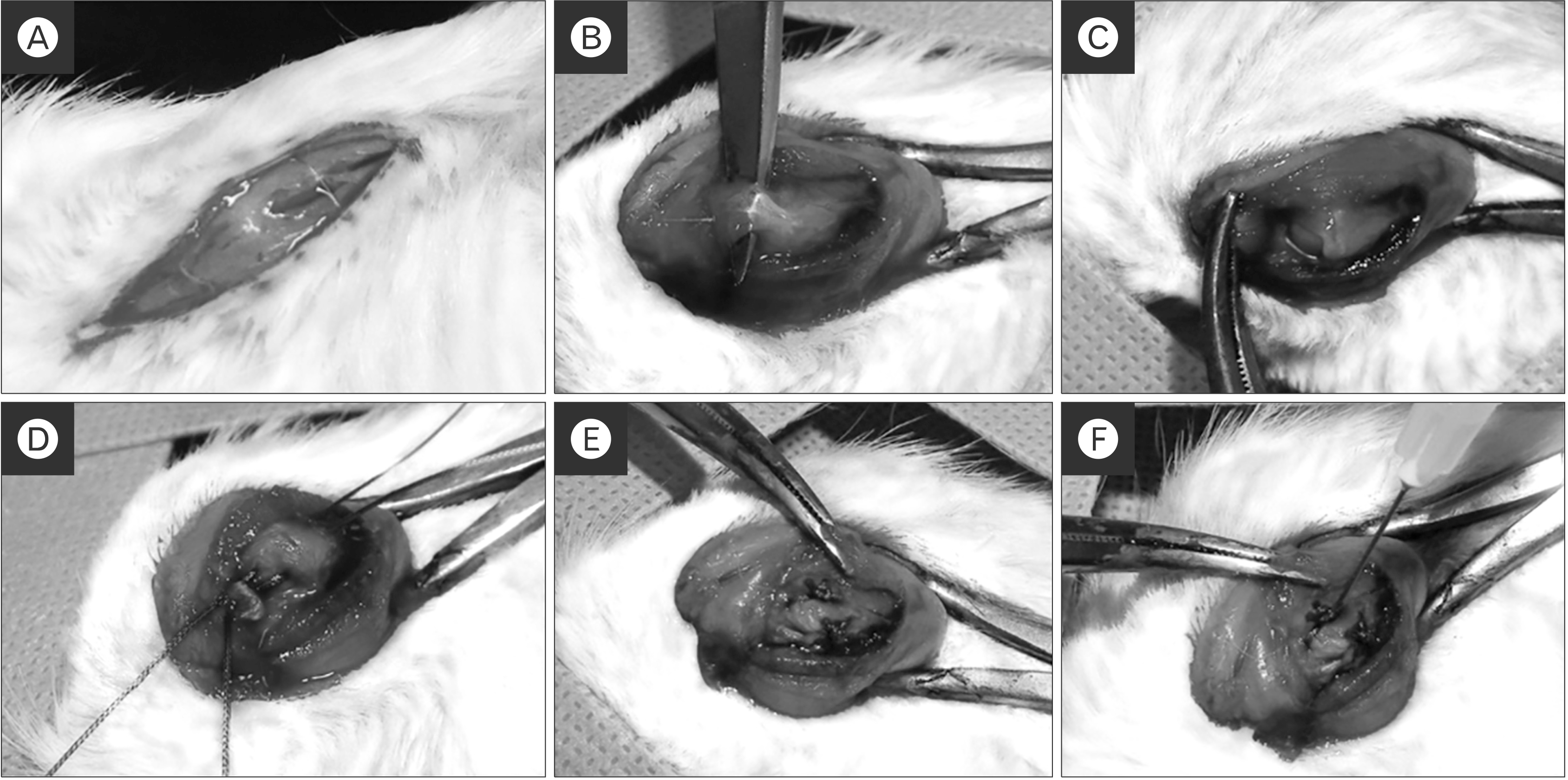
Twenty male Sprague Dawley rats (weight, 300–350 g; 12 weeks old) were divided into two groups. Ten rats were randomly assigned to each group; group 1 (repair only, n=10) and group 2 (KGN single injection, n=10). The rats were anesthetized by intramuscular injection of zolazepam (15 mg/kg, Zoletil; Virbac S.A., Carroscedex, France) and xylazine hydrochloride (5 mg/kg, Rompun; Bayer HealthCare, Leverkusen, Germany). Both shoulders of the rat were shaved and sterilized with iodophor for aseptic conditions. A 3-cm longitudinal incision was made along the scapular spine. The deltoid muscle was split and the supraspinatus tendon exposed at the greater tuberosity. The supraspinatus tendon was isolated and transected the end of the tendon insertion site. Decortication of the foot print was performed with burr. Two bone tunnels were made using a drill at the greater tuberosity of the humeral head. The supraspinatus tendon was repaired with a single row through the bone tunnel with 3.0 Ethibond (Ethicon, Cincinnati, OH, USA). The control group (group 1) was subjected exclusively to repair. The experimental group (group 2; 500 μM of KGN) was injected in the bone- to-tendon interface (Fig. 1). Rats in both groups were sacrificed at 8 weeks after surgery. The right shoulder of each rat was used for biomechanical analysis, and the left was used for histological analysis.
4. Biomechanical evaluation
At 8 weeks after surgery, each rat was euthanized with CO2 gas inhalation. The humeral head and proximal humeral-attached supraspinatus tendons were separated from both shoulders of each rat. We evaluated the biomechanical strength of the bone-to-tendon interface using a Universal Testing Machine (OTT-03; Oriental TM, Siheung, Korea). The specimen was measured to failure at a rate of 10 mm/sec with a 20-N load cell using a custom clamping system. The custom fixture clamping system for tensile test of the supraspinatus tendon consisted of two separate fixtures, namely a humeral head fixation unit, which rigidly fixed the humeral head and permitted the supraspinatus tendon and muscle attached to the humeral head to come out through a hole, and a cryogenic tendon fixation unit, which secured the myotendinous junction of the tendon using a sandpaper-attached clamp and liquid nitrogen to prevent slippage. The supraspinatus tendon was fixed to this system along its anatomic direction to allow tensile loading with the tendon-to-bone interface forming a right angle. The tensile load-to-failure data were automatically collected with a personal computer-based data acquisition system.
5. Histological evaluation
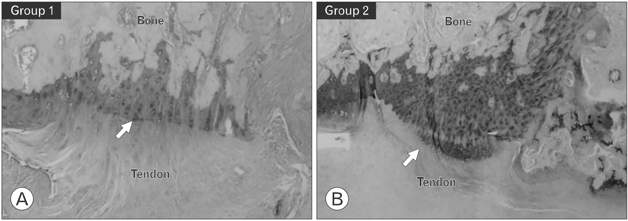
All specimens from both groups were fixed in 10 mL of sterile 10% formalin. After dehydration, they were embedded in paraffin and cut into 4-μm sections. The sections were stained with hematoxylin and eosin (H&E), Masson trichrome, picrosirius red, and toluidine blue staining.
We assessed the healing quality of the tendon-to-bone repair site using various aspects of tendon tissue. The score items included: (1) continuity of collagen fiber, (2) orientation of collagen fiber, (3) maturation of the tendon-to-bone interface, (4) collagen fiber density, (5) vascularity, and (6) cellularity. Each item was evaluated using a 4-point scoring system. For each item, the histological findings were classified semiquantitatively into four grades (grades 0–3). Grade 0 corresponded with the poorest appearance of the ruptured tendon, grade 1 indicated a poorer; grade 2 indicated a better grade, and grade 3 indicated a marked regeneration. Overall, the total score of a slide could range from 0 (ruptured tendon) to 18 (most marked regeneration)14. To eliminate observer error, all tissue slides were evaluated by a pathologist with at least 10 years of experience in a randomized, blinded fashion. All tissue slides were examined three times in the same position and area of rotator cuff tissue by microscope (Leica DMIL LED; Leica, Wetzlar, Germany) and image system (LAS V4.8; Leica) at ×50 magnification.
6. Statistical analysis
All statistical analyses were performed using SPSS ver. 12.0 software (SPSS Inc., Chicago, IL, USA). A p-value less than 0.05 indicated statistical significance. The Kruskal-Wallis test and post-hoc Mann-Whitney U-test were performed to evaluate the biomechanical and histological differences based on H&E staining between groups. Data are presented as the mean and standard deviation. Interclass correlation coefficients were used to assess intra- and interobserver reliability for histological analysis of the fat-to-muscle ratio.
Results
1. Gross inspection and biomechanical evaluation
Gross inspection showed no retear of the supraspinatus tendon-to-bone sites in both groups (p=0.531 at 8 weeks) (Table 1). However, in the biomechanical analysis, the prevalence of midsubstance tear was significantly higher in group 1 (p=0.01 at 8 weeks). Group 1 exhibited a greater tensile strain (group 1: 320.52±94.89%, group 2: 307.69±110.32%; p=0.784) and cross-section area (group 1: 4.23±1.17 mm2, group 2: 3.08±1.30 mm2; p=0.054) compared with group 1. However, there were no significant differences between the groups. Group 2 exhibited a greater ultimate failure load (group 1: 25.78±31.38 N, group 2: 55.64±36.02 N; p=0.011), ultimate stress (group 1: 5.96±5.95 N, group 2: 21.89±16.83 N; p=0.064), and stiffness (group 1: 3.44±5.59 N/mm, group 2: 6.62±4.21 N/mm; p=0.168). However, the ultimate failure load showed a significant difference (Table 2).
2. Histological evaluation
In the results of H&E staining (magnification, ×50), Masson’s trichrome staining, and picrosirius red staining (Fig. 2), group 2 exhibited a significantly greater total score at 8 weeks (group 1: 7.20±2.14, group 2: 9.50±1.84; p=0.019) compared with group 1. Collagen fiber continuity (group 1: 1.20±0.42, group 2: 1.70±0.48; p=0.024) and collagen fiber density (group 1: 1.50±0.52, group 2: 2.20±0.63; p=0.080) were significantly different in both groups. However, collagen fiber orientation (group 1: 1.30±0.48, group 2: 1.70±0.48; p=0.081), maturation of the tendon-to-bone interface structure (group 1: 1.30±0.48, group 2: 1.70±0.48; p=0.080), vascularity (group 1: 0.40±0.51, group 2: 0.10±0.31; p=0.135), and cellularity (group 1: 1.40±0.51, group 2: 1.70±0.82; p=0.342) demonstrated no significant differences between groups 1 and 2 (Table 3). The metachromasia area in toluidine blue staining was measured to confirm fibrocartilage at the tendon-to-bone junction. In group 2, the metachromasia appeared stronger and higher compared with group 1 (Fig. 3).
Discussion
The purpose of this study was to investigate the effect of KGN on tendon-bone healing in a rat rotator cuff tear model. In this study, the KGN group showed more improved biomechanical properties and a histologically better healing quality in the bone-to-tendon interface than the control group. Johnson et al.15 reported that KGN, a small synthetic heterocyclic compound, was found to promote robust chondrogenic differentiation of primary human mesenchymal stem cells. After that, when KGN was injected intra-articularly into a mouse model of osteoarthritis, regeneration of the cartilage and prevention of joint degeneration was observed11,16. In 2014, Zhang et al.9 reported that when injected into intact rat patellar tendons and injured rat Achilles in vivo, KGN induced cartilage-like tissue formation in the tendon-to-bone junctions. Zhang et al.9,13 have studied the use of KGN in combination with platelet-rich plasma to facilitate fibrocartilage formation at the interface between tendon and bone of rat Achilles tendons. In their studies, KGN treatment was found to enhance production of collagens I and II and increase expression of Sox-9 and scleraxis, which is consistent with the formation of fibrocartilage-like tissue13. In addition, KGN treatment resulted in more organized collagen fibers and chondrocytes at the tissue interface, and KGN-treated tendon-to-bone constructs demonstrated greater mean ultimate strengths compared with the control group9,13.
Two previous studies reported on the effects of KGN in a rotator cuff tear model. Wang et al.17 evaluated the effect of KGN in augmenting healing of the repaired enthesis after rotator cuff repair in a murine model. They reported that superior collagen fiber organization and higher ultimate failure loads were seen in the KGN group at 4 weeks. Huang et al.18 compared the effects of repair only (control), microfracture+repair, and microfracture+ repair augmentation with a KGN-loaded gelatin methacryloyl (GelMA) hydrogel scaffold (combined) in tendon-to-bone healing in a rabbit rotator cuff tear model. The authors reported that the KGN-loaded GelMA hydrogel scaffolds group exhibited greater ultimate load to failure and stiffness than the other groups at 8 and 12 weeks after repair. In histological evaluation, the tendon maturation score of the KGN-loaded GelMA hydrogel scaffolds group was significantly higher than that of the other groups at 8 and 12 weeks after repair.
Our results were similar to previous studies. The tendons of the KGN group showed a greater mean ultimate load to failure than the control group, and KGN treatment appeared to enhance the formation and organization of collagen fibrils at the healing enthesis. This suggests that KGN has significant effects on collagen formation and organization at the interface between tendon and bone, even in a rotator cuff tear model. In addition, the two previous studies used scaffolds to minimize the diffusion of KGN into surrounding tissues and maximize the retainment of KGN. However, in our study, only a single injection of KGN was applied in KGN group. Compared with the control group, there was significant biomechanical and histological improvement in KGN group. Clinically single injection of the drug is the simplest method that can be further performed for healing after rotator cuff repair. It is considered important information on the usefulness of KGN to show a significant difference in effect through the simplest method. Therefore, we suggest that our research can provide basic information in other studies based on KGN.
In our study, the metachromasia area in toluidine blue staining was measured to confirm fibrocartilage at the tendon-to-bone junction. In the KGN group, the metachromasia appeared stronger and higher than in the control group. This suggests that KGN, which induces cartilage differentiation, promotes the production of fibrocartilage. Huang et al.18 also reported better fibrocartilage formation in the KGN group at 4, 8, and 12 weeks in toluidine blue staining compared with the control and microfracture+repair groups. However, Wang et al.17 reported that the mean percentage of area of fibrocartilage at the tendon-to-bone interface in Alcian blue staining was higher in the control group compared with the KGN group at 4 weeks. Regarding this difference, it seems that the histological evaluation system of fibrocartilage formation is semiquantitative and difficult to quantify. In the rotator cuff tear model, a significant increase in fibrocartilage formation can be observed at least 8 weeks after KGN injection.
It is believed that our study can be a foundation for future research on the use of KGN in rotator cuff tears. In previous cartilage defect models, various modalities such as chitosan nano- and microparticles16, photo-cross-linked scaffold with KGN- encapsulated nanoparticles19, and polyethylene glycol-modified poly (amidoamine) dendrimer20 have been used to enhance the effects of KGN. Future research will require evaluation of other formulations that can improve the effects of KGN in a rotator cuff tear model.
The current study had several limitations. First, in the experimental group, only a single dose of KGN was applied at the concentration suggested in a previous experiment. Therefore, it is not possible to know the dose-dependent effect of KGN, and information on the optimal concentration of it is not known. Second, because the grading system applied for histological evaluation was semiquantitative, there is a disadvantage that it is not objectively reliable. Third, the 8-week evaluation period may not be long enough for the final analysis, as there may be further change after 8 weeks. Further study with a longer evaluation period may be needed. Fourth, our tendon damage method is more like an acute rotator cuff tear than the chronic degenerative rotator cuff tears observed in most clinical patients.
In conclusion, a single-dose injection of KGN reinforces the biomechanical and histological properties at the tendon-to-bone interface in a rat rotator cuff tear model. Our study confirmed that KGN improves tissue regeneration and mechanical strength of rotator cuff tear. In previous studies, KGN has been widely evaluated as a remedy for cartilage regeneration, but the results of the current study confirm the tendon regeneration effect in a rotator cuff tear model.

 XML Download