

 PDF
PDF Citation
 Print
Print


Introduction
The scapula acts as the kinetic link between the upper extremity and trunk and provides stable base for upper extremity during the high-speed movement of the throwing in overhead athletes. The stability of scapula is provided by periscapular muscle balance, in which the lower trapezius (LT) muscle and the serratus anterior (SA) muscle have been known to play an important role. Researchers demonstrated that periscapular muscle imbalance is associated with abnormal scapular position and loss of dynamic control of scapular motion, known as scapular dyskinesis. The scapular dyskinesis has been considered to be associated with a variety of shoulder injuries1-3. Therefore, shoulder rehabilitation should be integrated with balancing periscapular muscles, in which strengthening and coordinating the LT and SA play an important role.
Kibler et al.4 firstly introduced dynamic exercises engaging the kinetic chain to restore control of dynamic scapular motion. The kinetic chain play a role in transmitting energy and force from one part of the body to the other part. For example, in throwing motion, half of the energy and force are transferred from the leg and trunk to the upper extremity via the kinetic chain. Therefore, shoulder rehabilitation using the kinetic chain rather than a single joint strengthening exercise may be more effective in restoring normal motion of the scapula without excessive load on the rotator cuff or periscapular muscles. Two different views exist in the literature about the effect of shoulder rehabilitations adopting the kinetic chain. One view is that studies focuses on increasing of shoulder muscle activities depending on static and dynamic body positions. The other view is focusing on the effect of the exercise utilizing the kinetic chain on increasing shoulder muscle activities4-6.
Recently, Nakamura et al.7 reported higher shoulder muscle activities in the seated position compared with dynamic standing position during a free-motion robbery exercise. Therefore, further investigations about the effect of exercises adopting the kinetic chain on muscle activation are still required to be addressed. An elastic band has been commonly used during shoulder exercises and shoulder rehabilitation programs3,8-10. So, we tried to prove the effect of exercise adopting the kinetic chain using an elastic band.
In the present study, we mainly aimed to evaluate the shoulder muscle activities during shoulder external rotation exercises using an elastic band with arm at side or 90° of abduction in static and dynamic body positions. Static positions included the sitting, squat positions and static rotational squat (SRS), while, in dynamic positions, body extension and body extension combined with trunk rotation were included. The working hypothesis was that shoulder muscle activities would be different between the exercises with or without body movements regardless of shoulder position and shoulder external rotation exercises using kinetic chain concept would be some benefit to shoulder muscle exercises, which is required for the patients with weak periscapular muscles.
Go to : 
Methods
The Institutional Review Board of Konkuk University Hospital approved this study (IRB No. KUMC202007076), and informed consent was obtained from each subject.
1. Subject selection
In 2017, a total of 19 right-handed male subjects (mean±standard deviation: age, 22.4±1.6 years; height, 175.1±4.3 cm; weight, 72.5±6.9 kg; body mass index, 26.6±2.4 kg/m2) were included in this study. All tests were performed on the dominant arm. Subjects were excluded if they (1) had previous injury affecting the musculoskeletal system, (2) received rehabilitation treatments or orthopedic surgeries for any injuries, (3) had difficulty in following the exercise motion as indicated.
2. Electromyography
Surface electromyography (EMG) signals were recorded from the anterior deltoid (AD), middle deltoid (MD), upper trapezius (UT), LT, SA and infraspinatus (IS) muscle. The signals were amplified with a gain of 400, noise <1 μV and a common mode rejection ratio of 100 dB. They were sampled at 1,500 Hz and filtered with a bandwidth of 10–500 Hz. To minimize skin impedance, we prepared the skin surface by cleaning with an alcohol swab before placing the electrodes. Bipolar surface EMG electrodes (Noraxon, Scottsdale, AZ, USA) with a distance of 2 cm between active recording sites were used. The electrodes were placed at the center of the muscle belly in line with the muscle fibers according to the surface EMG for a non-invasive assessment of muscles guideline and previous studies as follows: AD, one finger width distal and anterior to the acromion; MD, from the acromion to the lateral epicondyle of the elbow (the greatest bulge of the muscle); UT, halfway between the C7 spinous process and the acromion process; LT, two-thirds on the line from the trigonum spinea to the eighth thoracic vertebra; SA, below the axilla between the latissimus dorsi and pectoralis major at the level of the seventh rib; IS, two fingerbreadths below the medial portion of the spine of the scapula.
3. Inertial measurement units
Three wireless inertial measurement units (IMUs) sensors (I2M Motion Tracking IMUs; NexGen Ergonomics, Pointe-Claire, QC, Canada) were placed on the scapular spine (halfway between acromion and medial border of the scapula), sternum (just below the sternal angle) and sacrum (between right and left posterior superior iliac spine) for evaluation of motion angle of the scapula in the static position. Because the validity of measurement of scapular motion during body movement has not been sufficiently established, we used this device only for calculation of scapular motion during the static position11. Sensors placed on the sternum and pelvis were for monitoring whether the static position was well maintained. The sensors were attached with a double-sided adhesive skin tape and fixed firmly with a transparent plaster. IMU signals were recorded with a sample frequency of 128 Hz. Signal acquisition was conducted with the I2M Motion Tracking IMUs software (TK Motion Manager 1.0.0.201508052305, NexGen Ergonomics), and data processing and analysis were performed using MATLAB R2013a (MathWorks Inc., Boston, MA, USA). The average value of scapular orientation was calculated from three repetitions of each exercise.
4. Procedures
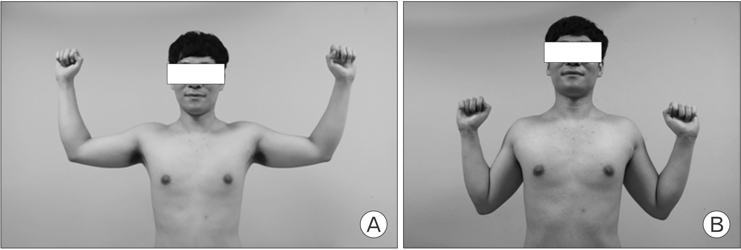
The subjects underwent the maximal voluntary isometric contraction (MVIC) testing of each muscle using the manual muscle test, which was conducted three sets of five exercises following the guideline of Kendall et al.12 Subjects were provided with verbal encouragement and visual feedback of signal amplitude to generate maximal force on each muscle. At least 2 minutes of rest between MVIC tests was given to minimize muscle fatigue. Each exercise protocol consisted of a series of five exercises in the 90/90 position or the W position (Fig. 1). For the W position, each subject rotated the investigated arm externally with arm at side and palm facing forward. To achieve the W position properly, the scapula should be squeezed toward the spine, and the elbow should be pulled down toward back pocket. The 90/90 position consisted of 90° of shoulder abduction and ER, similar to the late cocking phase of the throwing motion. Each exercise consisted of three phases: concentric, isometric and eccentric phase, and the speed of each phase was controlled by a smartphone metronome application at a tempo of 40 beats per minute. Subjects repeated each exercise three times with 4.5 seconds rest intervals using an elastic band of green color (Theraband; Hygenic, Akron, OH, USA). Before data collection, each subject performed all exercises for 10 minutes without load to become habituated to study procedures. Exercise protocols were selected in random order. Five exercises in each protocol were performed randomly with 2 minutes of rest between the exercises. For the sitting position which was designed to eliminate the influence of the kinetic chain of the lower extremity, each subject sit in the wheelless chair with back straight and flexed the shoulder with elbow in full extension and forearm in neutral rotation, grasping the TheraBand. Then, each subject was instructed to perform the W position or 90/90 position. Subjects performed the exercise in the static squat (SS) position with the trunk and knee flexed to 40° to 50° to cause static tension in the kinetic chain from the lower extremity to the trunk. For the SRS position, subjects were instructed to rotate the trunk laterally at 45° while keeping the pelvis and leg in SS position. For the dynamic squat to standing (DSS) position, each subject was instructed to extend the knee and hip joint concomitantly from the SS position to investigate the effects of engaging of the kinetic chain by extension of the lower extremity. As for the exercise in the dynamic squat to standing and trunk rotation (DSSR) position which was introduced as the lawnmower exercise in the study of Kibler et al.4, each subject was instructed to perform exercise in the DSS position and rotate the trunk at 45° externally simultaneously from the SS position. With the DSSR position we expected to observe the effect of trunk rotation combined with DSS position (Fig. 2).
 | Fig. 2Shoulder external rotation exercises using an elastic band with 90° of shoulder abduction and ER (90/90 position) in static (A−C, F−H) and dynamic body positions (D, E, I, J). (A, F) Sitting position. (B, G) Static squat position. (C, H) Static rotational squat position. (D, I) Dynamic squat to standing position. (E, J) Dynamic squat to standing and trunk rotation position.
|
5. Statistics
A one-way repeated-measures analysis of variance (ANOVA) was used for detecting differences in analyzing the normalized EMG amplitude of each muscle and the kinematic data of the scapula according to each shoulder position. A two-way repeated- measures ANOVA (postural position [sitting, SS, SRS, DSS, DSSR]×shoulder position [W position or 90/90 position]) was calculated to analyze EMG and kinematic data. The α level of p<0.05 was considered statistically significant. We used SPSS ver. 17.0 (SPSS Inc., Chicago, IL, USA) to analyze all data.
Go to :
Results
1. EMG data
Two-way interaction was not noted in all muscle activities investigated. However, differences in the muscle activities between the shoulder postures were found in the UT (F=13.218, p=0.001), SA (F=17.753, p<0.001) and IS (F=5.842, p=0.021). Muscle activities of AD, MD, UT, LT, SA and IS were obtained at each positions (Table 1, Fig. 3).
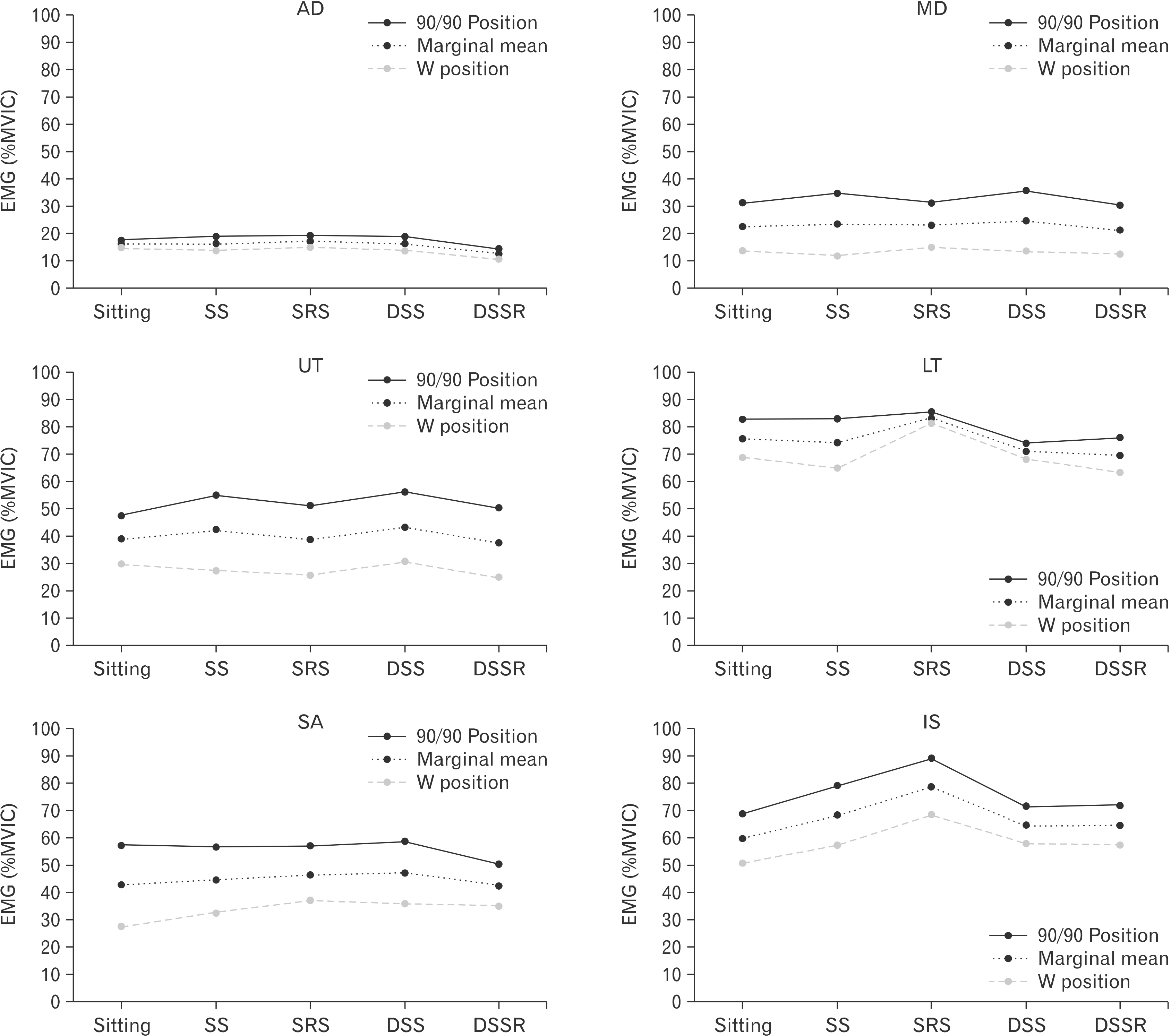 | Fig. 3Muscle activities of anterior deltoid (AD), middle deltoid (MD), upper trapezius (UT), lower trapezius (LT), serratus anterior (SA), and infraspinatus (IS). EMG: electromyography, MVIC: maximum voluntary isometric contraction, SS: static squat, SRS: static rotational squat, DSS: dynamic squat to standing, DSSR: dynamic squat to standing and trunk rotation.
|
Table 1
Electromyographic activities of six muscles according to the body and shoulder position
![]()
1) Anterior deltoid
The marginal mean (collapsed across shoulder position) in the DSSR position was less than that in all other positions (sitting, p=0.011; SS, p=0.005; DSS, p<0.001; DSSR, p=0.001). The marginal mean (collapsed across postural position) in the 90/90 position was greater than that in the W position (p=0.049). In the W position, one-way repeated measures ANOVA test indicated that the normalized amplitude in the SRS and DSS positions was greater than that in the DSSR position. In the 90/90 position, muscle activity in the DSSR position was lower than that in all other positions.
2) Middle deltoid
The marginal mean (collapsed across shoulder position) in the SRS position was greater than that in the SS position (p=0.009) and DSSR position (p=0.006). The marginal mean (collapsed across postural position) in the 90/90 position was greater than that in the W position (p<0.001). In the W position, statistical difference was found between the normalized amplitudes in the SRS and DSSR positions (p=0.046). In the 90/90 position, the normalized amplitude in the SRS position was lower than that in SS positions (p=0.025).
3) Upper trapezius
The marginal mean (collapsed across shoulder position) in the SRS position was greater than that in the SS (p=0.007), DSS (p=0.037) and DSSR (p=0.001) positions. The marginal mean (collapsed across postural position) in the 90/90 position was greater than that in the W position (p=0.001). In the W position, the normalized amplitude in the SRS position was lower than that in the SS (p=0.035) and DSS (p=0.004) positions. In the 90/90 position, the normalized amplitude in the SRS position was greater than that in the sitting (p=0.018) and DSSR (p=0.034) positions. Statistical difference was also found between the sitting and DSS positions in the 90/90 position (p=0.008).
4) Lower trapezius
The marginal mean (collapsed across shoulder position) in the SRS position was greater than that in the SS (p=0.005), DSS (p=0.006), and DSSR (p=0.003) positions. There was no statistical difference in the marginal mean (collapsed across postural position) between the 90/90 position and the W position. In the W position, the normalized amplitude in the SRS position was greater than that in the SS (p=0.005) and DSSR (p=0.027) positions. In the 90/90 position, the normalized amplitude in the SRS position was greater than that in the DSS (p=0.012) and DSSR (p=0.048) positions. We also noted significant difference in the normalized amplitude between in the sitting and DSS positions (p=0.038) in the 90/90 position.
5) Serratus anterior
There were no statistical differences in the marginal means (collapsed across shoulder position) between all postural positions. The marginal mean (collapsed across postural position) in the 90/90 position was greater than that in the W position (p=0.001). In the W position, the normalized amplitude in the sitting position was less than that in the SS (p=0.02), SRS (p=0.01), and DSS (p=0.023) positions. In the 90/90 position, statistical differences were not found in the normalized amplitudes between the postural positions.
6) Infraspinatus
The marginal mean (collapsed across shoulder position) in the SRS position was greater than that in all other positions (p< 0.001). Statistical difference was found in the marginal mean (collapsed across postural position) between the 90/90 position and the W position (p=0.021). In the W position, the normalized amplitude in the SRS position was greater than that in all other positions (sitting, p<0.001; SS, p=0.005; DSS, p=0.01) except DSSR position (p=0.066). Statistical difference was also noted in the normalized amplitude between in the sitting and DSS position (p=0.027) in the W position. In the 90/90 position, the normalized amplitude in the SRS position was greater than that in all other positions (sitting, p<0.001; SS, p=0.017; DSS, p=0.003; DSSR, p<0.001). In addition, the normalized amplitude in the SS position was greater than that in the sitting (p=0.02) and DSSR (p=0.022) positions in the 90/90 position.
Go to :
Discussion
Most studies about the effects of shoulder exercises evaluated various dynamic motions and reported which one was better than others in inducing more LT or SA activations or scapular posterior tilt or external rotation. However, our main interest was to observe any difference between static and dynamic postures to demonstrate whether body movements influence on shoulder muscle activities through the kinetic chain. Basically, by comparison of the sitting and SS positions, we intended to investigate the difference between disconnection and maintenance of the kinetic chain between the upper body and lower extremity. To evaluate specifically the influence of dynamic body motion, the exercises between in the SS and DSS positions and in the SRS and DSSR were compared. The main finding of this study was that shoulder external rotation exercise in the DSSR position was effective in reducing shoulder muscle activities except the SA of sitting position compared with SRS position.
Exercise in the sitting position blocks off the effects of the kinetic chain linked to the lower extremity, while the SS position permits functional continuity of the kinetic chain without energy transmission from dynamic movement of the trunk or lower extremity. In this study, comparison of exercises in the sitting and SS positions did not show any significant difference in the EMG and kinematic data, except that of the SA in the W position. This result is in line with the study of De Mey et al.2 in which the authors did not find differences in the UT and LT activities between the sitting and SS positions during shoulder retraction exercise. Meanwhile, Nakamura et al.7 reported increase of shoulder muscle activities in the sitting position than in the DSS position during shoulder ER exercise in the 90/90 position. Because in the sitting position, body balance and stability should be maintained without assistance of the lower extremity. Although this study did not demonstrate differences in muscle activities between the sitting and SS position, differences might be detected with higher load applied to the shoulder than that used in this study.
In comparison of the SS and DSS positions, we did not observe any effectiveness of dynamic motion on muscle activities regardless of the shoulder positions. Although shoulder external rotation exercises in the DSS position was reported as effective in reducing shoulder muscle activities, it should be considered that the shoulder muscle activities may vary depending on the pulling height. There is a possibility that the pulling effect at the chest height is not as effective as the pulling action from below the waist. In the study of De Mey et al.2, a difference was not also found between the SS and DSS positions at the head height with a pulley apparatus during shoulder retraction exercise.
When comparing between the SRS and DSSR positions, we noted that dynamic motion significantly decreased muscle activities. The trunk rotation would substantially increase the tension of the elastic band, which can be inferred by higher muscle activities in the SRS position than in any other position. Different from the exercise in the DSS position, changes both in the muscle activities and scapular motions were achieved with higher load in the DSSR position. The lawnmower exercise in which dynamic body extension and trunk rotation are incorporated was introduced as it activated the LT in larger amplitude compared with other closed kinetic exercises4. However, Tsuruike and Ellenbecker13 reported that the lawnmower exercise did not increase the LT and posterior deltoid muscle activities across three different intensities of 3%, 5%, and 7% of body weight. The authors speculated that the lower leg, hip, or trunk muscles would be progressively activated as the intensity was increased. Considering the results of this study, dynamic body extension combined with trunk rotation enables to control scapular motion effectively with less shoulder muscle activations. Correction of dyskinetic scapular motion has been a primary goal of shoulder rehabilitation in patients with subacromial pain syndrome. Dyskinetic scapular motion appears to be decreased in posterior tilt and upward rotation in accordance with decrease of SA and LT activities. Therefore, the kinetic chain exercises incorporated with lower extremity, hip, or trunk would be helpful in patients with weak periscapular muscles, in whom the LT activities were found to be frequently decreased14-18.
There are some limitations to our study. First, all the people who participated in this experiment were all healthy male subjects. However, we wanted to do a basic study to find out the activities of shoulder muscles depending on each positions for healthy adults. It’s a limitation that the female subjects were not included. Second, the lawnmower exercise or shoulder external rotation exercise is often performed when there is shoulder pathology such as rotator cuff tear, but it seems that there is a limit to revealing with the current study whether the same results can be obtained in the shoulder with pathology. Third, one cycle of shoulder external rotation exercise includes all three phases; concentric, isometric, and eccentric, but there may be some differences between individuals and between subjects. To reduce these differences, we trained the subjects to perform the test at a constant rhythm and speed for one cycle.
We found that shoulder ER exercise in the DSSR position was effective in reducing shoulder muscle activities except the SA compared with SRS position. Considering the results of this study, DSSR enabled to control scapular motion effectively with less shoulder muscle activations. Therefore, the kinetic chain exercises incorporated with lower extremity, hip, or trunk would be some benefit to shoulder muscle exercises, which is required for the patients with weak periscapular muscles, in whom the LT activities was found to be frequently decreased.
Go to :
 XML Download