Journal List > Korean J Orthod > v.51(1) > 1146328

Sivarajan, Mani, John, Mona M. Salah, Kook, and Wey: The global distribution of permanent canine hypodontia: A systematic review

Abstract

Objective

To systematically review studies on canine agenesis prevalence in different populations and continents, based on the jaw, sex, location, and associated dental anomalies.

Methods

Electronic and hand searches of English literature in PubMed, Web of Science, Scopus, OpenGrey, and Science Direct were conducted, and the authors were contacted when necessary. Observational studies (population-based, hospital/clinic-based, and cross-sectional) were included. For study appraisal and synthesis, duplicate selection was performed independently by two reviewers. Study quality was assessed using a modified Strengthening the Reporting of Observational Studies in Epidemiology checklist, with main outcome of prevalence of canine agenesis.

Results

The global population prevalence of canine agenesis was 0.30% (0.0−4.7%), highest in Asia (0.54%), followed by Africa (0.33%), and the least in Europe and South America (0.19% in both continents). Canine agenesis was more common in the maxilla (88.57%), followed by both maxilla and mandible (8.57%), and the least common was mandible-only presentation (2.86%). The condition was more common in females (femalemale ratio = 1.23), except in Asia (femalemale ratio = 0.88) and Africa (femalemale ratio = 1). In Asia, unilateral agenesis was almost twice as prevalent as bilateral, but in Europe, the bilateral form was more common.

Conclusions

The overall prevalence of canine agenesis is 0.30%, with the highest prevalence in Asia, followed by Africa, Europe, and South America. The condition is more common in the maxilla than the mandible, and in females than males (except in Asia and Africa), with unilateral agenesis being more common in Asia and the bilateral form showing a greater prevalence in Europe.

INTRODUCTION

Congenital absence of teeth, hypodontia,1 is the most prevalent craniofacial malformation and dental anomaly.2 Its reported prevalence varies across studies, continents, racial groups, dentitions, sexes, and jaws. The prevalence ranges widely among Caucasians (3.9% to 11.3%)3 and is higher among African populations (13.4%), followed by European (7%), Asian (6.3%), and Australian (6.3%) populations.4 Female subjects are more likely to show hypodontia than male.3-6 The occurrence of this condition can be classified as common, less common, and rare.3 Canine agenesis refers to failure of canine formation, which are considered to be the most stable teeth; agenesis of maxillary canines is less common, while that of mandibular canines is rarely observed.7 Nevertheless, the absence of canines complicates orthodontic treatment planning because of their esthetic and functional importance.8
Information regarding the global and regional distribution of canine agenesis is of paramount importance since it can elucidate the treatment need, complexity of treatment, and the resources required to manage these cases. Early detection may facilitate interventions to ameliorate the disease process, such as early primary tooth removal to enhance space closure or maintenance of the predecessor to ensure adequate alveolar bone for future replacement.9 Some degree of multidisciplinary combined management may be required, especially in cases of unilateral agenesis. Furthermore, the assessment of agenesis prevalence by continents can reflect the comparative frequency of missing teeth in different regions of the world. There is a paucity of studies on the prevalence of canine agenesis, with very few studies reporting the prevalence of agenesis exclusively, and most only superficially referring to individual studies without analyzing the combined prevalence, and instead only focused on the prevalence of hypodontia in general.
The aim of the current review was to summarize the available worldwide data on canine agenesis. The primary objective was to systematically evaluate the available evidence related to its prevalence in different general populations and continents. The secondary objectives were to report the prevalence by jaw (maxilla and mandible), sex (male and female), and location (unilateral or bilateral), and to report the associated dental anomalies. Identifying the overall prevalence and pattern can enhance management and better treatment planning of this condition.

MATERIALS AND METHODS

Protocol and registration

This systematic review was conducted and reported in accordance with the Cochrane Handbook for Systematic Reviews of Interventions and Preferred Reporting Items for Systematic Reviews and Meta-Analyses (PRISMA).10 The review protocol was registered in the National Institute of Health Research database (https://www.crd.york.ac.uk/prospero/; protocol no: CRD42019120204; registration Date: March 14, 2019). Ethical approval was unnecessary since we retrieved data from previously published studies in which informed consent had been obtained by the primary investigators.

Eligibility criteria

Inclusion criteria

  1. Participants: Male and female subjects with no age restriction; sample size of 50 participants or more

  2. Outcome measures

    • a. Primary outcome: Overall prevalence of canine agenesis

    • b. Secondary outcomes: Prevalence of canine agenesis in the maxilla and mandible, female:male ratio, ratio of unilateral to bilateral cases, dental anomalies associated with canine agenesis

  3. Study design: Observational studies (population-based studies, hospital/clinic-based studies, and cross-sectional studies), studies supported by radiographic imaging of the teeth or relevant history and records

  4. Published English studies with no publication-year restriction

Exclusion criteria

  1. Studies on syndromic patients (e.g., patients with a cleft involving the alveolus or those with Down’s syndrome)

  2. Case reports, case series, systematic reviews, or meta-analyses

  3. Studies that reported canine agenesis in specific samples of patients with teeth agenesis that cannot be generalized to the general population, e.g., canine agenesis in hypodontia patients with no relevance to the general population.

Information sources, search strategy, and study selection
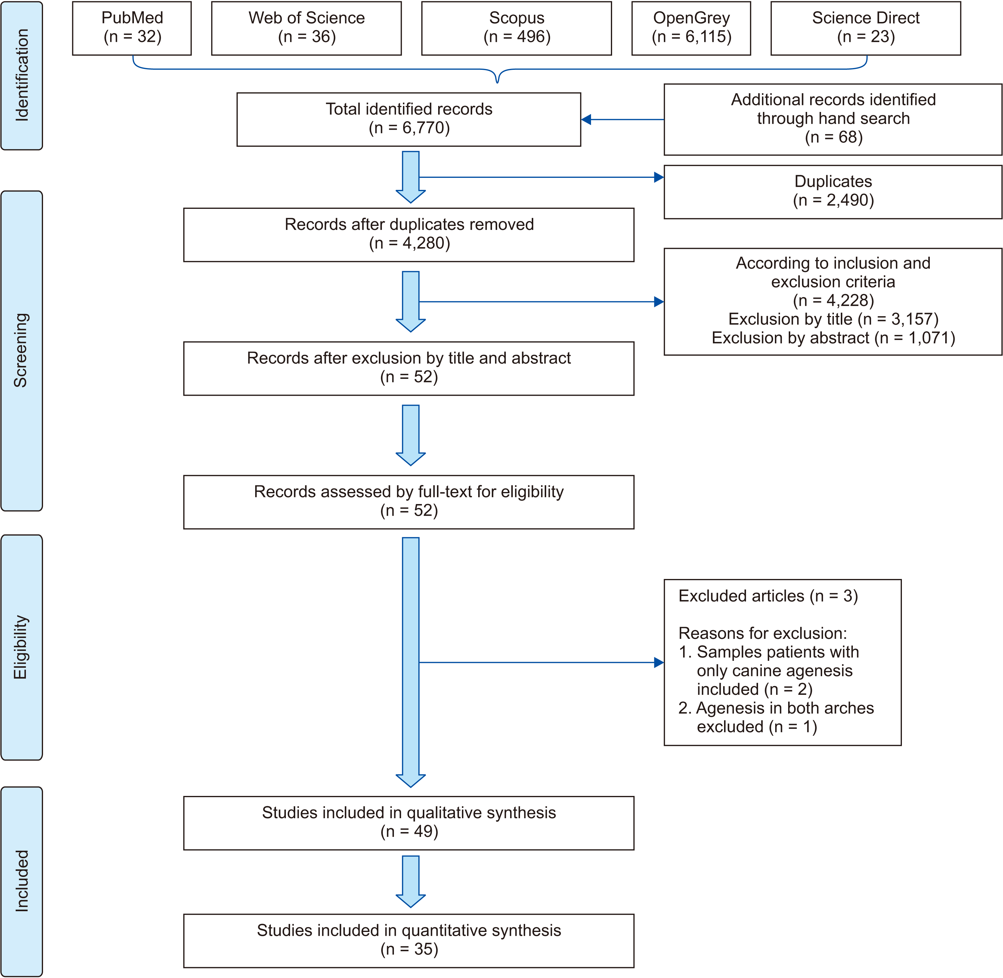
Comprehensive electronic database searches without publication-year restrictions were conducted for literature published until May 4, 2019 (Table 1 and Figure 1). Only articles in English were included from relevant databases such as PubMed, Web of Science, Scopus, OpenGrey, and Science Direct. In addition, hand searches of relevant journals, such as those listed in relevant systematic reviews, was performed. Articles and reference lists of the included studies were individually screened for additional relevant studies. The corresponding authors were contacted for obtaining clarifications or additional information when necessary.
The search strategy was implemented using a combination of Medical Subject Headings (MeSH) and free-text words for PubMed and optimized for each database (Table 1). Literature search, study inclusion, methodology quality assessment, and data extraction were carried out independently and in duplicate by two pairs of reviewers (S.S. & M.C.W. and S.A.M. & J.J.) who were not blinded to the authors, and the results were revised by the fifth author (M.M.S.F.).
Eligible articles were assessed in two phases. In the first phase, only titles and abstracts were screened. Full-text assessment was then conducted in the second phase to determine final eligibility. Articles were excluded when they did not meet one or more of the inclusion criteria. Any disagreements were resolved by discussion and consultation with the fifth author (M.M.S.F.) for consensus.

Data items

A standardized data extraction sheet was designed for data extraction by the two pairs of independent reviewers in duplicate (S.S. & M.C.W. and S.A.M. & J.J.). Data extraction included general information (the names of the authors, the year of publication, and the study setting), data pertaining to methods (study design), participant data (sample size, age, sex, country, region, race, and population) and outcome data (primary and secondary outcomes mentioned). Race referred to a group of people who shared similar physical characteristics.

Risk of bias across studies

Critical appraisal of the study was performed using a modified version of Strengthening the Reporting of Observational Studies in Epidemiology (STROBE) checklist11 consisting of seven items related to (1) study design, (2) study setting, (3) participant criteria, (4) sample size, (5) variable description, (6) outcome measurements, and (7) statistical analysis. The quality of the studies was categorized as weak (3 and less), moderate (4 or 5), or high (6 or more) by two pairs of independent reviewers in duplicate (S.S. & M.C.W. and S.A.M. & J.J.). Any disagreements were resolved by discussion and consultation with the fifth author for consensus (M.M.S.F.).

Summary measures and synthesis of results

Relevant prevalences from every study were recalculated and summed to be reported as overall percentages across all studies (Tables 2 and 3) and in terms of agenesis in the maxilla and mandible, female:male ratio, and unilateral and bilateral agenesis.

Additional analyses

No subgroup analysis was performed.

RESULTS

Study selection

Initial database and hand searches yielded a total of 6,770 studies, of which 2,490 duplicates were excluded (Figure 1). Subsequent exclusion by title (3,157) and abstract (1,071) yielded 52 remaining articles, which were considered for this review. This included 18 articles identified from database searches and 34 articles identified with hand searches.

Study characteristics

Full texts of all 52 articles were assessed for eligibility. Two studies12,13 were excluded since they did not report actual prevalence data or data that can be used to calculate the prevalence for their populations, and another study excluded samples with canine agenesis in both arches,14 which affected the accuracy of the overall prevalence estimation.

Risk of bias within studies

Critical appraisal of the remaining 49 articles was performed using the modified STROBE checklist (Table 4). After excluding 14 studies (28.6%) that were of moderate quality (modified STROBE score of 4 or 5), 35 (71.4%) high-quality studies5,15-48 (modified STROBE score of 6 or 7) were included in this systematic review.

Results of individual studies

Description of the studies

A total of 35 studies were included in this review. The demographic data are summarized in Table 5. Slightly less than two-thirds15-34 (57.1%) of the selected studies were retrospective, about a third35-46 (34.3%) were cross-sectional, while two studies29,47 were both retrospective and cross-sectional. Almost half (48.6%)17-21,24,29-35,42,45-47 of the studies were published after the year 2010. The sample size varied from 85 to 100,577, with a median of 1,622 participants. The study participants, aged 5–46 years, were recruited equally from orthodontic (36.1%) and dental clinics (36.1%), followed by the pediatric clinic (13.9%). One study45 recruited patients from both orthodontic and pediatric clinics, another44 recruited them from dental and orthodontic clinics, while another37 recruited participants from the Australian defense force. Only two studies39,43 were epidemiological surveys. Almost an equal number of studies were from Asia (n = 14, 40.0%) and Europe (n = 12, 34.3%), followed by South America (n = 4, 11.4%), North America (n = 2, 5.7%), Australia (n = 2, 5.7%), and Africa (n = 1, 2.9%). The global reported population prevalence of canine agenesis ranged from 0.0% to 4.7% (Table 6), with high variations among studies, and the median prevalence was 0.29%. The term population refers to the people living in a particular country. The prevalence of canine agenesis in the maxilla (between 0.03% and 4.7%) was higher than that in the mandible (between 0% and 1.12%).

Synthesis of results

Primary outcomes

Due to the high variation, the overall prevalence was calculated from studies that reported canine agenesis data either by individual (population prevalence based on the number of assessed individuals) or by the number of missing teeth (prevalence of canine agenesis based on the number of missing teeth). The overall prevalence of canine agenesis based on the total number of missing teeth (2.99%) (Table 3) was higher than the population prevalence based on the number of assessed individuals (0.30%) (Table 2). The prevalence in the general population excluding the orthodontic population was higher (0.38%) than that investigated among the orthodontic population (0.10%). The overall population prevalence was the highest in Asia (0.54%), followed by Africa (0.33%),23 and was the least in Europe and South America (0.19% in both continents). Similarly, the prevalence of agenesis as a percentage of missing teeth was the highest in Asia (7.40%), followed by Oceania (Australia) (7.14%), South America (4.51%), Africa (3.80%), and Europe (2.55%), with the lowest prevalence in North America (1.85%) (Table 6).

Secondary outcomes

Pooled prevalence based on studies that reported the prevalence in the maxilla and mandible using data for individuals revealed the same findings, with the maxilla-only prevalence being the highest (88.57%), followed by the prevalence in both the maxilla and mandible (8.57%), and the mandible-only prevalence being the least (2.86%) (Table 7). Similarly, the overall prevalence by teeth was greater in the maxilla (73.73%) than the mandible (26.27%). By continent, the prevalence in the maxilla was the highest in Asia (0.78%), followed by Africa (0.33%) based on one study,25 South America (0.19%) based on one study,14 and the least in Europe (0.13%) based on five studies.16,20,34,36,41 Meanwhile, the prevalence of agenesis in the mandible only and in both maxilla and mandible was only reported in Europe (0.01% and 0.03%, respectively; Table 7). Among all forms of canine agenesis, the overall prevalence of missing maxillary permanent canines was almost similar (35.60% and 39.63%, respectively). Likewise, in the mandible, the prevalence of missing mandibular permanent canines was almost similar (12.07% and 12.69%, respectively).
By sex, the overall ratio of canine agenesis was higher in females than in males, with a female:male ratio of 1.23. However, this was only true in Europe and South America, wherein females were twice as much affected than males. In Asia, the ratio was higher among males (ratio = 0.88), while the prevalence was the same for both sexes (ratio = 1) in Africa (Table 8). The overall bilateral:unilateral agenesis ratio was 1.13. The worldwide prevalence of unilateral agenesis was almost similar to that of the bilateral form (50.0% and 46.7%, respectively). However, in Asia, the prevalence of unilateral agenesis was almost double that of bilateral agenesis (66.7% and 33.3%, respectively). In Europe, the prevalence of bilateral (58.8%) agenesis was higher than that of unilateral agenesis (35.3%) (Table 9).
Common dental anomalies associated with canine agenesis were retained primary canines,16,30 agenesis of other permanent teeth,16,30 agenesis of the third molar,31,32,45,46 supernumerary teeth,16 ankylosis,28 taurodontism,28 enamel hypoplasia and conical incisor,28 and Class III malocclusion.31,45

DISCUSSION

This systematic review attempted to evaluate the global distribution of canine agenesis in isolation.3,49 We presented the population prevalence of canine agenesis in terms of individuals, which better reflected the actual treatment need, unlike another review4 that reported the prevalence in terms of the number of missing teeth. Reports based on individual prevalence without considering the population sample can be biased, since the bigger quantity of smaller-sized studies may overwhelm the smaller quantity of bigger-sized studies and distort the final summary. Therefore, we recalculated the prevalence in every included study to generate an overall prevalence.
In this review, an almost equal proportion of the included studies were conducted in Asia (37.1%) and Europe (37.1%); this was in contrast to the review on the overall prevalence of hypodontia, in which most studies were conducted in European countries (43.0%), followed by the Asian region (32.0%).4 The global distribution in this review ranged from 0.0% to 4.7%, with a pooled overall prevalence of 0.30%, which is much lower than that of hypodontia (6.4%).4 Polder et al.3 reported that canines are one of the rarely missing teeth after the first and the second molars.
In this review, the prevalence of canine agenesis was higher in the Asian region than in the African, European, and South American regions. In contrast, the overall prevalence of hypodontia was the highest in Africa (13.4%, 95% confidence interval [CI]: 9.7, 18.0), followed by Europe (7% CI: 6.0, 8.0%) and Asia (6.3% CI: 4.4, 9.1).4 This suggests that canine agenesis per se is more common in the Asian region than in the European region, possibly due to the racial differences between the two continents.
Our findings showing that canine agenesis was more common in the maxilla than the mandible are in agreement with the general pattern of hypodontia reported in two other systematic reviews addressing hypodontia, both of which reported marked differences between the jaws in relation to the frequency of agenesis of various tooth types.3,4 Similarly, the greater prevalence in females is in agreement with the findings of these two systematic reviews.3,4 However, in Asia, the higher prevalence in males may indicate a genetic inheritance of this trait among males. Bilateral agenesis was more prevalent than unilateral agenesis in Europe, similar to the general pattern of hypodontia except for the maxillary lateral incisors.3 However, in Asia, the prevalence of unilateral agenesis was double that of the bilateral form, indicating a genetic inheritance pattern among Asians. The overall information presented in this review could provide valuable guidance to clinicians for treatment planning and managing patients with canine agenesis.
Since we aimed to report the prevalence based on the number of individuals with missing canines, we could not include studies reporting the number of missing canines instead of the number of individuals with missing canines; this limited our ability to present the data in terms of combined prevalence by number of teeth. Most studies assessed either orthodontic patients,5,14,17,19,22-25,29,31,32,43 pediatric patients,21,26,27,33 both orthodontic and pediatric patients,15 both orthodontic and dental patients,44 or dental patients.16,18,20,28,30,35,39-41,45-47 Three studies were epidemiological surveys of school children,34,37,42 one enrolled defense force recruits,36 while only one study was a truly epidemiological study on a general population.38

CONCLUSION

  1. The global distribution of canine agenesis ranged from 0.0% to 4.7%, with a pooled overall population prevalence of 0.30%.

  2. The population prevalence of canine agenesis was the highest in Asia (0.54%), followed by Africa (0.33%); the least prevalence was observed in Europe and South America (0.19% for both continents).

  3. The highest prevalence was of the maxilla-only form (88.57%), followed by the presentation in both maxilla and mandible (8.57%), while the mandible-only form showed the lowest prevalence (2.86%).

  4. Canine agenesis was more common in females, with an overall female:male ratio of 1.23, except in Asia (0.88) and Africa (1).

  5. In Asia, the prevalence of unilateral agenesis was almost double that of bilateral agenesis, but in Europe, bilateral agenesis was more common.

With a clearer picture of the occurrence of canine agenesis and its accompanying predilection, management of the condition can be better predicted and planned. Future research on prevalence is suggested to report both in terms of missing teeth and individuals, also moving forward, research linked to its aetiology and genetic-based treatment can be considered.

Notes

CONFLICTS OF INTEREST

No potential conflict of interest relevant to this article was reported.

REFERENCES

1. Goodman JR, Jones SP, Hobkirk JA, King PA. 1994; Hypodontia: 1. Clinical features and the management of mild to moderate hypodontia. Dent Update. 21:381–4.
2. Lee JH, Yang BH, Lee SM, Kim YH, Shim HW, Chung HS. 2011; A study on the prevalence of dental anomalies in Korean dental-patients. Korean J Orthod. 41:346–53. DOI: 10.4041/kjod.2011.41.5.346.
3. Polder BJ, Van't Hof MA, Van der Linden FP, Kuijpers-Jagtman AM. 2004; A meta-analysis of the prevalence of dental agenesis of permanent teeth. Community Dent Oral Epidemiol. 32:217–26. DOI: 10.1111/j.1600-0528.2004.00158.x. PMID: 15151692.
pmid
4. Khalaf K, Miskelly J, Voge E, Macfarlane TV. 2014; Prevalence of hypodontia and associated factors: a systematic review and meta-analysis. J Orthod. 41:299–316. DOI: 10.1179/1465313314Y.0000000116. PMID: 25404667.
pmid
5. Endo T, Ozoe R, Kubota M, Akiyama M, Shimooka S. 2006; A survey of hypodontia in Japanese orthodontic patients. Am J Orthod Dentofacial Orthop. 129:29–35. DOI: 10.1016/j.ajodo.2004.09.024. PMID: 16443475.
pmid
6. Finkelstein T, Shapira Y, Pavlidi AM, Schonberger S, Shpack N. 2018; Agenesis of permanent canines in orthodontic patients: prevalence, location, treatment options and outcomes. J Dent Child (Chic). 85:133–8. PMID: 30869590.
pmid
7. Butler PM. 1939; Studies of the mammalian dentition differentiation of the post-canine dentition. Proc Zool Soc London. B109:1–36. DOI: 10.1111/j.1469-7998.1939.tb00021.x. PMID: 18760768. PMCID: PMC2981872.
8. Kokich VO Jr, Kinzer GA. 2005; Managing congenitally missing lateral incisors. Part I: canine substitution. J Esthet Restor Dent. 17:5–10. DOI: 10.1111/j.1708-8240.2005.tb00076.x. PMID: 15934680.
pmid
9. Cho SY, Lee CK, Chan JC. 2004; Congenitally missing maxillary permanent canines: report of 32 cases from an ethnic Chinese population. Int J Paediatr Dent. 14:446–50. DOI: 10.1111/j.1365-263X.2004.00571.x. PMID: 15525314.
pmid
10. Higgins J, Green S. 2011. Cochrane handbook for systematic reviews of interventions version 5. Cochrane Training;London:
11. von Elm E, Altman DG, Egger M, Pocock SJ, Gøtzsche PC, Vandenbroucke JP. STROBE Initiative. 2007; The Strengthening the Reporting of Observational Studies in Epidemiology (STROBE) statement: guidelines for reporting observational studies. Epidemiology. 18:800–4. DOI: 10.1097/EDE.0b013e3181577654. PMID: 18049194.
pmid
12. Gkantidis N, Katib H, Oeschger E, Karamolegkou M, Topouzelis N, Kanavakis G. 2017; Patterns of non-syndromic permanent tooth agenesis in a large orthodontic population. Arch Oral Biol. 79:42–7. DOI: 10.1016/j.archoralbio.2017.02.020. PMID: 28288390.
pmid
13. Peker I, Kaya E, Darendeliler-Yaman S. 2009; Clinic and radiographical evaluation of non-syndromic hypodontia and hyperdontia in permanent dentition. Med Oral Patol Oral Cir Bucal. 14:e393–7. PMID: 19300354.
14. Al-Abdallah M, AlHadidi A, Hammad M, Al-Ahmad H, Saleh R. 2015; Prevalence and distribution of dental anomalies: a comparison between maxillary and mandibular tooth agenesis. Am J Orthod Dentofacial Orthop. 148:793–8. DOI: 10.1016/j.ajodo.2015.05.024. PMID: 26522039.
pmid
15. Gomes RR, da Fonseca JA, Paula LM, Faber J, Acevedo AC. 2010; Prevalence of hypodontia in orthodontic patients in Brasilia, Brazil. Eur J Orthod. 32:302–6. DOI: 10.1093/ejo/cjp107. PMID: 19837747.
pmid
16. Rózsa N, Nagy K, Vajó Z, Gábris K, Soós A, Alberth M, et al. 2009; Prevalence and distribution of permanent canine agenesis in dental paediatric and orthodontic patients in Hungary. Eur J Orthod. 31:374–9. DOI: 10.1093/ejo/cjp018. PMID: 19474230.
pmid
17. Mani SA, Mohsin WS, John J. 2014; Prevalence and patterns of tooth agenesis among Malay children. Southeast Asian J Trop Med Public Health. 45:490–8. PMID: 24968691.
pmid
18. Souza-Silva BN, Vieira WA, Bernardino ÍM, Batista MJ, Bittencourt MAV, Paranhos LR. 2018; Non-syndromic tooth agenesis patterns and their association with other dental anomalies: A retrospective study. Arch Oral Biol. 96:26–32. DOI: 10.1016/j.archoralbio.2018.08.014. PMID: 30172942.
pmid
19. Patil S, Doni B, Kaswan S, Rahman F. 2013; Prevalence of dental anomalies in Indian population. J Clin Exp Dent. 5:e183–6. DOI: 10.4317/jced.51119. PMID: 24455078. PMCID: PMC3892239.
20. Kazanci F, Celikoglu M, Miloglu O, Ceylan I, Kamak H. 2011; Frequency and distribution of developmental anomalies in the permanent teeth of a Turkish orthodontic patient population. J Dent Sci. 6:82–9. DOI: 10.1016/j.jds.2011.03.003.
21. Aktan AM, Kara IM, Şener İ, Bereket C, Ay S, Çiftçi ME. 2010; Radiographic study of tooth agenesis in the Turkish population. Oral Radiol. 26:95–100. DOI: 10.1007/s11282-010-0049-2.
22. Küchler EC, Risso PA, Costa Mde C, Modesto A, Vieira AR. 2008; Studies of dental anomalies in a large group of school children. Arch Oral Biol. 53:941–6. DOI: 10.1016/j.archoralbio.2008.04.003. PMID: 18490001.
pmid
23. Tavajohi-Kermani H, Kapur R, Sciote JJ. 2002; Tooth agenesis and craniofacial morphology in an orthodontic population. Am J Orthod Dentofacial Orthop. 122:39–47. DOI: 10.1067/mod.2002.123948. PMID: 12142896.
pmid
24. Abu-Hussein M, Watted N, Watted A, Abu-Hussein Y, Yehia M, Awadi O, et al. 2015; Prevalence of tooth agenesis in orthodontic patients at Arab population in Israel. Int J Public Health Res. 3:77–82.
25. Ng'ang'a RN, Ng'ang'a PM. 2001; Hypodontia of permanent teeth in a Kenyan population. East Afr Med J. 78:200–3. DOI: 10.4314/eamj.v78i4.9063. PMID: 12002071.
pmid
26. Sisman Y, Uysal T, Gelgor IE. 2007; Hypodontia. Does the prevalence and distribution pattern differ in orthodontic patients? Eur J Dent. 1:167–73. DOI: 10.1055/s-0039-1698333. PMID: 19212561. PMCID: PMC2638243.
pmid pmc
27. Calvano Küchler E, De Andrade Risso P, De Castro Costa M, Modesto A, Vieira AR. 2008; Assessing the proposed association between tooth agenesis and taurodontism in 975 paediatric subjects. Int J Paediatr Dent. 18:231–4. DOI: 10.1111/j.1365-263X.2007.00876.x. PMID: 18384350.
pmid
28. Lai PY, Seow WK. 1989; A controlled study of the association of various dental anomalies with hypodontia of permanent teeth. Pediatr Dent. 11:291–6. PMID: 2639323.
pmid
29. Sheikhi M, Sadeghi MA, Ghorbanizadeh S. 2012; Prevalence of congenitally missing permanent teeth in Iran. Dent Res J (Isfahan). 9(Suppl 1):105–11. PMID: 23814548. PMCID: PMC3692187.
pmid pmc
30. Behr M, Proff P, Leitzmann M, Pretzel M, Handel G, Schmalz G, et al. 2011; Survey of congenitally missing teeth in orthodontic patients in Eastern Bavaria. Eur J Orthod. 33:32–6. DOI: 10.1093/ejo/cjq021. PMID: 20660130.
pmid
31. González-Allo A, Campoy MD, Moreira J, Ustrell J, Pinho T. 2012; Tooth agenesis in a Portuguese population. Int Orthod. 10:198–210. DOI: 10.1016/j.ortho.2012.03.001. PMID: 22541886.
pmid
32. Vahid-Dastjerdi E, Borzabadi-Farahani A, Mahdian M, Amini N. 2010; Non-syndromic hypodontia in an Iranian orthodontic population. J Oral Sci. 52:455–61. DOI: 10.2334/josnusd.52.455. PMID: 20881340.
pmid
33. Topkara A, Sari Z. 2011; Prevalence and distribution of hypodontia in a Turkish orthodontic patient population: results from a large academic cohort. Eur J Paediatr Dent. 12:123–7. PMID: 21668285.
pmid
34. Bernadette KM, Krisztina M, Melinda S. 2013; Prevalence and characteristics of tooth agenesis in permanent dentition of subjects from Tîrgu Mureș. Acta Medica Marisiensis. 59:187–90. DOI: 10.2478/amma-2013-0043.
35. Alsoleihat F, Khraisat A. 2014; Hypodontia: prevalence and pattern amongst the living Druze population - a Near Eastern genetic isolate. Homo. 65:201–13. DOI: 10.1016/j.jchb.2014.03.003. PMID: 24780113.
pmid
36. Bäckman B, Wahlin YB. 2001; Variations in number and morphology of permanent teeth in 7-year-old Swedish children. Int J Paediatr Dent. 11:11–7. DOI: 10.1046/j.1365-263x.2001.00205.x. PMID: 11309867.
pmid
37. Lynham A. 1990; Panoramic radiographic survey of hypodontia in Australian Defence Force recruits. Aust Dent J. 35:19–22. DOI: 10.1111/j.1834-7819.1990.tb03021.x. PMID: 2322168.
pmid
38. Magnússon TE. 1977; Prevalence of hypodontia and malformations of permanent teeth in Iceland. Community Dent Oral Epidemiol. 5:173–8. DOI: 10.1111/j.1600-0528.1977.tb01635.x. PMID: 268264.
pmid
39. Muller TP, Hill IN, Peterson AC, Blayney JR. 1970; A survey of congenitally missing permanent teeth. J Am Dent Assoc. 81:101–7. DOI: 10.14219/jada.archive.1970.0151. PMID: 5267852.
pmid
40. Nordgarden H, Jensen JL, Storhaug K. 2002; Reported prevalence of congenitally missing teeth in two Norwegian counties. Community Dent Health. 19:258–61. PMID: 12489841.
pmid
41. Locht S. 1980; Panoramic radiographic examination of 704 Danish children aged 9--10 years. Community Dent Oral Epidemiol. 8:375–80. DOI: 10.1111/j.1600-0528.1980.tb01311.x. PMID: 6937285.
pmid
42. Gokkaya B, Kargul B. 2016; Prevalence and pattern of non-syndromic hypodontia in a group of Turkish children. Acta Stomatol Croat. 50:58–64. DOI: 10.15644/asc50/1/8. PMID: 27688427. PMCID: PMC5017276.
pmid pmc
43. Rølling S, Poulsen S. 2009; Agenesis of permanent teeth in 8138 Danish schoolchildren: prevalence and intra-oral distribution according to gender. Int J Paediatr Dent. 19:172–5. DOI: 10.1111/j.1365-263X.2008.00958.x. PMID: 19207735.
pmid
44. Chung CJ, Han JH, Kim KH. 2008; The pattern and prevalence of hypodontia in Koreans. Oral Dis. 14:620–5. DOI: 10.1111/j.1601-0825.2007.01434.x. PMID: 18248591.
45. Zhang J, Liu HC, Lyu X, Shen GH, Deng XX, Li WR, et al. 2015; Prevalence of tooth agenesis in adolescent Chinese populations with or without orthodontics. Chin J Dent Res. 18:59–65. PMID: 25815384.
46. Al-Abdallah M. 2015; Prevalence and gender distribution of permanent tooth agenesis among Jordanian dental patients: a cross-sectional survey. Jordan Med J. 49:241–51. DOI: 10.12816/0028175.
47. Afify AR, Zawawi KH. 2012; The prevalence of dental anomalies in the Western region of Saudi Arabia. ISRN Dent. 2012:837270. DOI: 10.5402/2012/837270. PMID: 22778974. PMCID: PMC3388344.
48. Nik-Hussein NN. 1989; Hypodontia in the permanent dentition: a study of its prevalence in Malaysian children. Aust Orthod J. 11:93–5. PMID: 2639661.
pmid
49. Lombardo C, Barbato E, Leonardi R. 2007; Bilateral maxillary canines agenesis: a case report and a literature review. Eur J Paediatr Dent. 8:38–41. PMID: 17359213.
pmid

Figure 1
Preferred Reporting Items for Systematic Reviews and Meta-Analyses (PRISMA) flowchart of the study selection process.
kjod-51-1-55-f1.tif
Table 1
Search engines, keywords, dates of searches, and the data retrieved
Search engines Keywords Date Results Duplicates Exclusion by title Exclusion by abstract Exclusion by full text Final
PubMed ‘Canine Or Cuspid’ AND ‘Agenesis OR missing OR hypodontia’ AND ‘Prevalence Or Incidence Or Association’ AND ‘Maxillary Or Mandibular’ AND ‘Population or Hospital’ AND ‘Dental anomalies’ 2.5.19 32 2,490 20 6 0 1
Web of Science ‘Canine Or Cuspid’ AND ‘Agenesis OR missing OR hypodontia’ AND ‘Prevalence Or Incidence Or Association’ AND ‘Maxillary Or Mandibular’ AND ‘Population or Hospital’ AND {Dental anomalies} 2.5.19 36 20 4 1 2
Scopus Same as above 2.5.19 496 413 54 1 9
OpenGrey Same as above 2.5.19 6,115 2,695 1,001 1 1
Science Direct 'canine' AND 'agenesis' AND 'prevalence' AND 'maxillary or mandibular' AND 'population' AND 'dental anomalies' 2.5.19 23 9 6 3
Hand-searched articles 68 0 0 33
Total 6,770 2,490 4,280 1,123 52 49

The process of exclusion that led to the final list of included studies is presented.

Table 2
Population prevalence of canine agenesis among the assessed individuals
No. Study Number of individuals with canine agenesis Study size (n) Prevalence of agenesis by individual (%)
1 Mani et al.17 (2014) 5 834 0.60
2 Alsoleihat and Khraisat35 (2014) 4 85 4.71
3 Patil et al.19 (2013) 18 4,133 0.44
4 Afify and Zawawi47 (2012) 5 878 0.57
5 Rózsa et al.16 (2009) 13 4,417 0.29
6 Kazanci et al.20 (2011) 1 3,165 0.03
7 Bäckman and Wahlin36 (2001) 0 739 0.00
8 Locht41 (1980) 1 704 0.14
9 Bernadette et al.34 (2013) 2 947 0.21
10 Gomes et al.15 (2010) 2 1,049 0.19
11 Ng’ang’a and Ng’ang’a25 (2001) 2 615 0.33
Total 53 17,566 0.30
Table 3
Prevalence of canine agenesis based on the total number of missing teeth
No. Study Number of cases of canine agenesis Number of cases of tooth agenesis Prevalence of agenesis by number of teeth (%)
1 Mani et al.17 (2014) 8 508 1.57
2 Alsoleihat and Khraisat35 (2014) 4 14 28.57
3 Endo et al.5 (2006) 56 696 8.05
4 Abu-Hussein et al.24 (2015) 3 167 1.80
5 Nik-Hussein48 (1989) 2 81 2.47
6 Sisman et al.26 (2007) 9 182 4.95
7 Sheikhi et al.29 (2012) 27 454 5.95
8 Chung et al.44 (2008) 25 329 7.60
9 Vahid-Dastjerdi et al.32 (2010) 10 197 5.08
10 Zhang et al.45 (2015) 106 941 11.26
11 Al-Abdallah46 (2015) 21 584 3.60
12 Kazanci et al.20 (2011) 2 153 1.31
13 Aktan et al.21 (2010) 87 3,147 2.76
14 Bäckman and Wahlin36 (2001) 0 89 0.00
15 Magnússon38 (1977) 3 167 1.80
16 Nordgarden et al.40 (2002) 14 834 1.68
17 Locht41 (1980) 1 93 1.08
18 Rølling and Poulsen43 (2009) 17 1,070 1.59
19 Behr et al.30 (2011) 42 693 6.06
20 González-Allo et al.31 (2012) 3 298 1.01
21 Topkara and Sari33 (2011) 9 375 2.40
22 Bernadette et al.34 (2013) 2 136 1.47
23 Gomes et al.15 (2010) 2 108 1.85
24 Souza-Silva et al.18 (2018) 9 114 7.90
25 Küchler et al.22 (2008) 4 99 4.04
26 Calvano Küchler et al.27 (2008) 3 78 3.85
27 Tavajohi-Kermani et al.23 (2002) 2 226 0.88
28 Muller et al.39 (1970) 18 940 1.91
29 Lai and Seow28 (1989) 26 314 8.28
30 Lynham37 (1990) 3 92 3.26
31 Ng’ang’a and Ng’ang’a25 (2001) 3 79 3.80
Total 397 13,258 2.99
Table 4
Quality analysis of the 49 studies based on a modified STROBE checklist
No. Author Study design Setting Participant criteria Sample size Variable description Outcome measurement Statistical test Total score
1 Gomes et al.15 (2010) 7
2 Rózsa et al.16 (2009) 7
3 Finkelstein et al.6 (2018) X X 5
4 Mani et al.17 (2014) 7
5 Alsoleihat and Khraisat35 (2014) X 6
6 Souza-Silva et al.18 (2018) 7
7 Patil et al.19 (2013) X 6
8 Kazanci et al.20 (2011) 7
9 Aktan et al.21 (2010) 7
10 Edward et al. (2008) X X 5
11 Küchler et al.22 (2008) X 6
12 Tavajohi-Kermani et al.23 (2002) 7
13 Shafi et al. (2008) X X 5
14 Endo et al.5 (2006) 7
15 Aasheim and Ogaard (1993) X X 5
16 Abu-Hussein et al.24 (2015) 7
17 Afify and Zawawi47 (2012) 7
18 Altug-Atac and Erdem (2007) X X X 4
19 Bäckman and Wahlin36 (2001) 7
20 Bergstnou (1977) X X 5
21 Davis (1987) X X X 4
22 Fekonja (2005) X X 5
23 Fukuta et al. (2004) X X X 4
24 Nik-Hussein48 (1989) X 6
25 Ng’ang’a and Ng’ang’a25 (2001) 7
26 Sisman et al.26 (2007) X 6
27 Calvano Küchler et al.27 (2008) X 6
28 Lai and Seow28 (1989) X 6
29 Lynham37 (1990) X 6
30 Magnússon38 (1977) X 6
31 Muller et al.39 (1970) X 6
32 Nordgarden et al.40 (2002) X 6
33 Locht41 (1980) X 6
34 da Cunha Coelho et al. (2012) X X 5
35 Gokkaya et al.42 (2016) X 6
36 Sheikhi et al.29 (2012) 7
37 Rølling and Poulsen43 (2009) X 6
38 Rose (1966) X X X 4
39 Behr et al.30 (2011) 7
40 Chung et al.44 (2008) X 6
41 González-Allo et al.31 (2012) 7
42 Vahid-Dastjerdi et al.32 (2010) 7
43 Zhang et al.45 (2015) X 6
44 Topkara and Sari33 (2011) 7
45 Shetty et al. (2012) X X 5
46 Bernadette et al.34 (2013) 7
47 Al-Abdallah46 (2015) 7
48 Raju et al. (2011) X X 5
49 O'Dowling and McNamara (1990) X X 5

STROBE, Strengthening the Reporting of Observational Studies in Epidemiology.

Table 5
Demographic characteristics of the high-quality studies
No. Author Year of study Study design Study settings Sample size Age (yr) Sex (M:F) Country Region Race Population
1 Endo et al.5 (2006) NA Retrospective study Pediatric and orthodontic clinics of Nippon Dental University 3,358 5–15 1:1.3 Japan Niigata Japanese Orthodontic patients
2 Gomes et al.15 (2010) 1998–2000 Retrospective study Orthodontic patient at the Federal District Brazil (16 orthodontic clinics) 1,049 10–15 1:1.07 Brazil Brasília NA Orthodontic patients
3 Rózsa et al.16 (2009) NA Retrospective study Department of Pediatric Dentistry and Orthodontics, University Budapest 4,417 6–18 1:1 Hungary Budapest Hungarian Orthodontic and pediatric patients
4 Mani et al.17 (2014) 2004–2010 Retrospective study Radiology department of the dental clinic of the University Sains Malaysia 834 12–16 1:1.32 Malaysia Kelantan Malay Dental patients
5 Souza-Silva et al.18 (2018) 2014–2016 Retrospective study Radiographic centre 3,400 8–30 1:1.17 Brazil Northeast region NA Orthodontic patients
6 Patil et al.19 (2013) 2008–2012 Retrospective study Department of Oral Medicine and Radiology, Jodhpur Dental College General Hospital 4,133 13–38 1:0.93 India Jodhpur Indian Dental patients
7 Kazanci et al.20 (2011) 1996–2008 Retrospective study Department of Orthodontics, Faculty of Dentistry, Ataturk University 3,165 9–25 1:1.58 Turkey NA Turkish Orthodontic patients
8 Aktan et al.21 (2010) NA Retrospective study Eight clinics 100,577 5–37 1:2.37 Turkey 6 regions
- Samsun, Gaziantep, Kayseri, Tokat, Konya, Bolu
Turkish Dental patients
9 Küchler et al.22 (2008) 1999–2007 Retrospective study Federal University of Rio de Janeiro’s Continuing Education Clinical Program in Pediatric Dentistry 1,167 6–12 1:1.24 Brazil Northern Rio de Janeiro NA Pediatric patients
10 Tavajohi-Kermani et al.23 (2002) NA Retrospective study Department of Orthodontics, University of Tennessee, and orthodontic practices in Memphis 1,016 8–18 1:2.3 USA Pittsburgh NA Orthodontic patients
11 Abu-Hussein et al.24 (2015) 2006–2013 Retrospective study Hypodontia patients to Center for Dentistry, Research & Aesthetics 2,200 10.2–39.5 1:1.6 Israel Jatt, Almothalat Arab Orthodontic patients
12 Ng’ang’a and Ng’ang’a25 (2001) 2001 Retrospective case study Private orthodontic practice 615 8–15 1:0.86 Kenya Nairobi 30% from other parts of thecountry Kenyans of African descent Orthodontic patients
13 Sisman et al.26 (2007) 2007 Retrospective study Orthodontic patient files Department of Orthodontics of Erciyes University, Kayseri and Kırıkkale University 2,413 9–36 1:1.82 Turkey Kirikkale NA Orthodontic patients
14 Calvano Küchler et al.27 (2008) 2008 Retrospective study Those attending the Federal University of Rio De Janeiro’s continuing Education Clinical program in Pediatric Dentistry 975 patients 6–12 1:1.01 Brazil Rio de Janeiro NA Pediatric patients
15 Lai and Seow28 (1989) 1989 Retrospective study Current patient records kept at the Pediatric Dentistry Unit of the Dental School, University of Queensland 1,032 patients obtained after screening 6–19 1:0.95 Australia Queensland, dental school Caucasian Pediatric patients
16 Sheikhi et al.29 (2012) 2012 Retrospective and cross-sectional Faculty of dentistry and dental clinics 2,422 7–35 1:1.74 Iran 8provinces Iranians Dental patients
17 Behr et al.30 (2011) 1994– 2006 Retrospective study Regensburg University Medical Centre 1,4421,353 final 5–44 1:1.13 Germany Eastern Bavaria Caucasian Orthodontic patients
18 González-Allo et al.31 (2012) 2005– 2009 Retrospective study Clinical files from dental clinic 2,888 panoramic radiographs 7–21 1:1.06 Portugal NA Portuguese Dental patients
19 Vahid-Dastjerdi et al.32 (2010) 2010 Retrospective study Records of Iranian orthodontic patients treated at two schools 1,751 9–27 1:0.99 Iran Tehran Iranians Orthodontic patients
20 Topkara and Sari33 (2011) 2011 Retrospective study Department of Orthodontics of the Faculty of Dentistry of the Selcuk University 2,761 patients 9–46 1:1.55 Turkey Konya Caucasian patients Orthodontic patients
21 Bernadetteet al.34 (2013) 2004– 2012 Retrospective study Patient’s dental records from belonging to a Pediatric dental office 947 9–34 1:1.54 Romania Tîrgu Mureș NA Pediatric patients
22 Alsoleihat and Khraisat35 (2014) 2011 Cross-sectional Un-admixed Druze school children (schoolchildren of two schools) 85 14–18 1:0.89 Jordan (East Jordan) Al-Azraq Druze practising consanguin eous marriages and endogamy School children
23 Bäckman and Wahlin36 (2001) 1976 Cross-sectional Department of Odontology/Pedodontics, UmeaÊ University 739 7 1:1 Sweden Umeå, northern Sweden Swedish Dental patients
24 Lynham37 (1990) 1990 Cross-sectional Australian defense force recruits 662 obtained after screening 16–26 1:0.24 Australia NA NA Australian defense force
25 Magnússon38 (1977) 1977 Cross-sectional School children 1,116 final sample 8–16 1:1.14 Iceland Reykjavik NA School children
26 Muller et al.39 (1970) 1970 Cross-sectional Children part of a large survey 14,9401. White – 13,4592. African American – 1,481 11–15 Overall 1:1.01White 1:1.01African American1:0.91 USA Illinois White and African American Epidemiological study
27 Nordgarden et al.40 (2002) 2002 Cross-sectional 97 public clinics 9,532 18 1:0.95 Norway Oslo and Akershus counties Norwegians Dental patients
28 Locht41 (1980) 1980 Cross-sectional One school district 704 9–10 1:0.88 Denmark Arhus municipality Danish Dental patients
29 Gokkaya and Kargul42 (2016) 2016 Cross-sectional Department of Pediatric Dentistry, Dental School of Marmara University 1,658 7–12 1:1.11 Turkey Istanbul Turkish Dental patients
30 Rølling and Poulsen43 (2009) 2009 Cross-sectional One district, all children examined as part of a systematic oral health care 8,138 9–12 1:1 Denmark Arhus municipality Danish All children, epidemiological
31 Chung et al.44 (2008) 2008 Cross-sectional Department of Orthodontics, Yongdong Severance Dental Hospital, Yonsei University 883 1:1.65 Korea Seoul Koreans Orthodontic patients
32 Zhang et al.45 (2015) 2008 Cross-sectional General group enrolled in three university in Hebei province
Orthodontic group visiting the Department of Orthodontics, Peking University
6,015 -general
3,481 -orthodontic
10–26 General 1:0.89
Orthodontic1:1.5
China Hebei province Han origin Dental and orthodontic patients
33 Al-Abdallah46 (2015) 2011 Cross-sectional Department of Oral Medicine and Radiology, Institute of Dental Sciences 8,225 12–18 1:1.19 India Bareilly, UP North Indian Dental patients
34 Afify and Zawawi47 (2012) 2002–2011 Retrospective and cross-sectional study Faculty of Dentistry, King Abdul Aziz University 878 12–30 1:1.04 Saudi Arabia Western region Saudi patients Dental patients
35 Nik-Hussein48 (1989) 1989 NA Patients attending Faculty of Dentistry, University of Malaya 1,583 6–15 1:1.01 Malaysia Kuala Lumpur NA Dental patients

M, male; F, female; NA, not available.

Table 6
Overall prevalence of canine agenesis in different geographic locations
Studies/continents Prevalence of canine agenesis (%) Prevalence of canine agenesis by individual (%) Prevalence of canine agenesis by number of teeth (%) Prevalence of canine agenesis in the general population, excluding the orthodontic group (%) Prevalence of canine agenesis in the orthodontic group (%)
Overall
Based on studies which reported the outcome of interest 0.30 2.99 0.38 0.10
Asia
Endo et al.5 (2006) NA* 0.54 7.40 - -
Mani et al.17 (2014) 0.6
Patil et al.19 (2013) 0.44
Abu-Hussein et al.24 (2015) NA*
Sisman et al.26 (2007) NA*
Sheikhi et al.29 (2012) NA*
Vahid-Dastjerdi et al.32 (2010) NA*
Alsoleihat and Khraisat35 (2014) 4.7
Gokkaya and Kargul42 (2016) NA
Chung et al.44 (2008) NA*
Zhang et al.45 (2015) NA*
Al-Abdallah46 (2015) NA*
Afify and Zawawi47 (2012) 0.57
Nik-Hussein48 (1989) NA*
Europe
Rózsa et al.16 (2009) 0.29 0.19 2.55 - -
Kazanci et al.20 (2011) 0.03
Aktan et al.21 (2010) NA*
Behr et al.30 (2011) NA*
González-Allo et al.31 (2012) NA*
Topkara and Sari33 (2011) NA*
Bernadette et al.34 (2013) 0.21
Bäckman and Wahlin36 (2001) 0
Magnússon38 (1977) 0.27
Nordgarden et al.40 (2002) 0.10
Locht41 (1980) 0.14
Rølling and Poulsen43 (2009) NA*
South America
Gomes et al.15 (2010) 0.19 0.19 4.51 - -
Souza-Silva et al.18 (2018) NA*
Küchler et al.22 (2008) NA
Calvano Küchler et al.27 (2008) NA*
North America
Tavajohi-Kermani et al.23 (2002) NA* NA* 1.85 - -
Muller et al.39 (1970) NA*
Australia
Lai and Seow28 (1989) NA* NA* 7.14 - -
Lynham37 (1990) NA*
Africa
Ng’ang’a and Ng’ang’a25 (2001) 0.33 0.33 3.80 - -

NA, not available.

*Study reported the number of cases of canine agenesis, but did not report the number of individuals with canine agenesis.

Table 7
Prevalence of canine agenesis by jaw in different geographic locations
Studies/continents Prevalence in maxilla alone (%) Prevalence in mandible alone (%) Prevalence in both maxilla and mandible (%) Continent
prevalence in maxilla alone (%)
Continent
prevalence in mandible alone (%)
Prevalence in both maxilla and mandible in the same individuals (%)
Overall
Based on studies which reported the outcome of interest 88.57 2.86 8.57
Asia
Endo et al.5 (2006) NA* NA* NA* 0.78 0.00 0.00
Mani et al.17 (2014) 0.6 0 0
Patil et al.19 (2013) NA NA NA
Abu-Hussein et al.24 (2015) NA* NA* NA
Sisman et al.26 (2007) NA NA* NA
Sheikhi et al.29 (2012) 0.58 0.25 NA
Vahid-Dastjerdi et al.32 (2010) NA* NA* NA*
Alsoleihat and Khraisat35 (2014) 4.7 0 0
Gokkaya and Kargul42 (2016) NA 0 0
Chung et al.44 (2008) NA* NA* NA*
Zhang et al.45 (2015) NA* NA* NA*
Al-Abdallah46 (2015) NA* NA* NA
Afify and Zawawi47 (2012) 0.57 0 NA
Nik-Hussein48 (1989) NA* 0 NA*
Europe
Rózsa et al.16 (2009) 0.20 0.02 0.07 0.13 0.01 0.03
Kazanci et al.20 (2011) 0.03 0 0
Aktan et al.21 (2010) NA* NA* NA
Behr et al.30 (2011) NA* NA* NA*
González-Allo et al.31 (2012) NA* NA* NA*
Topkara and Sari33 (2011) NA* NA* NA*
Bernadette et al.34 (2013) 0.21 0.00% 0.00%
Bäckman and Wahlin36 (2001) 0% 0 0
Magnússon38 (1977) 0.27 0 NA
Nordgarden et al.40 (2002) 0.09 0.01 NA
Locht41 (1980) 0.14 0 0.14
Rølling and Poulsen43 (2009) NA* NA* NA*
South America
Gomes et al.15 (2010) 0.19 0 0 0.19 0 0
Souza-Silva et al.18 (2018) NA* NA* NA
Küchler et al.22 (2008) NA NA NA
Calvano Küchler et al.27 (2008) NA NA NA
North America
Tavajohi-Kermani et al.23 (2002) NA NA NA NA NA NA
Muller et al.39 (1970) 0.06 0.01 NA
Australia
Lai and Seow28 (1989) NA* NA* NA NA* NA* NA*
Lynham37 (1990) NA NA* NA
Africa
Ng’ang’a and Ng’ang’a25 (2001) 0.33 0 0 0.33 0 0

NA, not available.

*Study reported the number of cases of canine agenesis, but did not report the number of individuals with canine agenesis.

Table 8
Prevalence of canine agenesis by sex
Continents Male:female Overall male:female ratio
Overall
Based on studies which reported the outcome of interest 1:1.23
Asia
Endo et al.5 (2006) NA 1:0.88
Mani et al.17 (2014) 1:0.67
Patil et al.19 (2013) 1:0.8
Abu-Hussein et al.24 (2015) NA
Sisman et al.26 (2007) NA
Sheikhi et al.29 (2012) NA
Vahid-Dastjerdi et al.32 (2010) NA
Alsoleihat and Khraisat35 (2014) 1:3
Gokkaya and Kargul42 (2016) NA
Chung et al.44 (2008) NA
Zhang et al.45 (2015) NA
Al-Abdallah46 (2015) NA
Afify and Zawawi47 (2012) 1:0.67
Nik-Hussein48 (1989) NA
Europe
Rózsa et al.16 (2009) 1:2.25 1:2.25
Kazanci et al.20 (2011) NA
Aktan et al.21 (2010) NA
Behr et al.30 (2011) NA
González-Allo et al.31 (2012) NA
Topkara and Sari33 (2011) NA
Bernadette et al.34 (2013) NA
Bäckman and Wahlin36 (2001) 0:0
Europe
Magnusson38 (1977) NA
Nordgarden et al.40 (2002) NA
Locht41 (1980) NA
Rølling and Poulsen43 (2009) NA
South America
Gomes et al.15 (2010) 0:2 0:2
Souza-Silva et al.18 (2018) NA
Küchler et al.22 (2008) NA
Calvano Küchler et al.27 (2008) NA
North America
Tavajohi-Kermani et al.23 (2002) NA NA
Muller et al.39 (1970) NA
Australia
Lai and Seow28 (1989) NA NA
Lynham37 (1990) NA
Africa
Ng’ang’a and Ng’ang’a25 (2001) 1:1 1:1

NA, not available.

Table 9
Prevalence of canine agenesis by location
Continents Unilateral:bilateral Prevalence of individuals with unilateral missing canine only (Individuals with unilateral missing canine only, excluding combined unilateral + bilateral in same individual/all individuals with missing canines) (%) Prevalence of individuals with bilateral missing canine only (Individual with bilateral missing canine only, excluding combined unilateral + bilateral in same individual/all individuals with missing canines) (%) Prevalence of individuals with combined unilateral and bilateral missing canines in the maxilla and mandible (%)
Overall
Based on studies which reported the outcome of interest 1:1.13 50.0 46.7 3.3
Asia
Endo et al.5 (2006) NA 66.7 33.3 0.0
Mani et al.17 (2014) 1:1.5
Patil et al.19 (2013) NA
Abu-Hussein et al.24 (2015) NA
Sisman et al.26 (2007) NA
Sheikhi et al.29 (2012) NA
Vahid-Dastjerdi et al.32 (2010) NA
Alsoleihat and Khraisat35 (2014) 4:0
Gokkaya and Kargul42 (2016) NA
Chung et al.44 (2008) NA
Zhang et al.45 (2015) NA
Al-Abdallah46 (2015) NA
Afify and Zawawi47 (2012) NA
Nik-Hussein48 (1989) NA
Europe
Rózsa et al.16 (2009) 1:3 35.3 58.8 5.9
Kazanci et al.20 (2011) 0:1
Aktan et al.21 (2010) NA
Behr et al.30 (2011) NA
González-Allo et al.31 (2012) NA
Topkara and Sari33 (2011) NA
Bernadette et al.34 (2013) 2:0
Bäckman and Wahlin36 (2001) 0:0
Magnusson38 (1977) N
Nordgarden et al.40 (2002) NA
Locht41 (1980) 1:0
Rølling and Poulsen43 (2009) NA
South America
Gomes et al.15 (2010) 2:0 100.0 0.0 0.0
Souza-Silva et al.18 (2018) NA
Küchler et al.22 (2008) 1:0.5
Calvano Küchler et al.27 (2008) NA
North America
Tavajohi-Kermani et al.23 (2002) NA NA NA NA
Muller et al.39 (1970) NA
Australia
Lai and Seow28 (1989) NA NA NA NA
Lynham37 (1990) NA
Africa
Ng’ang’a and Ng’ang’a25 (2001) 1:1 50.0 50.0 0

NA, not available.

TOOLS
Similar articles