This article has been
cited by other articles in ScienceCentral.
Abstract
Objective
To investigate the cephalometric predictors of the future need for orthognathic surgery in Korean patients with unilateral cleft lip and palate (UCLP) despite long-term use of facemask with miniplate (FMMP).
Methods
The sample consisted of 53 UCLP patients treated by a single orthodontist using an identical protocol. Lateral cephalograms were taken before commencement of FMMP therapy (T0; mean age, 10.45 years), after FMMP therapy (T1; mean age, 14.72 years), and at follow-up (T2; mean age, 18.68 years). Twenty-eight cephalometric variables were measured. At T2 stage, the subjects were divided into FMMP-Nonsurgery (n = 33, 62.3%) and FMMP-Surgery (n = 20, 37.7%) groups according to cephalometric criteria (point A-nasion-point B [ANB] < –3°; Wits-appraisal < –5 mm; and Harvold unit difference [HUD] > 34 mm for FMMP-Surgery group). Statistical analyses including discrimination analysis were performed.
Results
In FMMP-Surgery group, the forward position of the mandible at T0 stage was maintained throughout the whole stages and Class III relationship worsened with significant growth of the mandibular body and ramus and counterclockwise rotation of the maxilla and mandible at the T1 and T2 stages. Six cephalometric variables at T0 stage including ANB, anteroposterior dysplasia indicator, Wits-appraisal, mandibular body length, HUD, and overjet were selected as effective predictors of the future need for surgical intervention to correct sagittal skeletal discrepancies.
Conclusions
Despite long-term use of FMMP therapy, 37.7% of UCLP patients became candidates for orthognathic surgery. Therefore, differential diagnosis is necessary to predict the future need for orthognathic surgery at early age.
Go to :

Keywords: Unilateral cleft lip and palate, Cephalometric predictors, Orthognathic surgery, Facemask with miniplate
INTRODUCTION
Due to inherited growth deficiencies in the midface and post-surgical scar tissues in the lip and palate, cleft patients often develop specific skeletodental growth pattern.
1-3 When the degree of cleft involvement increases from the primary palate to the secondary palate, cleft patients exhibit the predominance of the Class III relationship and a hyperdivergent pattern.
2,3 For adolescent cleft patients, facemask with miniplate (FMMP) therapy using skeletal anchorage has been applied to treat the maxillary hypoplasia.
4-10 FMMP therapy has two main advantages as follows
4,5,8: (1) The miniplates can be installed and removed under local anesthesia; and (2) The FMMP therapy can produce the maximum orthopedic effect and reduce unwanted side effects. However, some cleft patients still need orthognathic surgery or distraction osteogenesis (DO) to correct their sagittal skeletal discrepancy.
11,12
Several previous studies have reported that a wide range (20% to 76.5%) of patients with unilateral (UCLP) and bilateral cleft lip and palate (BCLP) need orthognathic surgery or DO.
1,13-15 Antonarakis et al.
16 implied that Asian patients with UCLP might have a higher frequency of orthognathic surgery requirement compared to other populations. Recently, Park et al.
15 reported that the frequency of surgical intervention to correct the sagittal skeletal discrepancy was significantly higher in the BCLP group, followed by the UCLP group and the cleft lip and alveolus (CLA) group (30.0%, 21.4%, and 8.5%, respectively;
p < 0.05; [CLA, UCLP] < [UCLP, BCLP]).
Meazzini et al.
17 reported that children with UCLP showed a significant decrease in maxillary prominence from the age of 5 years to the end of growth. Furthermore, Heliövaara and Rautio
18 suggested that UCLP children with an point A-nasion-point B (ANB) angle of less than 1° at the age of 6 years would be candidates for orthognathic surgery in adulthood. However, there is no study that investigated that how many percentage of cleft patients would need orthognathic surgery or DO despite long-term use of FMMP therapy and which cephalometric parameters at the initial stage were related to the future need for orthognathic surgery or DO after completion of facial growth.
Yun-Chia Ku et al.
19 suggested that if an unfavorable prognosis is expected in cleft patients, extraction and camouflage orthodontic treatment should be delayed. However, since treatment of cleft patients has a variety of confounding factors to interpret the data, it is necessary to use relatively strict inclusion criteria for increasing the sample purity as follows
15,20,21: (1) The cleft type of the samples should be confined; (2) Treatment protocols for the timing and methods of cheiloplasty, palatoplasty and alveolar bone grafting (ABG) and the methodology of orthodontic and orthopedic treatment should be identical; and (3) The racial and ethnic backgrounds of the samples should be identical. Therefore, the purpose of this retrospective study was to investigate the cephalometric predictors of the future need for orthognathic surgery or DO in Korean UCLP patients despite long-term use of FMMP.
Go to :
MATERIALS AND METHODS
The samples consisted of 53 Korean patients with nonsyndromic UCLP (40 males and 13 females), who were treated at the Departments of Orthodontics and Oral and Maxillofacial Surgery, Seoul National University Dental Hospital (SNUDH) and the Department of Pediatric Plastic and Reconstructive Surgery, Seoul National University Children's Hospital (SNUCH). This retrospective study was reviewed and approved by the Institutional Review Board of SNUDH (ERI20020).
The inclusion criteria were as follows: (1) patients who were diagnosed as UCLP, (2) patients who were born between 1993 and 2004, (3) patients who were treated with an identical treatment protocol by a single orthodontist (SHB), (4) patients whose lateral cephalograms were taken just before secondary ABG and commencement of FMMP therapy (T0; mean age, 10.45 years), just after stop using the FMMP (T1; mean age, 14.72 years), and at a minimum of 15 years of age (T2; mean age, 18.68 years). The exclusion criteria were as follows: (1) patient with syndromic cleft including Van der Woude syndrome; (2) patient with craniofacial anomalies; (3) patient with previously fractured face; and (4) patient with endocrine diseases such as diabetes mellitus and hyperparathyroidism.
The treatment protocol used in SNUDH and SNUCH is summarized as follows: (1) Primary cheiloplasty (Millard rotation and advancement flap) at the age of 3–5 months; (2) Palatoplasty (Furlow double opposing Z-plasty or V-Y pushback method for one-stage palatorrhaphy) at the age of 12–18 months; (3) If needed, the maxillary arch was expanded before performing the secondary ABG; (4) During the mixed dentition stage, secondary ABG with particulate cancellous bone and marrow from the iliac crest was conducted; (5) If patients had moderate-to-severe maxillary hypoplasia at the prepubertal growth stage, two curvilinear surgical miniplate (KLS Martin, Tuttlingen, Germany) were installed on the infrazygomatic crest area. Then, the FMMP therapy was applied to correct the anterior crossbite and protract the maxilla with a Petit type facemask (Kwang Myung DAICOM, Seoul, Korea; downward and forward force vector, 500 g per each side, 12 to 14 hours of wearing per day;
Figure 1); and (6) Fixed orthodontic treatment was performed during the permanent dentition stage.
4,5,8,15
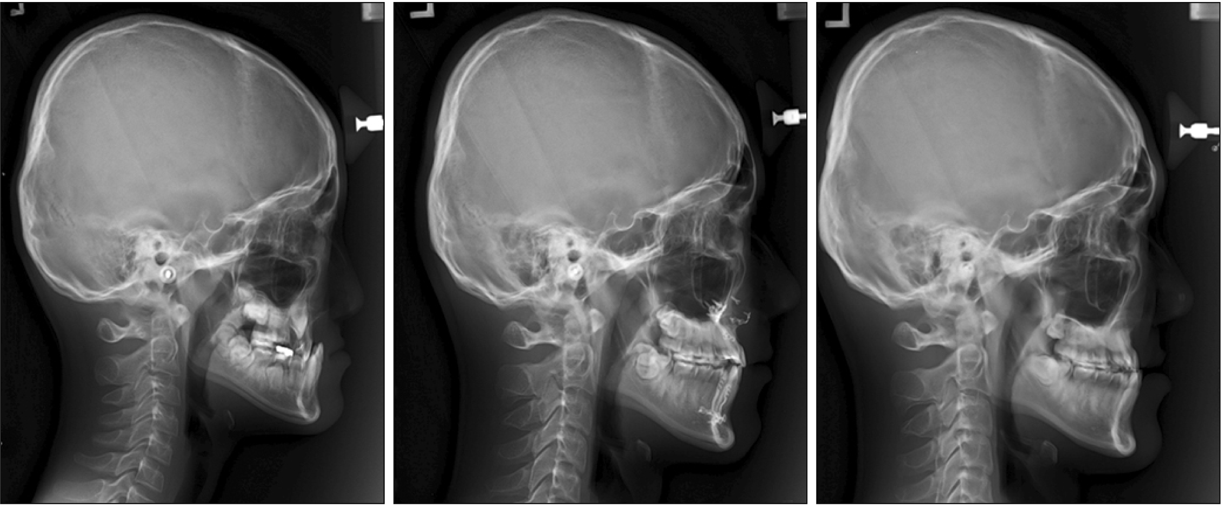
 | Figure 1Serial lateral cephalograms taken before facemask with miniplate (FMMP) therapy (T0), after FMMP therapy (T1), and follow-up (T2) in the FMMP-Nonsurgery group (A) and FMMP-Surgery group (B). 
|
A total of 28 cephalometric variables were measured to evaluate the status and amount of changes in the skeletal, dental, and soft tissue relationship. To evaluate the actual anteroposterior change of point A with consideration of the forward growth of Nasion during FMMP therapy, the vertical and horizontal reference planes, and a linear variable were defined as follows: (1) Horizontal reference plane, a horizontal line passing through the Sella at an angle of 7 degree clockwise with the Sella-Nasion (SN) plane
22; (2) Vertical reference plane, a plane perpendicular to the horizontal reference plane passing through the Sella; (3) A-VRP, the horizontal distance from the vertical reference plane to the point A. Cephalometric tracing and measurement were conducted by a single operator (SWO) utilizing the V-ceph program (version 7.0, Osstem Implant Co., Seoul, Korea).
At the T2 stage, the subjects were divided into the FMMP-Surgery and FMMP-Nonsurgery groups according to two criteria: (1) If patients met the cephalometric criteria with ANB < –3°, Wits appraisal < –5 mm, and Harvold unit difference (ΔCoGn-CoSn) > 34 mm, they were classified into the FMMP-Surgery group.
15,16,20,23 (2) If patients had been treated with preoperative orthodontic treatment or had already undergone orthognathic surgery or DO, they were automatically classified into the FMMP-surgery group.
15,16,20 As a result, UCLP patients were divided into the FMMP-Nonsurgery group (n = 33, 62.3%) and the FMMP-Surgery group (n = 20, 37.7%) (
Table 1 and
Figure 1).
Table 1
Demographic data of the FMMP-Nonsurgery and FMMP-Surgery groups
|
UCLP patient |
FMMP-Nonsurgery group (n = 33) |
|
FMMP-Surgery group (n = 20) |
p-value |
|
|
|
Mean |
SD |
Mean |
SD |
|
Sex (M:F)†
|
24:9 |
16:4 |
0.6534 |
|
Side involvement‡
|
12 cases with right side and 21 cases with left side |
6 cases with right side and 14 cases with left side |
0.7678 |
|
Age§ (yr) |
T0 |
10.27 |
0.63 |
10.75 |
0.97 |
0.0368*
|
|
T1 |
14.76 |
0.94 |
14.65 |
0.99 |
0.9534 |
|
T2 |
18.42 |
1.54 |
19.10 |
1.33 |
0.1791 |
|
Duration§ (mo) |
T0–T1 |
53.79 |
11.65 |
45.65 |
15.63 |
0.0718 |
|
T1–T2 |
44.45 |
18.72 |
53.05 |
17.41 |
0.1743 |
|
T0–T2 |
98.76 |
18.56 |
99.10 |
20.82 |
0.9634 |

All cephalometric variables in 10 randomly selected samples were reanalyzed to verify the intra-examiner reliability with an interval of one-month using the Dahlberg formula.
24 The differences in the angular measurement ranged from 0.25° to 0.49°, and those in the linear measurement ranged from 0.14 to 0.49 mm. As a result, the first measurement was adopted for further statistical analysis.
Mann–Whitney test, Wilcoxon signed-rank test, chi-square test, Fisher’s exact test, and discrimination analysis were performed using SPSS ver. 22.0 (IBM Corp., Armonk, NY, USA). p-values of < 0.05 were considered statistically significant.
Go to :
RESULTS
Demographic data of the two groups (Table 1)
The distributions of sex and cleft side were not different between the FMMP-Nonsurgery and FMMP-Surgery groups (all p > 0.05).
Differences in the mean ages at T0 stage of the two groups were statistically significant but clinically insignificant (10.27 years in FMMP-Nonsurgery group vs. 10.75 years in the FMMP-Surgery group, p < 0.05). There were no significant differences in the mean ages at T1 and T2 stages between the two groups (all p > 0.05).
There were also no significant differences in the mean duration of FM-MP therapy (T0–T1), follow-up (T1–T2), and total duration (T0–T2) between the FMMP-Nonsurgery and FMMP-Surgery groups (all p > 0.05).
Comparison of missing teeth and space management between the two groups (Table 2)
The frequency of missing maxillary lateral incisor was higher in the FMMP-Nonsurgery group than the FMMP-Surgery group (83.3% [n = 25/30] vs. 45.0% [n = 9/20], p < 0.05). However, there was no significant difference in the space management (opening and closure) of missing maxillary lateral incisor between the FMMP-Nonsurgery and FMMP-Surgery groups (48.0% [n = 12/25] and 52.0% [n = 13/25] vs. 44.4% [n = 4/9] and 55.6% [n = 5/9]; p > 0.05).
Table 2
Comparison of missing teeth, extraction, and space management between FMMP-Nonsurgery and FMMP-Surgery groups
|
UCLP patient |
FMMP-Nonsurgery group (n = 33) |
FMMP-Surgery group (n = 20) |
p-value |
|
Missing of the maxillary lateral incisor†
|
25 cases with missing and 8 cases without missing (including 8 peg lateralis) |
9 cases with missing and 11 cases without missing (including 10 peg lateralis) |
0.0236*
|
|
Space management of missing maxillary lateral incisor‡
|
12 cases with space opening and 13 cases with space closure |
4 cases with space opening and 5 cases with space closure |
1.0000 |
Comparison of the cephalometric parameters between the two groups at each stage (Table 3)
At the T0 stage, the FMMP-Surgery group showed more severe Class III relationship (ANB, anteroposterior dysplasia indicator [APDI], and Wits appraisal; p < 0.05; Harvold unit difference, p < 0.01), longer mandibular body length (p < 0.05), longer ramus height (p < 0.05), more acute point A-point B (AB) to mandibular plane angle (p < 0.05), and more negative overjet (p < 0.05) than the FMMP-Nonsurgery group.
At the T1 stage, the FMMP-Surgery group still showed more severe Class III relationship (Wits appraisal and Harvold unit difference, p < 0.05; pogonion [Pog] to N-perpendicular [N-Perp] and APDI, p < 0.01; ANB, p < 0.001), longer mandibular body length (p < 0.05), more acute AB to mandibular plane angle (p < 0.01), and more negative overjet (p < 0.001) than the FMMP-Nonsurgery group. At this stage, the FMMP-Surgery group appeared to show more counterclockwise rotation of the occlusal plane to SN angle (p < 0.05), smaller overbite depth indicator (ODI) (p < 0.01), and more dental compensation of the mandibular incisor (incisor mandibular plane angle [IMPA] and interincisal angle, p < 0.01) compared to the FMMP-Nonsurgery group.
At the T2 stage, the FMMP-Surgery group still showed more severe Class III relationship (sella-nasion-point B [SNB], p < 0.05; Pog to N-Perp and Wits appraisal, p < 0.01; ANB, APDI, and Harvold unit difference, p < 0.001), longer mandibular body length (p < 0.01), more counterclockwise rotation of the occlusal plane to SN angle (p < 0.05), more acute AB to mandibular plane angle (p < 0.01), smaller ODI (p < 0.001), and more negative overjet (p < 0.001) than the FMMP-Nonsurgery group. At this stage, the FMMP-Surgery group appeared to show more counterclockwise rotation of the mandible (articular angle, p < 0.05) compared to the FMMP-Nonsurgery group.
Throughout the whole stages, seven cephalometric variables, including ANB, APDI, Wits appraisal, mandibular body length, AB to mandibular plane angle, Harvold unit difference, and overjet, exhibited significant differences between the two groups. At both T1 and T2 stages, Pog to N-Perp, occlusal plane to SN angle, and ODI appeared to have significant differences between the two groups.
Table 3
Comparison of the cephalometric parameters between the FMMP-Nonsurgery and FMMP-Surgery groups at each stage
|
Variables |
T0 |
|
T1 |
|
T2 |
|
|
|
FMMP-
Nonsurgery
group |
|
FMMP-
Surgery
group |
p-value |
FMMP-
Nonsurgery
group |
|
FMMP-
Surgery
group |
p-value |
FMMP-Nonsurgery group |
|
FMMP-
Surgery
group |
p-value |
|
|
|
|
|
|
|
Mean |
SD |
Mean |
SD |
Mean |
SD |
Mean |
SD |
Mean |
SD |
Mean |
SD |
|
SNA (°) |
75.97 |
3.60 |
75.03 |
3.08 |
0.4409 |
|
76.57 |
3.70 |
75.29 |
3.73 |
0.2872 |
|
75.99 |
3.55 |
75.55 |
3.98 |
0.7831 |
|
SNB (°) |
75.64 |
3.49 |
77.31 |
3.87 |
0.1745 |
|
75.08 |
3.75 |
77.34 |
4.31 |
0.1188 |
|
75.18 |
3.84 |
78.70 |
4.76 |
0.0179*
|
|
ANB (°) |
0.33 |
2.63 |
−2.29 |
3.39 |
0.0119*
|
|
1.48 |
2.40 |
−2.05 |
3.11 |
0.0002***
|
|
0.82 |
2.16 |
−3.15 |
3.30 |
< 0.0001***
|
|
A to N-Perp (mm) |
−3.84 |
3.45 |
−5.54 |
3.50 |
0.0896 |
|
−4.87 |
3.95 |
−5.73 |
4.00 |
0.3129 |
|
−4.39 |
3.56 |
−5.48 |
4.00 |
0.1421 |
|
A-VRP (mm) |
53.60 |
4.83 |
55.27 |
4.35 |
0.2914 |
|
55.89 |
4.50 |
56.86 |
5.74 |
0.8257 |
|
56.78 |
4.88 |
56.99 |
6.61 |
0.7136 |
|
Pog to N-Perp (mm) |
−7.38 |
5.75 |
−5.72 |
6.94 |
0.2121 |
|
−10.59 |
6.79 |
−5.15 |
7.34 |
0.0082**
|
|
−8.40 |
7.96 |
−2.42 |
6.08 |
0.0036**
|
|
APDI (°) |
86.96 |
6.23 |
91.73 |
6.79 |
0.0184*
|
|
83.90 |
5.95 |
91.02 |
6.76 |
0.0010**
|
|
84.81 |
5.80 |
92.38 |
7.25 |
0.0009***
|
|
Wits appraisal (mm) |
−2.89 |
3.20 |
−5.33 |
3.94 |
0.0297*
|
|
−1.18 |
3.13 |
−4.35 |
4.69 |
0.0154*
|
|
−2.06 |
2.60 |
−6.11 |
4.85 |
0.0017**
|
|
Mandibular body length (mm) |
59.64 |
3.95 |
62.52 |
3.52 |
0.0147*
|
|
65.04 |
4.65 |
68.12 |
4.72 |
0.0381*
|
|
65.16 |
5.32 |
70.16 |
5.03 |
0.0029**
|
|
Saddle angle (°) |
124.83 |
4.55 |
124.24 |
3.82 |
0.5571 |
|
124.80 |
4.41 |
123.75 |
4.29 |
0.4409 |
|
124.43 |
4.23 |
124.17 |
4.34 |
0.9634 |
|
Articular angle (°) |
147.24 |
5.19 |
144.81 |
5.07 |
0.2055 |
|
150.07 |
6.40 |
147.35 |
6.12 |
0.1926 |
|
150.22 |
6.32 |
146.32 |
6.97 |
0.0435*
|
|
Gonial angle (°) |
127.19 |
5.09 |
128.24 |
5.78 |
0.4630 |
|
124.67 |
6.58 |
126.62 |
6.46 |
0.1990 |
|
124.15 |
6.73 |
125.41 |
6.44 |
0.3447 |
|
Bjork sum (°) |
399.25 |
5.07 |
397.29 |
5.54 |
0.2830 |
|
399.55 |
6.35 |
397.72 |
6.49 |
0.4742 |
|
398.79 |
6.67 |
395.90 |
7.22 |
0.2224 |
|
Ramus height (mm) |
36.84 |
3.56 |
38.84 |
3.32 |
0.0455*
|
|
42.71 |
5.21 |
44.23 |
5.15 |
0.1864 |
|
45.05 |
5.64 |
48.26 |
6.56 |
0.0613 |
|
Facial height ratio (PFH/AFH) |
62.07 |
3.50 |
63.04 |
3.79 |
0.3589 |
|
63.28 |
4.49 |
63.77 |
4.85 |
0.8544 |
|
64.37 |
4.76 |
65.58 |
5.83 |
0.6464 |
|
FMA (°) |
29.28 |
5.37 |
27.79 |
5.19 |
0.4463 |
|
29.35 |
6.65 |
27.59 |
6.81 |
0.4972 |
|
28.61 |
7.15 |
25.75 |
7.04 |
0.2403 |
Mandibular plane angle
(Go-Gn to SN) (°) |
37.04 |
4.27 |
35.34 |
4.98 |
0.2366 |
|
37.09 |
5.57 |
35.69 |
5.82 |
0.6074 |
|
36.62 |
5.69 |
34.29 |
6.08 |
0.2514 |
|
Occlusal plane to SN angle (°) |
19.51 |
4.69 |
17.77 |
4.84 |
0.0781 |
|
18.82 |
4.65 |
15.84 |
4.52 |
0.0283*
|
|
18.99 |
4.70 |
15.42 |
5.72 |
0.0208*
|
|
Palatal plane angle (°) |
1.74 |
3.77 |
1.56 |
2.45 |
0.7691 |
|
0.74 |
3.27 |
0.77 |
2.82 |
0.8544 |
|
0.68 |
2.99 |
−0.80 |
3.14 |
0.0751 |
|
AB to mandibular plane (°) |
65.49 |
4.39 |
62.05 |
7.41 |
0.0399*
|
|
67.49 |
4.57 |
62.16 |
8.30 |
0.0045**
|
|
67.26 |
4.71 |
61.08 |
7.81 |
0.0013**
|
|
ODI (°) |
67.23 |
5.33 |
63.61 |
8.66 |
0.0846 |
|
68.23 |
5.67 |
62.94 |
7.84 |
0.0023**
|
|
67.95 |
5.94 |
60.28 |
8.72 |
0.0005***
|
|
Harvold unit difference (mm) |
25.93 |
3.41 |
29.14 |
4.93 |
0.0087**
|
|
30.71 |
4.33 |
35.14 |
5.69 |
0.0102*
|
|
32.51 |
4.75 |
38.50 |
6.16 |
0.0007***
|
|
U1 to SN (°) |
98.89 |
8.06 |
99.88 |
10.24 |
0.6730 |
|
102.46 |
5.51 |
102.83 |
8.96 |
0.4972 |
|
103.36 |
5.24 |
105.28 |
7.37 |
0.4463 |
|
IMPA (°) |
85.59 |
8.44 |
84.68 |
6.36 |
0.7412 |
|
88.49 |
7.59 |
82.61 |
5.84 |
0.0028**
|
|
89.82 |
8.21 |
85.99 |
5.80 |
0.1104 |
|
Interincisal angle (°) |
136.27 |
12.24 |
138.15 |
14.19 |
0.9561 |
|
129.50 |
8.67 |
136.85 |
6.04 |
0.0019**
|
|
128.02 |
9.77 |
132.83 |
7.82 |
0.0638 |
|
Overbite (mm) |
1.01 |
2.05 |
2.02 |
2.85 |
0.2709 |
|
0.60 |
1.22 |
0.45 |
2.09 |
0.7136 |
|
0.24 |
0.88 |
0.31 |
1.63 |
0.6268 |
|
Overjet (mm) |
−0.31 |
2.58 |
−2.47 |
3.36 |
0.0381*
|
|
2.46 |
1.70 |
−0.72 |
3.59 |
0.0006***
|
|
1.94 |
1.50 |
−2.18 |
3.38 |
< 0.0001***
|
|
Nasolabial angle (°) |
79.01 |
12.38 |
79.48 |
14.38 |
0.5448 |
|
76.91 |
10.82 |
72.73 |
14.99 |
0.3784 |
|
76.75 |
10.05 |
71.58 |
14.57 |
0.2790 |
Comparison of the amount of change in the cephalometric parameters between the two groups (Table 4)
When compared the two groups, during T0–T1, there was no difference in the cephalometric parameters except ΔIMPA (p < 0.05). These findings suggested that the effects of FMMP therapy might be similar between the two groups. However, considering the values at the T0 and T1 stages, more severe Class III relationship, longer mandibular body length, more acute AB to mandibular plane angle, and more negative overjet in the FMMP-Surgery group were not fully corrected despite long-term use of FMMP.
During T1–T2, the FMMP-Surgery group showed significant pubertal and late mandibular growth (ΔSNB, ΔMandibular body length, ΔRamus height, and ΔHarvold unit difference, p < 0.05) and counterclockwise rotation of the maxilla and mandible (ΔPalatal plane angle, ΔODI, and ΔBjork sum, p < 0.05) compared to the FMMP-Nonsurgery group.
During T0–T2, the FMMP-Surgery group exhibited a more worsening of Class III relationship (ΔANB, ΔHarvold unit difference, ΔAB to Mandibular plane angle, and ΔODI, p < 0.05; ΔSNB, p < 0.01) and less increase in overjet (p < 0.01) compared to the FMMP-Nonsurgery group.
Table 4
Comparison of the amount of change in the cephalometric parameters between the FMMP-Nonsurgery and FMMP-Surgery groups
|
Variables |
ΔT0–T1 |
|
ΔT1–T2 |
|
ΔT0–T2 |
|
|
|
FMMP-
Nonsurgery
group |
|
FMMP-
Surgery
group |
|
Compa-
rison of the two groups |
FMMP-
Nonsurgery
group |
|
FMMP-
Surgery
group |
|
Compa-
rison of
the two
groups |
FMMP-
Nonsurgery
group |
|
FMMP-
Surgery
group |
|
Compa-rison of the two groups |
|
|
|
|
|
|
|
|
|
|
Mean |
SD |
p-value†
|
Mean |
SD |
p-value†
|
p-value‡
|
Mean |
SD |
p-value†
|
Mean |
SD |
p-value†
|
p-value‡
|
Mean |
SD |
p-value†
|
Mean |
SD |
p-value†
|
p-value‡
|
|
SNA (°) |
0.59 |
1.95 |
0.1098 |
0.26 |
2.54 |
0.5256 |
0.5448 |
−0.57 |
1.32 |
0.0217*
|
0.27 |
2.05 |
0.3045 |
0.0399*
|
0.02 |
2.11 |
1.0000 |
0.53 |
2.35 |
0.2959 |
0.3042 |
|
SNB (°) |
−0.56 |
2.40 |
0.3214 |
0.03 |
2.27 |
0.7089 |
0.3686 |
0.09 |
1.15 |
0.7818 |
1.36 |
1.93 |
0.0137*
|
0.0270*
|
−0.47 |
2.67 |
0.3127 |
1.39 |
2.95 |
0.0152*
|
0.0064**
|
|
ANB (°) |
1.16 |
2.43 |
0.0163*
|
0.24 |
2.34 |
0.7510 |
0.1990 |
−0.67 |
1.18 |
0.0018**
|
−1.10 |
1.37 |
0.0043**
|
0.3589 |
0.49 |
2.32 |
0.2312 |
−0.86 |
2.12 |
0.1126 |
0.0465*
|
|
A to N-Perp (mm) |
−1.02 |
3.96 |
0.0815 |
−0.19 |
4.16 |
0.6274 |
0.4856 |
0.47 |
4.14 |
0.3897 |
0.25 |
4.34 |
0.9553 |
0.6597 |
−0.55 |
4.03 |
0.4584 |
0.06 |
2.98 |
0.8960 |
0.5820 |
|
A-VRP (mm) |
2.29 |
3.08 |
0.0002***
|
1.59 |
4.07 |
0.1365 |
0.4142 |
0.90 |
2.67 |
0.0725 |
0.13 |
3.09 |
0.8813 |
0.2998 |
3.18 |
3.84 |
0.0001***
|
1.72 |
4.72 |
0.1354 |
0.1446 |
|
Pog to N-Perp (mm) |
−3.21 |
5.94 |
0.0068**
|
0.57 |
7.15 |
0.7089 |
0.0797 |
2.19 |
7.12 |
0.0426*
|
2.72 |
7.64 |
0.1913 |
0.7551 |
−1.02 |
7.95 |
0.6616 |
3.30 |
7.31 |
0.0522 |
0.0813 |
|
APDI (°) |
−3.06 |
5.69 |
0.0049**
|
−0.71 |
4.97 |
0.3507 |
0.1990 |
0.90 |
3.14 |
0.0238*
|
1.36 |
3.27 |
0.1213 |
0.7831 |
−2.16 |
6.22 |
0.0463*
|
0.64 |
4.87 |
0.4897 |
0.0588 |
|
Wits appraisal (mm) |
1.71 |
3.06 |
0.0080**
|
0.97 |
2.99 |
0.2180 |
0.4742 |
−0.88 |
2.08 |
0.0115*
|
−1.76 |
2.49 |
0.0100*
|
0.1804 |
0.83 |
2.97 |
0.0847 |
−0.78 |
3.37 |
0.3507 |
0.1324 |
Mandibular body
length (mm) |
5.40 |
3.60 |
< 0.0001***
|
5.60 |
3.89 |
0.0002***
|
0.8978 |
0.11 |
2.65 |
0.7075 |
2.04 |
2.77 |
0.0064**
|
0.0203*
|
5.52 |
3.54 |
< 0.0001***
|
7.64 |
4.15 |
0.0001***
|
0.0781 |
|
Saddle angle (°) |
−0.02 |
2.53 |
0.9005 |
−0.50 |
2.16 |
0.5503 |
0.4972 |
−0.38 |
1.52 |
0.2419 |
0.42 |
2.66 |
0.9108 |
0.6074 |
−0.40 |
2.98 |
0.4584 |
−0.07 |
3.05 |
0.7938 |
0.9123 |
|
Articular angle (°) |
2.83 |
4.85 |
0.0011**
|
2.54 |
4.52 |
0.0304*
|
0.8616 |
0.15 |
2.14 |
0.5435 |
−1.03 |
3.23 |
0.1403 |
0.2022 |
2.98 |
5.43 |
0.0028**
|
1.51 |
5.56 |
0.2250 |
0.5448 |
|
Gonial angle (°) |
−2.51 |
3.02 |
0.0001***
|
−1.62 |
3.57 |
0.0793 |
0.4090 |
−0.53 |
1.67 |
0.0784 |
−1.21 |
1.79 |
0.0177*
|
0.3173 |
−3.04 |
3.38 |
0.0001***
|
−2.83 |
3.64 |
0.0057**
|
1.0000 |
|
Bjork sum (°) |
0.30 |
3.36 |
0.9501 |
0.42 |
2.83 |
0.3317 |
0.4630 |
−0.75 |
1.74 |
0.0140*
|
−1.81 |
2.15 |
0.0025**
|
0.0311*
|
−0.46 |
3.97 |
0.3042 |
−1.39 |
3.87 |
0.1084 |
0.4463 |
|
Ramus height (mm) |
5.87 |
3.22 |
< 0.0001***
|
5.39 |
3.14 |
0.0001***
|
0.6932 |
2.33 |
2.81 |
0.0001***
|
4.02 |
3.10 |
0.0003***
|
0.0390*
|
8.20 |
4.16 |
< 0.0001***
|
9.42 |
4.82 |
0.0001***
|
0.1804 |
Facial height ratio
(PFH/AFH) |
1.21 |
2.30 |
0.0036**
|
0.74 |
2.16 |
0.0569 |
0.1958 |
1.09 |
1.48 |
0.0007***
|
1.81 |
1.84 |
0.0012**
|
0.1472 |
2.30 |
2.96 |
0.0004***
|
2.54 |
3.45 |
0.0036**
|
0.8472 |
|
FMA (°) |
0.07 |
2.85 |
0.8372 |
−0.20 |
3.42 |
0.7089 |
0.7691 |
−0.74 |
2.05 |
0.1078 |
−1.84 |
3.30 |
0.0177*
|
0.1188 |
−0.67 |
3.56 |
0.3042 |
−2.04 |
3.70 |
0.0366*
|
0.2055 |
Mandibular
plane angle
(Go-Gn to SN) (°) |
0.05 |
3.18 |
0.7075 |
0.35 |
2.74 |
0.6407 |
0.4518 |
−0.47 |
1.75 |
0.2757 |
−1.40 |
2.29 |
0.0228*
|
0.1124 |
−0.42 |
3.55 |
0.4476 |
−1.05 |
3.04 |
0.1004 |
0.5207 |
Occlusal plane to
SN angle (°) |
−0.70 |
3.27 |
0.1244 |
−1.93 |
3.73 |
0.0064**
|
0.2189 |
0.17 |
2.39 |
0.8722 |
−0.43 |
3.93 |
0.1454 |
0.0736 |
−0.53 |
4.31 |
0.3482 |
−2.36 |
5.00 |
0.0569 |
0.1372 |
Palatal plane
angle (°) |
−1.00 |
2.49 |
0.0188*
|
−0.79 |
3.74 |
0.1084 |
0.6730 |
−0.06 |
2.89 |
0.5201 |
−1.57 |
3.19 |
0.0383*
|
0.0356*
|
−1.06 |
2.93 |
0.0390*
|
−2.36 |
2.55 |
0.0015**
|
0.0862 |
AB to mandibular
plane (°) |
2.00 |
3.87 |
0.0052**
|
0.12 |
4.55 |
0.8228 |
0.1232 |
−0.23 |
2.30 |
0.3528 |
−1.08 |
2.10 |
0.0569 |
0.2914 |
1.77 |
4.39 |
0.0217*
|
−0.96 |
4.68 |
0.2627 |
0.0318*
|
|
ODI (°) |
1.00 |
4.32 |
0.2312 |
−0.67 |
5.99 |
0.4781 |
0.1926 |
−0.29 |
3.79 |
0.7275 |
−2.65 |
3.88 |
0.0051**
|
0.0318*
|
0.72 |
4.76 |
0.5858 |
−3.33 |
5.60 |
0.0169*
|
0.0132*
|
Harvold unit
difference (mm) |
4.78 |
3.03 |
< 0.0001***
|
6.00 |
4.04 |
0.0001***
|
0.2330 |
1.80 |
2.47 |
0.0004***
|
3.36 |
2.24 |
0.0002***
|
0.0184*
|
6.58 |
3.36 |
< 0.0001***
|
9.36 |
4.84 |
0.0001***
|
0.0240*
|
|
U1 to SN (°) |
3.57 |
7.88 |
0.0217*
|
2.95 |
8.66 |
0.0365*
|
0.8400 |
0.91 |
3.23 |
0.0697 |
2.45 |
5.30 |
0.0930 |
0.5327 |
4.48 |
7.46 |
0.0031**
|
5.40 |
9.41 |
0.0206*
|
0.8115 |
|
IMPA (°) |
2.90 |
6.29 |
0.0202*
|
−2.07 |
5.45 |
0.0859 |
0.0102*
|
1.33 |
3.14 |
0.0227*
|
3.38 |
4.50 |
0.0051**
|
0.0914 |
4.23 |
6.53 |
0.0019**
|
1.31 |
5.24 |
0.4553 |
0.1472 |
|
Interincisal angle (°) |
−6.77 |
10.47 |
0.0016**
|
−1.30 |
10.61 |
0.5016 |
0.0638 |
−1.48 |
4.25 |
0.0548 |
−4.02 |
5.70 |
0.0036**
|
0.1104 |
−8.25 |
10.43 |
0.0003***
|
−5.32 |
10.36 |
0.0438*
|
0.2088 |
|
Overbite (mm) |
−0.41 |
2.38 |
0.3127 |
−1.57 |
3.08 |
0.0479*
|
0.2790 |
−0.36 |
1.12 |
0.0837 |
−0.14 |
1.71 |
0.6813 |
0.5632 |
−0.77 |
2.13 |
0.0984 |
−1.70 |
2.75 |
0.0169*
|
0.2553 |
|
Overjet (mm) |
2.77 |
3.00 |
< 0.0001***
|
1.76 |
2.20 |
0.0051**
|
0.1958 |
−0.52 |
1.76 |
0.0631 |
−1.47 |
2.30 |
0.0169*
|
0.1005 |
2.26 |
2.63 |
0.0001***
|
0.29 |
1.95 |
0.5503 |
0.0053**
|
|
Nasolabial angle (°) |
−2.10 |
9.32 |
0.1138 |
−6.75 |
12.39 |
0.0478*
|
0.0613 |
−0.16 |
7.90 |
0.7141 |
−1.15 |
12.33 |
0.6542 |
0.7136 |
−2.27 |
11.63 |
0.2641 |
−7.90 |
9.76 |
0.0057**
|
0.0986 |
Prediction cephalometric variables at the T0 stage for the future need for orthognathic surgery (Tables 5 and 6)
From the simultaneous estimation-discriminant analysis, the discriminant function affecting the future need for orthognathic surgery is as follows: D = 5.288 + [0.268 × ANB (°)] + [0.049 × APDI (°)] – [0.084 × Wits appraisal (mm)] – [0.148 × mandibular body length (mm)] – [0.022 × Harvold unit difference (mm)] + [0.154 × overjet (mm)]. The centroids of the FMMP-Nonsurgery group and FMMP-surgery group were 0.412 and –0.680, respectively (
Table 5). The percentage of correctly classified original grouped cases was 69.8%, with a sensitivity of 69.7% and specificity of 70.0% (
Table 6).
Table 5
Discriminant analysis to identify cephalometric predictors of future need for orthognathic surgery
Canonical discriminant
function coefficients |
Function 1 |
|
Predictable variable |
ANB |
0.268 |
|
APDI |
0.049 |
|
Wits appraisal |
–0.084 |
|
Mandibular body length |
–0.148 |
|
Harvold unit difference |
–0.022 |
|
Overjet |
0.154 |
|
Constant |
5.288 |
|
Functions at group centroids |
Function 1 |
|
Group |
FMMP-Nonsurgery group |
0.412 |
|
FMMP-Surgery group |
–0.680 |
Table 6
Classification results in discrimination analysis
|
Number of cases |
Predicted membership (%) |
|
FMMP-Nonsurgery group
(n = 29) |
FMMP-Surgery group
(n = 24) |
|
Actual group |
FMMP-Nonsurgery group (n = 33) |
23 (69.7) |
10 (30.3) |
|
FMMP-Surgery group (n = 20) |
6 (30.0) |
14 (70.0) |
Go to :
DISCUSSION
Although this study was performed with a single university hospital-based data, this study might have some originalities. First, we used relatively strict inclusion criteria to increase the purity of the samples. Second, the identical treatment protocol was used to avoid confounding factors in interpreting the results. Third, to the authors’ knowledge, it is the first study to report the cephalometric parameters at the initial stage related to the risk of future orthognathic surgery or DO despite long-term use of FMMP therapy.
Candidates for orthognathic surgery or distraction osteogenesis
In the present study, 37.7% of UCLP patients became candidates for orthognathic surgery or DO despite long-term use of FMMP therapy (
Table 1). It was higher than Park et al’s result (21.4%).
15 The reasons might be (1) the UCLP subjects used in Park et al’s study
15 were a combination of patients with various degrees of maxillary hypoplasia and (2) in the present study, FMMP therapy was applied to subjects with moderate-to-severe maxillary hypoplasia.
Comparison of the cephalometric parameters at each stage and the amount of change between the two groups (Tables 3 and 4)
In the FMMP-Surgery group, a forward position of the mandible at the T0 stage was maintained throughout the whole stages and Class III relationship worsened with significant pubertal and late mandibular growth and counterclockwise rotation of the maxilla and mandible at the T1 and T2 stages. These findings were similar to those in Meazzini et al.
17 and Yun-Chia Ku et al.,
19 who reported that a significant late mandibular growth in UCLP children played an important role in deciding the need for orthognathic surgery. Scheuer et al.
25 also reported that UCLP patients showed more significant amounts of change in SNA and SNB from 12 to 16 years compared to that of 8 to 12 years.
Previous studies insisted that the vertical cephalometric variables including palatal plane inclination, mandibular plane angle, and facial height would not be the determining factors for orthognathic surgery.
17,19,21 However, it is necessary to investigate the effects of the rotation of the maxilla and mandible and the changes in the vertical dimension on worsening of Class III relationship in future studies.
Prediction cephalometric variables at the T0 stage for the future need for orthognathic surgery (Tables 5 and 6)
Zemann et al.
26 reported that UCLP children with negative values of ANB angle at the age of 6 years showed no improvement after a 4-year period. Antonarakis et al.
16 and Meazzini et al.
21 suggested that ANB would be a powerful predictor in cleft patients at the age of 5–6 years. These findings were similar to the result of this study, which selected ANB as one of the major predictors from the discriminant analysis. However, since accurate tracing and cephalometric analysis of the anterior maxilla (including point A) might be difficult at the age of 5–6 years, it would be better to use the samples with completely erupted permanent maxillary incisors.
15 Therefore, the mean age at the T0 stage in the present study was around 10 years (
Table 1).
Comparison of the cephalometric predictors in previous studies (Table 7)
The present study selected six cephalometric variables at the T0 stage related to the size of the mandible (mandibular body length), intermaxillary sagittal relationship (ANB, APDI, Wits appraisal, and Harvold unit difference) and dental compensation (overjet). These findings were similar to those reported by Park et al.
15 and Yun-Chia Ku et al.
19 However, there were some differences in the cephalometric parameters. Park et al.
15 included more cephalometric parameters such as position of the maxilla (A to N-Perp), shape of the mandible (gonial angle), intermaxillary sagittal and vertical relationship (ODI and AB to mandibular plane angle), and dental compensation (IMPA) compared to the present study. Yun-Chia Ku et al.
19 also added more cephalometric parameters including size of the maxilla (maxillary length), position of the mandible (SNB), intermaxillary vertical relationship (overbite), anterior cranial base length (SN), and dental compensation (IMPA); while they did not use the cephalometric variables explaining the intermaxillary sagittal relationship (APDI, Wits appraisal, and Harvold unit difference) compared to the present study.
The reasons for these differences might be as follows: First, Park et al.
15 and Yun-Chia Ku et al.
19 did not confine the subjects who were treated with long-term use of FMMP. Second, in the present study, because the amounts of change during T0–T1 in the two groups were similar (
Table 4), Class III relationship at the T0 stage was not fully corrected at the T1 stage in the FMMP-Surgery group (
Table 3). Third, in the present study, the FMMP-Surgery group showed significant pubertal and late growth of the mandibular body and ramus and counterclockwise rotation of the maxilla and mandible at the T1 and T2 stages, resulting in worsening of the skeletal Class III relationship (
Tables 3 and
4).
In the present study, accuracy, sensitivity and specificity (69.8%, 69.7%, and 70.0%) were relatively lower than those in previous studies.
15,19 It is assumed that this difference came from the sample size, statistical method, various treatment responses of patients, need of orthognathic surgery for correction of transverse or other dimensional problems, and the patient’s desire for facial esthetics. Therefore, it is necessary to investigate the effects of aforementioned issues on the accuracy, sensitivity and specificity in future studies.
Table 7
Comparison of the cephalometric predictors in previous studies
|
Park et al.15 (2015) |
Yun-Chia Ku et al.19 (2018) |
The present study |
|
Anterior cranial base length |
|
SN |
|
|
Position and size of the maxilla |
A to N-perpendicular |
Maxillary length |
|
|
Position, size and shape of the mandible |
Gonial angle |
SNB |
Mandibular body length |
|
Mandibular body length |
|
Intermaxillary sagittal relationship |
ANB |
ANB |
ANB |
|
APDI |
APDI |
|
Wits appraisal |
Wits appraisal |
|
Harvold unit difference |
Harvold unit difference |
|
Intermaxillary vertical relationship |
|
Overbite |
|
|
Intermaxillary sagittal and vertical relationship |
ODI |
|
|
|
AB to mandibular plane angle |
|
Dental compensation |
IMPA |
L1-MP (IMPA) |
Overjet |
|
Overjet |
Overjet |
|
Method |
Machine learning (Feature Wrapping method with support vector machine/sequential forward search algorithms) |
Receiver operating characteristic analysis with a scoring system model based on 3 dichotomized variables |
Discrimination analysis |
|
Predictors |
10 cephalometric variables |
ANB, ≤ –0.45° |
6 cephalometric variables |
|
Overjet, ≤ −2.00 mm |
|
Maxillary length, ≤ 47.25 mm |
|
Diagnostic accuracy (%) |
77.3 |
86.9 (sum of score: 2) |
69.8 |
|
Sensitivity (%) |
99.0 |
90.0 |
69.7 |
|
Specificity (%) |
74.1 |
83.9 |
70.0 |
Clinical implications
The results from this study indicated that, although Class III relationship would not be fully corrected in the FMMP-Surgery group, the two groups did not exhibit the difference in the absolute amount of sagittal change in the cephalometric parameters by FMMP therapy (
Tables 3 and
4). Therefore, it can be stated that the FMMP therapy has a possibility of reducing the amount of maxillary advancement in orthognathic surgery or DO in UCLP patients.
As clinical suggestions, when a future need for orthognathic surgery is expected in children with UCLP (
Tables 5 and
6), there might be three recommendations for using the FMMP therapy. First, it is necessary to inform the parents about the possibility of poor prognosis and the possibility of orthognathic surgery after completion of facial growth. Second, after placing two miniplates in the mandibular symphysis area as well as two miniplates in the infrazygomatic crest of the maxilla, use of Class III elastics between the maxillary and mandibular miniplates at day time and FMMP at night time would be recommended (
Figure 2).
7,9,10 Third, it might be better to install two additional miniplates on the lateral nasal wall in the maxilla or combine the intraoral anchorage in the maxillary dentition to increase the orthopedic effect.
 | Figure 2An example of installation of two additional miniplates on the mandibular symphysis and use of Class III elastics between the maxillary and mandibular miniplates at day time and facemask with miniplate at night time.
|
Limitations of this study and suggestions for future studies
Although this is the first study that investigated the cephalometric predictors for the future need for orthognathic surgery, despite long-term use of FM-MP, in Korean adolescent patients with UCLP, the present study has some limitations as follows: First, this study had a retrospective study design with a relatively small sample size. Second, the samples were not subdivided according to the amount and degree of scar tissue on the lip and palate. Third, since patient’s compliance is one of the important factors in obtaining the successful treatment outcome, especially in long-term FMMP therapy, it is necessary to identify a method to objectively measure patient’s compliance. In the near future, it is necessary to perform a nationwide multi-center study with a large sample size and more sophisticated statistical analysis methods.
Go to :
CONCLUSION
• Despite long-term use of FMMP therapy, 37.7% of UCLP patients became candidates for orthognathic surgery or DO. Therefore, differential diagnosis is necessary to predict the future need for orthognathic surgery at early age.
• A total of six cephalometric variables including ANB, APDI, Wits appraisal, mandibular body length, Harvold unit difference, and overjet at the age of 10 years were selected as predictors of the future need for orthognathic surgery in Korean UCLP patients.
Go to :


 PDF
PDF Citation
 Print
Print


 XML Download