

 PDF
PDF Citation
 Print
Print


INTRODUCTION
Spontaneous intracerebral hemorrhage (SICH) is a severe disease associated with high morbidity and mortality rates and accounts for 10%–15% of all cases of stroke.1 In Korea, cerebrovascular disease was the fourth most common cause of death in 2018, and 44.7 deaths were recorded per 100,000 population deaths.23 Furthermore, unlike the decreasing mortality rates associated with subarachnoid hemorrhage and arteriovenous malformation (AVM) attributed to improvements in surgical and critical care strategies, the morbidity and mortality rates associated with SICH is still unacceptably high and has not been changed up to the recent.4
SICH can affect the brain parenchyma anywhere, but more commonly affects specific locations, including the basal ganglia, thalamus, pons, and cerebellum.1 Among them, thalamic hemorrhage accounts for 8.3%–15% of all cases of SICH, secondary to SICH originating from the basal ganglia.5 A prospective Japanese cohort study on SICH in 2014 revealed an increasing incidence of thalamic hemorrhage in contrast to a decreasing incidence of putaminal hemorrhage with time, especially in older patients.6 The thalamus is an oval-shaped area of gray matter located above the mesencephalon and consists of multiple nuclei with shared connections to the cortex, brainstem, basal ganglia, cerebellum, and spinal cord. The dorsal side of the thalamus is under the cerebral hemisphere, while the ventral side is covered by the hypothalamus. The medial side is adjacent to the 3rd ventricle, and the lateral side contacts the limb of the internal capsules.7 Due to these anatomical characteristics, thalamic hemorrhage may frequently be accompanied by hemorrhage extending into the ventricle.
Although several studies on spontaneous thalamic hemorrhage (STH) report that volume of hemorrhage and intraventricular extension are strong predictors of poor patient prognosis,8 few studies have examined the importance of intraventricular hemorrhage (IVH) in patients with STH, especially with respect to therapeutic strategies for IVH originating from STH. In practice, the therapeutic approach can be diverse based on the features of the hemorrhage and status of the patients, as follows: 1) conservative treatment is administered if the patient is stable and the hemorrhage is small; 2) craniotomy with decompression is performed if the patient is unstable and the volume of the hematoma is large; 3) stereotactic evacuation of hematoma alone or followed fibrinolysis using urokinase; 4) endoscopic evacuation of STH or IVH alone followed by external ventricular drainage (EVD) is performed; 5) EVD alone or followed fibrinolysis using urokinase; or 6) combined therapy using the aforementioned approaches. However, as STH is located deep in the brain, there is no marked benefit of surgical treatment, such as craniotomy, with the removal of hematoma, except for large volume hemorrhages, making a much shift of the midline.9 Owing to the intolerable neurological sequelae of craniotomy, minimally invasive techniques, such as endoscopic removal of IVH or EVD followed by intraventricular fibrinolysis (IVF), are usually considered the first therapeutic option. A systemic review suggested that applying neuroendoscopic evacuation of IVH with EVD may be a better management option for IVH secondary to spontaneous supratentorial hemorrhage than EVD followed by IVF.10
With respect to IVH itself, IVH, which originated from STH, is a proven risk factor for increased mortality and poor functional outcome, especially in patients with STH.8 During the long-term follow-up of patients with STH, especially those with concurrent IVH, the development of chronic hydrocephalus is commonly noted. This secondary normal pressure hydrocephalus (NPH) can affect morbidity at a later stage in patients with STH.11 Hence, the clearance of blood from the ventricles should be an additional therapeutic goal with the evacuation of STH. In cases of acute hydrocephalus, certain approaches for removing the IVH, such as EVD or endoscopic evacuation, are mandatory steps but are often prove insufficient.
This study aimed to investigate the characteristics of patients with STH with concurrent IVH and compare them with those of patients to without IVH. The second objective was to examine the incidence of delayed NPH among the patients with STH accompanied by IVH according to the therapeutic modalities. Additionally, this study attempted to validate the data of the cohort by determining the 30-day mortality and 6-month functional recovery and comparing the results with those reported in the available literature.
Go to : 
METHODS
Data collection
We retrospectively studied consecutive patients diagnosed with STH admitted to the Center for Cerebrovascular Disease at Samsung Changwon Hospital during the recent 19 years to December 2018. Patients aged ≥ 20 years were included because childhood patients are likely to have a secondary etiology such as AVM. In addition, patients with an infratentorial hemorrhage were excluded because the small changes in hemorrhage size may have a stronger impact on survival than that noted in patients with supratentorial hemorrhage. The clinical information of the patients were independently obtained from a computerized database (PACS; m-view, Marosis Corporation, Seoul, Korea) by three investigators using a uniformly designed report form. All indexes which can identify the patients were hidden from the collated data set to maintain patient confidentiality. The inclusion criteria applied were a diagnosis of STH diagnosed by computed tomography (CT) or magnetic resonance imaging and admission to the Center for Cerebrovascular Disease within 24 hours of neurological symptom onset. As recommended by the Stroke Council of the American Heart Association (AHA),512 conventional angiography was performed to differentiate secondary causes of STH in the candidate patients, and following patients were excluded from this study; patients with intracranial hemorrhage after head trauma, patients with a ruptured cerebral aneurysm in the CT angiography, patients with an cerebral AVM, patients with a brain tumor, patients with bleeding diathesis, and patients with a hemorrhagic infarction.
Determination of the prognostic factor
Of the baseline clinical features recorded, the characteristics were selected as the most relevant potential prognostic indicators based on previous reports on SICH.1314151617181920 The demographic variables included age, sex, cigarette smoking and alcohol intake history, body mass index (BMI), level of consciousness, pupillary abnormalities, limb weakness, and underlying disease. The smoking status and alcohol intake was classified as those of previous study.21 The levels of consciousness at admission were assessed using the Glasgow Coma Scale (GCS).22 Motor weakness of the extremities was classified as present and absent, and aggravation of limb weakness was defined as a decrease in the motor grade of ≥ 1 in patients with underlying hemiparesis. The blood pressure at presenting was not included in a potential prognostic factors because it is often markedly elevated during the first 1–2 days after a SICH onset.
Radiologic and laboratory characteristics
The radiologic characteristics on CT, namely, location of main hemorrhage in the thalamus, extra-thalamic extension, ventriculomegaly at present, volume of hematoma in the thalamus, and lateral shift of the cerebral midline structures, were also included in the analysis. The hematoma volume in the thalamus was estimated from the CT scans using the formula of previous studies by Kothari et al.23 IVH was diagnosed as high density in the ventricle on the CT scan. The location of the hematoma was classified as anterior, posterior, medial, lateral, and global, according to the study by Teramoto et al.24 The radiological evaluation of aforementioned variables was performed by two different neurosurgeons (TM Nam and SH Kim) who were blinded to the clinical and demographic parameters. If there was a discrepancy in the radiological interpretation between the two investigators, they determined the more precise value after repeated examination and discussion.
The laboratory parameters obtained at admission were dichotomized during the analysis based on cutoff values; these were leukocyte count, platelet count, C-reactive protein level, aspartate aminotransferase level, alanine aminotransferase level, blood plasma glucose level, and hemoglobin A1c level.
Treatment modalities
Patients were treated following the institutional protocol established by the base on the AHA guideline.5 The treatment was administered according to the current practices at the Center for Cerebrovascular Disease as previous report by our center.20 However, the treatment was not rigidly administered, and the primary attending neurosurgeons were allowed to exercise their medical judgment.
The surgical intervention was undertaken in selected patients, such as those exhibiting clinical deterioration with a moderate or large amount of STH with extra-thalamic extension, including intense IVH, those with a thalamic hematoma volume ≥ 30 mL, those with an expanding hematoma, and those with evidence of progressive neurological deterioration. The surgical approach was individualized based on the STH site, size, and surgeon's preference. Craniotomy, craniectomy, and CT-guided stereotactic hematoma evacuation were used for the removal of STH. Patients without extra-thalamic extension usually underwent stereotactic evacuation when the hematoma volume was 30–60 mL, as assessed on preoperative CT. Immediately after initial hematoma aspiration or evacuation, patients underwent repeat CT to assess catheter placement. When repeat CT showed a remnant thalamic hematoma, urokinase (6,000 U) injection through the catheter was considered to facilitate aspiration, and a CT scan was repeated 12 hours later. Patients with a hemorrhage volume of 10–30 mL and severe neurological deficit (limb weakness) were also treated by stereotactic evacuation. When the hematoma volume was ≥ 60 mL, evacuation was performed via craniotomy or decompressive craniectomy. The attending neurosurgeon decided whether to implant a bone flap (craniotomy or craniectomy) depending on the intraoperative presence of cerebral swelling after the removal of STH. Moreover, EVD and endoscopic evacuation of IVH were used for the removal of concurrent IVH. In patients with both severe deterioration of neurological status and intense IVH combined with acute ventriculomegaly detected on the CT scan, endoscopic evacuation followed by EVD was primarily considered the first-choice treatment.
Conservative treatment primarily was undertaken for management of hypertension and intracranial pressure. Several medications were used to control the blood pressure. The mean arterial pressure was maintained in the range of 100 to 140 mm Hg using an antihypertensive drugs via intravenous injection or oral administration.25 In addition, a hypertonic agent (glycerol, mannitol, and other such agents) was administered when CT illustrated a mass effect or when the clinical symptoms suggesting an increased intracranial pressure. Time from ictus to medication was calculated and recorded. If all family members of the patients with STH with a GCS score of 3 did not want any medical care for life prolongation, treatment modalities to maintain the vital sign were not applied.
Assessments of the outcomes
Patients were followed-up for at least 6 months after STH. The outcomes were assessed based on mortality within 30 days after diagnosis of STH by CT and functional status (Modified Rankin Scale [MRS] and Modified Barthel Index [MBI]) in patients who survived for more than 90 days after STH onset. The MRS26 and MBI4 scores were defined as the previous report.
For statistical purposes, the patients were divided into two outcome category groups, namely, the “good functional recovery” group (an MRS score of 0–2 and an MBI score of ≥ 60), members of which were functionally independent, and the “poor functional recovery” group (an MRS score of 3–5 or an MBI score of < 60). Information on mortality and functional outcome was collected primarily from the medical records; information was collected from the family members by telephone interviews; and information from patients was collected by direct examinations at our outpatient clinics after discharge.
Delayed NPH was diagnosed by the clinical manifestation and radiological features detected on CT scans at 3 months after STH diagnosis. Findings of one or more clinical features of Hakim's triad for the diagnosis of NPH, such as symmetric gait disturbance, urinary incontinence, and memory impairment,26 and ventriculomegaly with periventricular edema on the CT scan can diagnose delayed NPH after STH.
Statistical analysis
As the extension of STH into the ventricle is commonly noted, the effect of concurrent IVH on the clinical outcomes of the STH patients was primarily investigated. To validate our cohort, we planned to evaluate the 30-day mortality rate and functional recovery at 6 months after ictus in STH patients. Accordingly, we aimed to identify the variables associated with 30-day mortality and functional recovery at 6 months after STH development.
The medical records with respect to the clinical history at presentation, radiographic findings, and laboratory reports of all study subjects were analyzed. The date of death was confirmed by telephone interview with a family member, in case of incomplete medical records. The survival period was defined as the time from the date of diagnosis of STH till death. The date of the CT scan at the Center for Cerebrovascular Disease was recorded as the date of diagnosis.
For the statistical analysis, the software SPSS (version 20.0; IBM, Armonk, NY, USA) was applied, and the Student t-test and a nonparametric test were applied to analyze the continuous and ordinal variables (such as mean age), respectively. Discrete variables (such as sex, smoking status, chronic alcohol intake, BMI, GCS score, pupillary abnormality, new onset of hemiparesis, underlying disease, and family history of stroke) were analyzed using Pearson χ2 test. Univariate analysis was initially performed to determine the possible association between the potential prognostic factors and outcomes using the χ2 test. Subsequently, multivariate logistic regression analysis was performed to identify the variables independently associated with functional recovery and mortality. Kaplan-Meier survival curve analysis was used to assess the cumulative events. Statistical significance was accepted for P-values of < 0.05, and all tests were 2-tailed.
Ethics statement
The Institutional Review Board of the Samsung Changwon Hospital approved this study (SCMC 2019-05-002). All analyses were conducted according to the guidelines of the Declaration of Helsinki for biomedical research. The requirement of obtaining informed consent was waived owing to the retrospective nature of the study and minimal hazard to the participants.
Go to :
RESULTS
From January 2000 to December 2018, 2,389 patients with SICH were treated at the Center for Cerebrovascular Diseases in our institute. Of these, 254 (10.6%) patients with STH were selected for this study. However, 21 patients were excluded owing to the lack of medical records or incomplete follow-up data. Finally, 233 (9.8%) patients were included in this study, and all patients had experienced the development of STH for the first time. The patients' mean age was 65.4 ± 14.9 years (range, 34.3–88.3 years), and 128 (54.9%) of the patients were female. There were 74 (31.8%) patients without IVH and 159 (68.2%) patients with concurrent IVH.
Comparison of the baseline characteristics between the patients with IVH and those without IVH
In terms of the demographic features, no significant intergroup differences were observed with respect to age, sex, smoking history, chronic alcohol intake, pupillary abnormality, or new onset of motor weakness. However, the STH patients with IVH had a higher BMI (P = 0.037), a lower GCS score (P = 0.032), a higher prevalence of underlying diseases (P = 0.007), and family history of stroke (P = 0.040) compared to those without IVH (Table 1).
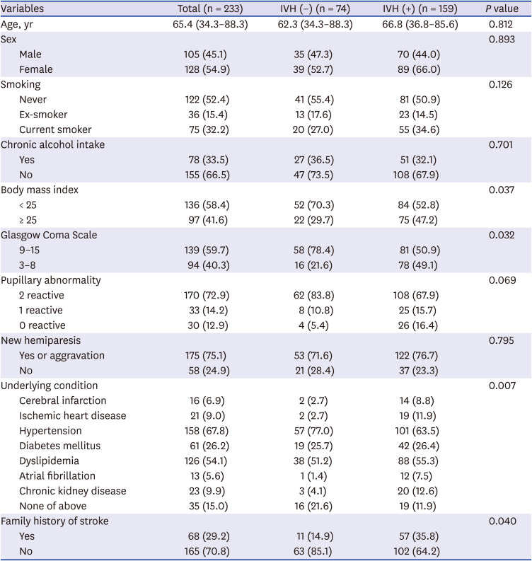
Table 1
Clinical features of the patients with spontaneous thalamic hemorrhage
![]()
Radiologically, the location of STH was markedly different between the patients with IVH and those without IVH. The STH patients with concurrent IVH showed that STH was commonly present in the posterior (34.5% vs. 2.7%, P = 0.007), medial (37.1% vs. 2.7%, P = 0.003), and global (23.3% vs. 1.3%, P = 0.008) regions than those without IVH. However, the STH patients without concurrent IVH showed that STH was commonly present in the anterior (31.1% vs. 3.1%, P = 0.016), lateral (44.6% vs. 0.0%, P = 0.005), and central (17.6% vs. 1.9%, P = 0.042) regions than those with IVH. The following radiological features were more frequently detected in patients with STH with concurrent IVH than in those without IVH: ventriculomegaly (59.7% vs. 4.1%, P = 0.006), a greater STH volume of ≥ 60 mL (31.4% vs. 1.3%, P = 0.013), and midline shift of ≥ 5 mm (49.7% vs. 17.6%, P = 0.041). However, the frequency of extra-thalamic extension was not different between the STH patients without IVH and STH patients with concurrent IVH (Table 2).
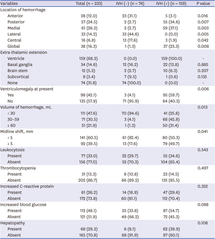
Table 2
Radiological and laboratory features of the patients with spontaneous thalamic hemorrhage
![]()
The laboratory findings of chemical analysis of plasma, including the leucocyte count and thrombocyte count and levels of C-reactive protein and blood glucose and hepatic enzymes were not statistically different between the STH patients without IVH and patients with IVH (Table 2).
Treatment modalities for the patients with STH
Among 233 STH patients, 73 (31.3%) patients underwent conservative treatment, and 160 (68.7%) patients underwent surgical treatment. A higher proportion of STH patients with IVH underwent surgical treatment than that of those without IVH (86.2% vs. 13.8%, P = 0.004). In the terms of the main surgical modalities, 19 (11.9%) patients underwent EVD alone; 45 (28.1%) patients, endoscopic evacuation of IVH; 73 (45.6%) patients, stereotactic evacuation of STH; and 23 (14.4%) patients, craniotomy with removal of STH (Table 3). All 45 patients who underwent endoscopic evacuation of IVH also underwent EVD through the route of endoscopic insertion. Twenty-two out of 73 (30.1%) patients who underwent stereotactic evacuation of STH were also treated concomitantly with EVD, and 6 of 23 (26.1%) patients who under craniotomy with the removal of STH were treated concomitantly with EVD. Additionally, among 73 patients who underwent stereotactic evacuation of STH, 66 (90.4%) patients received fibrinolysis treatment through a drainage catheter inserted in the cavity of the STH.
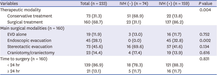
Table 3
Treatment modalities of the patients with spontaneous thalamic hemorrhage
Values are presented as number (%).
EVD = external ventricular drainage, IVH = intraventricular hemorrhage.
![]()
Treatment outcome of the patients with STH
Overall, 33 (14.2%) patients died within 1 month and 47 patients (20.2%) within 3 months of the onset of STH (Table 4). The 1- and 3-month mortality rates were 5.4% and 8.1%, respectively, for the STH patients without IVH and 18.2% and 25.8%, respectively, for those with IVH.
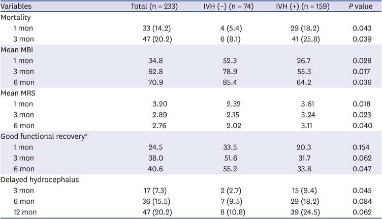
Table 4
Treatment outcome of the patients with spontaneous thalamic hemorrhage
Values are presented as number (%) unless otherwise indicated.
IVH = intraventricular hemorrhage, MBI = Modified Barthel Index, MRS = Modified Rankin Scale.
aGood functional recovery means the patients with a MRS score of 0 to 2 and a MBI score of ≥ 60.
![]()
The mean MBI score at 1 month, 3 months, and 6 months were 52.3, 78.9, and 85.4, respectively, for the STH patients without IVH and 26.7, 55.3, and 64.2, respectively, for those with IVH (Table 4). There was statistically significant differences between the two groups (P = 0.028 at 1 month, P = 0.017 at 3 months, and P = 0.036 at 6 months). The mean MRS scores at 1 month, 3 months, and 6 months were 2.32, 2.15, and 2.02, respectively, for the STH patients without IVH and 3.61, 3.24, and 3.11, respectively, for those with IVH (Table 4). There were statistically significant differences between the two groups (P = 0.018 at 1 month, P = 0.023 at 3 months, and P = 0.040 at 6 months). Delayed hydrocephalus was detected in 17 (7.3%) patients at 3 months, 36 (15.5%) patients at 6 months, and 47 (20.2%) patients at 12 months (Table 4). The rate of good functional recovery was 33.5% and 20.3% at 3 months (P = 0.154), 51.6% and 31.7% at 6 months, and 55.2% and 33.8% at 6 months for the STH patients with IVH and those without IVH, respectively (Table 4).
The overall mortality rates according to the therapeutic modalities were 3.1% (2 of 64 patients) for the STH patients who underwent conservative treatment, 100% (9 of 9 patients) for the STH patients who renounced active treatment owing to the poor condition such as GCS score of 3, and 22.5% (36 of 160) for the STH patients who underwent surgical intervention (Table 5). In term of the cohorts who were treated surgically, 4 of 19 (21.0%) patients who underwent EVD alone, 5 of 45 (11.1%) patients who underwent endoscopic evacuation of IVH, 19 of 73 (26.0%) patients who underwent stereotactic evacuation of STH, and 8 of 23 34.8%) patients who underwent craniotomy with the removal of STH died within 3 months after STH development. The mortality rate was not different according to the time to surgery (Table 5).
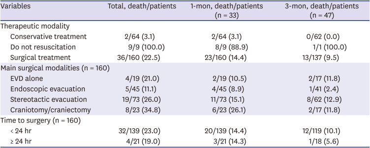
Table 5
Mortality according to the treatment modalities of the patients with spontaneous thalamic hemorrhage (n = 233)
![]()
Predictors of 3-month mortality of the patients with STH
In the univariate analysis, the GCS score at presentation (3–8 vs. 9–15; hazard ratio [HR], 3.327; 95% confidence interval [CI], 1.928–4.726), pupillary reactivity (0 reactive vs. 2 reactive; HR, 4.058; 95% CI, 2.274–5.842), location of hemorrhage (global vs. non-global; HR, 2.994; 95% CI, 1.843–4.145), extra-thalamic extension of STH (yes vs. no; HR, 3.052; 95% CI, 1.243–4.861), initial IVH (yes vs. no; HR, 3.641; 95% CI, 2.643–4.639), underlying disease (present vs. absent; HR, 2.477; 95% CI, 1.196–3.758), and treatment modality (surgery vs. conservative; HR, 3.805; 95% CI, 2.241–5.369) were associated with the 3-month mortality in the patients with STH (Table 6).
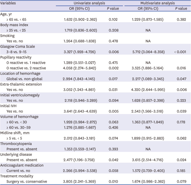
Table 6
Predictors of 3-month mortality in the patients with spontaneous thalamic hemorrhage
![]()
In the multivariate analysis, the GCS score at presentation (3–8 vs. 9–15; HR, 5.712; 95% CI, 3.064–8.358), pupillary reactivity (0 reactive vs. 2 reactive; HR, 3.525; 95% CI, 1.886–5.164), location of hemorrhage (global vs. non-global; HR, 2.217; 95% CI, 1.089–3.345), extra-thalamic extension of STH (yes vs. no; HR, 4.320; 95% CI, 2.644–5.995), initial IVH (yes vs. no; HR, 2.543; 95% CI, 1.566–3.518), and underlying disease (present vs. absent; HR, 3.615; 95% CI, 2.514–4.716) were independently associated with the 3-month mortality in the patients with STH. However, treatment modality, which showed an association with 3-month mortality in the univariate analysis was not statistically associated with the 3-month mortality in the multivariate analysis (HR, 1.674; 95% CI, 0.986–2.362; P = 0.079) (Table 6).
Predictors of poor functional recovery at 6 months of the patients with STH
In the univariate analysis, the GCS score at presentation (3–8 vs. 9–15; HR, 3.426; 95% CI, 2.624–4.228), pupillary reactivity (0 reactive vs. 2 reactive; HR, 3.163; 95% CI, 1.909–4.417), location of hemorrhage (global vs. non-global; HR, 2.348; 95% CI, 1.365–3.331), extra-thalamic extension of STH (yes vs. no; HR, 2.718; 95% CI, 1.219–4.217), initial IVH (present vs. absent; HR, 2.569; 95% CI, 1.483–3.655), and volume of STH (≥ 60 vs. < 30; HR, 4.016; 95% CI, 2.645–5.387) were associated with functional recovery at 6 months in the patients with STH (Table 7).
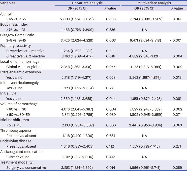
Table 7
Predictors of poor functional recovery at 6 months in the patients with spontaneous thalamic hemorrhage
![]()
In the multivariate analysis, all factors that showed an association with functional recovery at 6 months in the univariate analysis were also independently associated with functional recovery as follows: GCS score at presentation (3–8 vs. 9–15; HR, 6.471; 95% CI, 3.624–9.318), pupillary reactivity (0 reactive vs. 2 reactive; HR, 4.882; 95% CI, 2.643–7.121), location of hemorrhage (global vs. non-global; HR, 4.153; 95% CI, 2.316–5.989), extra-thalamic extension of STH (yes vs. no; HR, 3.262; 95% CI, 1.667–4.857), and volume of STH (≥ 60 vs. < 30; HR, 5.897; 95% CI, 2.942–8.852). However, initial IVH, which was associated with functional recovery at 6 months in the univariate analysis did not show an independent association in the multivariate analysis (Table 7).
The two factors, which were independently associated with the 3-month mortality, such as initial IVH and underlying disease did not show an association with functional recovery at 6 months after STH development. Consequently, the following factors were associated with both the 3-month mortality and 6-month functional recovery: GCS score and pupillary reactivity at presentation, location of hemorrhage, and extra-thalamic extension of STH.
Delayed hydrocephalus after STH
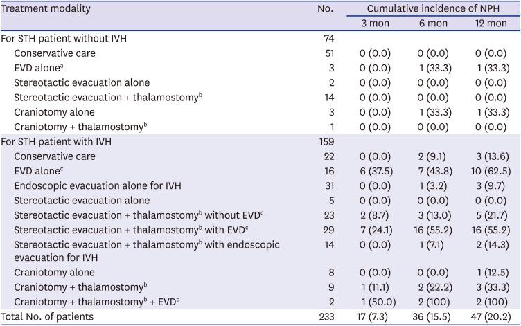
All surviving patients were followed-up for more than 12 months. The mean duration of the follow-up was 27 months and ranged from 12.3 months to 68.9 months. Among the 73 patients who were treated conservatively (51 patients did not have IVH and 22 patients had IVH), 3 (1.3%) patients presented with development of delayed NPH; none of the 51 STH (0.0%) patients without IVH and 3 of the 22 (13.6%) STH patients with IVH showed development of delayed NPH.
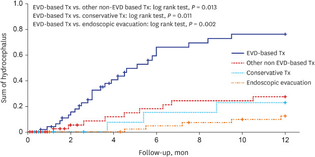
Among the 159 STH patients with concurrent IVH, 22 (13.8%) patients were managed with conservative treatment and 45 (28.3%) patients were treated with endoscopic evacuation of IVH (endoscopic evacuation alone in 31 patients and endoscopic evacuation in combination with surgery for STH in 14 patients). Forty-seven (29.6%) patients were treated with EVD of IVH (EVD alone in 16 patients and EVD in combination with stereotactic evacuation or craniotomy of STH in 31 patients). The other 45 (28.3%) patients underwent non-EVD combined surgical treatment (stereotactic evacuation alone or stereotactic evacuation followed by fibrinolysis in 28 patients and craniotomy in combination with ventriculostomy alone or craniotomy in combination with ventriculostomy with fibrinolysis in 17 patients. The patients who underwent EVD-based treatment for IVH showed a higher incidence of delayed NPH than those who underwent endoscopy-based treatment for IVH. The cumulative incidence of delayed NPH was 59.6% (28 of 47 patients) and 11.1% (5 of 45 patients) at the 12-month follow-up among the patients who underwent EVD-based treatment of IVH and those who underwent endoscopy-based treatment of IVH, respectively (P = 0.007) (Fig. 1). Among 45 STH patients, including those with concurrent IVH who underwent surgical treatment, such as stereotactic evacuation with or without followed fibrinolysis or craniotomy with removal of the hematoma, 9 (20.0%) patients experienced the development of delayed NPH (Table 8).
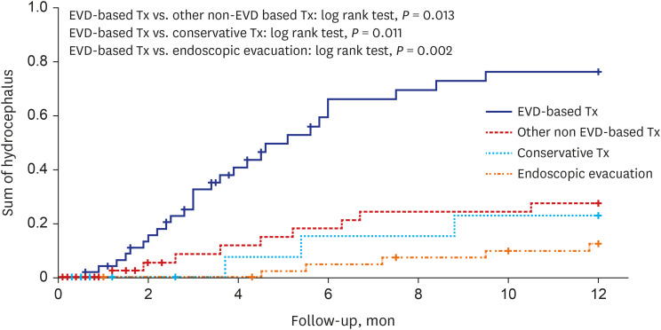 | Fig. 1Cumulative incidence of delayed normal pressure hydrocephalus in 159 patients with spontaneous thalamic hemorrhage accompanied with intraventricular hemorrhage according to the treatment modalities.EVD = external ventricular drainage, Tx = treatment.
|
Table 8
The patients with delayed hydrocephalus after spontaneous thalamic hemorrhage
Values are presented as number (%).
NPH = normal pressure hydrocephalus, STH = spontaneous thalamic hemorrhage, IVH = intraventricular hemorrhage, EVD = external ventricular drainage.
aThe EVD was performed in order to manage ventriculomegaly and treat the increased intracranial pressure; bThe thalamostomy was performed in order to fibrinolysis of the thalamic hematoma; cThe EVD was performed in order to drainage of intraventricular hemorrhage.
![]()
In the multivariate analysis using logistic regression model, several factors that showed an association with delayed development of NPH at 12 months in the univariate analysis were also independently associated with delayed NPH as follows: initial ventriculomegaly (yes vs. no; HR, 3.685; 95% CI, 1.291–6.079), initial IVH (yes vs. no; HR, 8.363; 95% CI, 4.867–11.859), and endoscopic evacuation vs. other surgical treatment (HR, 0.514; 95% CI, 0.188–0.841). However, extra-thalamic extension, volume of hemorrhage, surgical treatment vs. conservative treatment, EVD-based treatment vs. other surgical treatment, and other non EVD-based treatment vs. other surgical treatment, which were associated with delayed development of NPH at 12 months in the univariate analysis did not show an independent association in the multivariate analysis (Table 9).
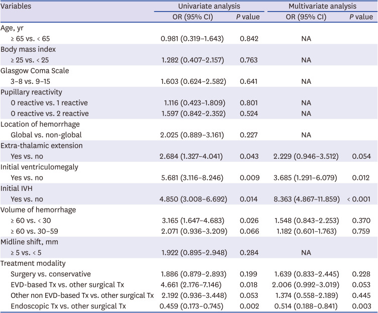
Table 9
Predictors for delayed hydrocephalus in the patients with spontaneous thalamic hemorrhage
OR = odds ratio, CI = confidence interval, IVH = intraventricular hemorrhage, EVD = external ventricular drainage, NA = not assessed, Tx = treatment.
![]()
Go to :
DISCUSSION
In this retrospective analysis of the STH patients, the clinical features and outcomes of the patients with IVH and those without IVH were compared. To the best of our knowledge, a comprehensive study reporting on the predisposing factors for the occurrence of IVH in STH patients has not been published previously. Although the rate of IVH concurrent with STH is approximately 45% among STH patients,527 this study showed a relatively high rate of concurrent IVH in STH patients at 68.2%. As our institute is an official regional emergency medical center in our province serving a population of 3.5 million people, we tend to receive more severe patients. The patients with stable neurological status, stable vital signs, and smaller volume of STH are usually treated in the general community hospitals. Therefore, the STH patients who were admitted to our hospital may have had a more severe status than those reported in other studies, which may account for the higher rate of accompanying IVH in STH patients in our study than that reported in the available literature.
This study suggested that several clinical factors, such as high BMI, low GCS score, underlying disease, and family history of stroke may be more frequently detected in the STH patients with concurrent IVH. However, these clinical factors were usually associated with a worse outcome in the whole SICH patient cohort. Additionally, we found that there were differences in certain radiological factors, such as location of STH, ventriculomegaly, and midline shifting between the STH patients with IVH and those without IVH. Ventriculomegaly and more midline shifting were considered to be the result of concurrent IVH in STH patients rather than the cause of concurrent development of IVH and STH. Global location was the only independent factor associated with mortality and functional recovery. In terms of location of hemorrhage in the thalamus, several studies have reported on the association between location of hemorrhage in the thalamus and IVH occurrence in STH patients.72428 Neisewander et al.28 reported that IVH was detected in 76.8% of the STH patients (129 of 168 STH patients), and the anterior location was the most common origin site of IVH from STH (94.4%). The posterior and lateral location of STH was associated with better survival compared to the global or medial location (P = 0.003 and P = 0.013, respectively).28 In this study, the medial location was the most common site of origin of IVH from STH (37.1%), and the posterior location was the second most common (34.6%). In contrast to the study by Neisewander et al.,28 the anterior location of IVH originating from STH was relatively rare at 3.1% in this study. The mortality at 1-month among the patients with STH accompanied by IVH was somewhat different at 18.2% in our study and 30.2% in the study by Neisewander et al.28; the different locations of STH in the two studies may have accounted for this difference. However, these results must be validated with multicenter randomized clinical trials.
Similar to that reported in the previous studies, the STH patients with accompanying IVH in this study also showed a worse outcome than those without accompanying IVH. The presence of IVH was considered a strong predictive factor of SICH prognosis, as reported previously, and controlling the increased intracranial pressure was an important therapeutic strategy.8,29,30 A worse outcome in STH patients with ruptured hemorrhage into the ventricle may be noted owing to obstructive hydrocephalus and mass effect exerted by the blood clot, which can lead to increased intracranial pressure and brain herniation. The mortality rate associated with the intraventricular expansion of STH was reported to be 40%–52%.272831 The 3-month mortality rate was 25.8% in this study, which is relatively low. The lower mortality rate in this study may be due to the different number of patients, different characteristics, and complex therapeutic methods used for STH patients with IVH compared to those reported in previous studies.
Owing to the high rates of mortality and morbidity in SICH patients with concurrent IVH, the management of IVH is another issue when planning the treatment. Without a specific treatment for IVH, the risk of poor outcome is 90%, and the risk of death is 78%.8 Therefore, the placement of an EVD catheter and natural drainage of IVH is commonly considered.32 However, EVD may be complicated by occlusion of the catheter with blood clots.32 In recent years, the administration of fibrinolytic agents, such as recombinant tissue-plasminogen activator (rt-PA) or urokinase, through the EVD catheter into the ventricles has been proposed as an effective way to maintain catheter patency, increase blood clearance and clot removal, decrease ventricular enlargement and delayed hydrocephalus, and ameliorate inflammation caused by blood and its toxic products.33 However, the efficacy of IVF in IVH is still debatable. Although 47 of 159 patients with STH accompanied by IVH underwent EVD catheter insertion and natural drainage of IVH in this study, none of the patients were treated with IVF after EVD. Among these 47 patients who underwent EVD for IVH, 12 (25.5%) patients succumbed to STH with IVH, which was similar to the rate noted for all the STH patients with IVH (25.8%). Even 31 of 47 patients who underwent insertion of the EVD catheter and natural drainage of IVH also stereotactic evacuation or craniotomy with the removal of STH, but the mortality of them was not worse than that of the whole STH patients with IVH. This suggests that there may be certain benefits of simple drainage of IVH, followed by STH, even without IVF. However, our results are different from the results of an outstanding randomized multi-central clinical trial showing the positive effect of IVF using rt-PA for IVH on the 6-month mortality.34 This discrepancy might be due to differences in the patient populations. This study included patients with IVH originating from only thalamic hemorrhage, but their study included IVH patients with non-traumatic intracerebral hemorrhage.34 Their study also suggested that the location of hemorrhage in the thalamus is a poor prognostic factor in the IVF treatment for IVH.34
In terms of endoscopic evacuation as another therapeutic modality for IVH of STH, a recent randomized clinical trial showed a significantly lower rate of shunt-dependent hydrocephalus. Their stay in the intensive care unit was shorter compared with EVD surgery for IVH caused by STH.35 The study did not report any difference in the 90-day mortality rate between the two groups (16.6% in the EVD group vs. 20.8% in the endoscopy group, P = 0.710). On comparison with Chen's study,35 we noted a statistical difference in the 3-month mortality rate in this study (25.5% for 47 patients who underwent EVD of IVH vs. 11.1% for 45 patients who underwent endoscopic evacuation of IVH, P = 0.047). Although this study was not a randomized prospective study, the number of patients was greater than their study (48 patients vs. 92 patients in this study).
From the literature review, both IVH and thalamic hemorrhagic were strong predictors for a permanent ventriculoperitoneal (VP) shunt, and approximately 66% of all patients with thalamic hemorrhage need a shunt.36 Especially in patients with IVH caused by thalamic hemorrhage, the shunt-dependent hydrocephalus rate was considered to be higher than 66%. Chen et al.35 reported that 33 (68.8%) patients of 48 with IVH caused by thalamic hemorrhage needed a permanent VP shunt. However, only 47.62% of patients in the endoscopic surgery group needed a VP shunt. In contrast, 90.48% of those in the EVD group needed shunts. Endoscopic surgery had a significantly lower prevalence of shunt-dependent hydrocephalus (P = 0.002). In this study, 45 (28.3%) patients of 159 STH patients with IVH received a VP shunt during the follow-up period owing to delayed NPH. The rate of use of VP shunt for delayed NPH was 11.1% (5 of 45 patients) among the patients who underwent endoscopic treatment and 59.6% (28 of 47 patients) among the patients who underwent EVD This discrepancy originated from a different definition of shunt-dependent hydrocephalus vs. delayed NPH. Shunt-dependent hydrocephalus was defined on the basis of the Graeb Score.37 However, delayed NPH in this study was diagnosed by the clinical presentation (Hakim's triad) and ventriculomegaly detected in CT scan (Evan's ratio > 35%). Regardless of the different incidence of hydrocephalus, which must be treated clinically, both studies suggested that the endoscopic evacuation could be effective for reducing the onset of hydrocephalus in STH patients, especially in those with concurrent IVH.
Although this study suggests that several clinical factors, such as high BMI, low GCS score, underlying disease, and family history of stroke are associated with the occurrence of IVH in STH patients, and endoscopic evacuation of IVH has a better outcome than EVD-based treatment, several important limitations must be noted. First, the most important limitation is the inherent bias introduced by the retrospective nature of the study. We attempted to reduce this bias by collecting patient data from the complete medical and radiological records and by recruiting patients treated using concrete protocols. As individual attending neurosurgeons choose the neurosurgical modalities according to their preference based on the AHA guidelines for SICH, the introduction of selection bias could not be avoided. Although multiple investigators, without any prior information of the patients, independently reviewed the radiological images, we cannot clearly claim that there was no bias in this retrospective study. Specifically, it can be difficult to define the location of STH in the thalamus simply using CT images, especially in patients with STH with accompanying IVH. Despite these efforts, however, the conclusions drawn from the study must be validated by prospective and randomized clinical trials.
Second, although this study suggested the benefit of endoscopic evacuation of IVH in STH patients with respect to mortality, the therapeutic modalities could not be homogeneously categorized. As the analysis was performed basically among subjects with STH, the major effects of characteristics of STH on the clinical outcome could influence the results when evaluating the role of endoscopic evacuation or EVD for IVH. The treatment modality for IVH was not an independent factor for predicting outcomes in STH patients. When planning surgical treatment for STH accompanied with IVH, the therapeutic strategies should be considered individually for managing STH and IVH simultaneously. Therefore, multiple combinations of conservative treatment, EVD and natural drainage of IVH, endoscopic removal of IVH, stereotactic evacuation of STH, and craniotomy with the removal of STH could be used for STH patients with IVH according to the neurological status of the patients and radiological features. Additionally, we did not perform endoscopic third ventriculostomy (ETV) and IVF to reduce the need for shunt surgery. The alternative method, involving EVD (with lysis) for a short duration followed by endoscopy, if possible, with ETV can perhaps reduce the need for shunt surgery and improve the prognosis of STH patients. Further studies are necessary to confirm this.
Finally, there may be a selection bias with respect to STH patients. The STH patients in our cohort could have different clinical and radiological characteristics owing to the unique position of our institute as the official regional emergency medical center in the province. Compared with the community hospitals, the specific cerebrovascular center of a university hospital tends to care for more patients with vascular lesions and advanced critical care needs. The patients at our institution exhibit a relatively high rate of comorbidities and poor socioeconomic status, which are associated with increased risk for stroke and adverse outcomes. Therefore, this cohort may not represent the characteristics of general STH patients. To validate the results of this study, it is necessary to perform the multicentre global studies.
Conclusively, we found several differences in the characteristics of STH patients with IVH and those without IVH and suggested that concurrent IVH should be independently associated with mortality. Therefore, we suggest that active removal of IVH originating from STH using endoscopy may improve the mortality of patients compared to the mortality rates associated with other surgical modalities. Despite the lack of association with 6-month morbidity, delayed NPH may develop more frequently in STH patients with IVH than in those without IVH. Therefore, it may be important to prevent the development of delayed NPH by performing endoscopic evacuation of IVH. However, it is mandatory to validate the data with multicentre prospective randomized clinical trials to overcome the inherit limitations of a retrospective study at a single center.
Go to :
 XML Download