Journal List > J Korean Med Sci > v.36(2) > 1146293

Park, Lee, Park, Kim, Jang, Bae, Yang, Park, Cho, Lee, Nam, Lee, Kim, Chae, and the Daegu COVID-19 Research Project: Impact of Cardiovascular Risk Factors and Cardiovascular Diseases on Outcomes in Patients Hospitalized with COVID-19 in Daegu Metropolitan City

Abstract

Background

Data regarding the association between preexisting cardiovascular risk factors (CVRFs) and cardiovascular diseases (CVDs) and the outcomes of patients requiring hospitalization for coronavirus disease 2019 (COVID-19) are limited. Therefore, the aim of this study was to investigate the impact of preexisting CVRFs or CVDs on the outcomes of patients with COVID-19 hospitalized in a Korean healthcare system.

Methods

Patients with COVID-19 admitted to 10 hospitals in Daegu Metropolitan City, Korea, were examined. All sequentially hospitalized patients between February 15, 2020, and April 24, 2020, were enrolled in this study. All patients were confirmed to have COVID-19 based on the positive results on the polymerase chain reaction testing of nasopharyngeal samples. Clinical outcomes during hospitalization, such as requiring intensive care and invasive mechanical ventilation (MV) and death, were evaluated. Moreover, data on baseline comorbidities such as a history of diabetes, hypertension, dyslipidemia, current smoking, heart failure, coronary artery disease, cerebrovascular accidents, and other chronic cardiac diseases were obtained.

Results

Of all the patients enrolled, 954 (42.0%) had preexisting CVRFs or CVDs. Among the CVRFs, the most common were hypertension (28.8%) and diabetes mellitus (17.0%). The prevalence rates of preexisting CVRFs or CVDs increased with age (P < 0.001). The number of patients requiring intensive care (P < 0.001) and invasive MV (P < 0.001) increased with age. The in-hospital death rate increased with age (P < 0.001). Patients requiring intensive care (5.3% vs. 1.6%; P < 0.001) and invasive MV (4.3% vs. 1.7%; P < 0.001) were significantly greater in patients with preexisting CVRFs or CVDs. In-hospital mortality (12.9% vs. 3.1%; P < 0.001) was significantly higher in patients with preexisting CVRFs or CVDs. Among the CVRFs, diabetes mellitus and hypertension were associated with increased requirement of intensive care and invasive MV and in-hospital death. Among the known CVDs, coronary artery disease and congestive heart failure were associated with invasive MV and in-hospital death. In multivariate analysis, preexisting CVRFs or CVDs (odds ratio [OR], 1.79; 95% confidence interval [CI], 1.07–3.01; P = 0.027) were independent predictors of in-hospital death after adjusting for confounding variables. Among individual preexisting CVRF or CVD components, diabetes mellitus (OR, 2.43; 95% CI, 1.51–3.90; P < 0.001) and congestive heart failure (OR, 2.43; 95% CI, 1.06–5.87; P = 0.049) were independent predictors of in-hospital death.

Conclusion

Based on the findings of this study, the patients with confirmed COVID-19 with preexisting CVRFs or CVDs had worse clinical outcomes. Caution is required in dealing with these patients at triage.

Graphical Abstract

jkms-36-e15-abf001.jpg

INTRODUCTION

Since the first report of coronavirus disease 2019 (COVID-19) in Hubei Province, China, in December 2019, it has been spreading rapidly worldwide, and the World Health Organization had declared the disease a pandemic on March 11, 2020.12 Studies on a small sample of patients with COVID-19 in China reported that preexisting cardiovascular risk factors (CVRFs) or cardiovascular diseases (CVDs) increase the risk of COVID-19.23456 However, studies on a large sample of patients with COVID-19 in China presented that the prevalence rate of preexisting CVRFs or CVDs in patients with COVID-19 is not higher than those in the general population.789 Therefore, it remains unclear whether CVRFs or known CVDs are causally linked to COVID-19.101112 Furthermore, it is uncertain whether patients with preexisting CVRFs or CVDs are more likely to progress to severe disease requiring intensive care and invasive mechanical ventilation (MV). Moreover, although several studies have reported on the association between preexisting CVRFs or CVDs and mortality,234567 a comprehensive analysis considering the demographic characteristics, initial presentation, and multiple comorbidities has not yet been conducted. Therefore, this study investigated the impact of preexisting CVRFs or CVDs on the outcomes of patients with COVID-19 hospitalized in a Korean healthcare system.

METHODS

Study population

The Daegu COVID-19 Research Project is an observational multicenter registry of patients with COVID-19 hospitalized in a Korean healthcare system in Daegu City. All data about the patients and management details were obtained at each hospital.
Between February 15, 2020, and April 24, 2020, 2,269 consecutive patients (814 male; mean age, 55.5 ± 20.2 years) admitted to 10 hospitals (viz., Kyungpook National University Hospital, Kyungpook National University Chilgok Hospital, Yeungnam University Hospital, Keimyung University Dongsan Medical Center, Keimyung University Daegu Dongsan Hospital, Daegu Catholic University Hospital, Daegu Fatima Hospital, Daegu Medical Center, Daegu Veterans Hospital, and Korea Workers' Compensation and Welfare Service Daegu Hospital in Daegu City) for confirmed severe acute respiratory syndrome coronavirus 2 (SARS-CoV-2) based on the positive results on polymerase chain reaction testing of nasopharyngeal samples were included. The data obtained included patient demographic information, initial vital signs, presenting symptoms, comorbidities, and history of medical illnesses, laboratory findings at baseline and during hospitalization, radiological findings, daily clinical course, inpatient medications, treatments (including intensive care unit [ICU] admission, invasive MV, hemodialysis, and extracorporeal membrane oxygenation), and outcomes (including the length of stay, readmission, and mortality). All variables were available for all admitted patients except for presenting symptoms and laboratory findings (available in 9 of the 10 hospitals). Baseline comorbidities were available for all patients in the 10 hospitals. Among the comorbidities, CVRF was defined as a history of hypertension, diabetes mellitus, dyslipidemia, and current smoking. Known CVD was defined as a history of coronary artery disease, congestive heart failure, cerebrovascular accidents, and other chronic cardiac diseases. Moreover, chronic cardiac disease was defined as other cardiac conditions excluding coronary artery disease and congestive heart failure.

Statistical analysis

The data were expressed as mean ± standard deviation for continuous variables and as percentages for categorical variables. Comparisons between the baseline variables were performed using Student's t-test for continuous variables and Pearson's χ2 test for categorical variables. The patients were categorized into two groups: group with preexisting CVRFs or CVDs and group without preexisting CVRFs or CVDs. Demographics, vital signs at admission, clinical presentation at admission, comorbidities, laboratory findings, treatment, and outcomes were compared between the two groups. Moreover, the prevalence rates of preexisting CVRFs or CVDs in hospitalized patients, treatments, and outcomes were compared by 10-year age intervals. We compared the prevalence rates of diabetes mellitus and hypertension between patients with COVID-19 and general population. The data regarding prevalence rates of diabetes mellitus and hypertension in general population were collected from Korea National Health And Nutrition Examination Survey (KNHANES) 2018. In addition, data regarding prevalence rates of diabetes mellitus and hypertension in general population of Daegu Metropolitan City were collected from Community Health Survey (CHS) 2019. The data are available in Korean Statistical Information Service (https://kosis.kr) and Community Health Survey (https://chs.cdc.go.kr), respectively. To determine the predictors of in-hospital death, multivariate logistic regression models were used to provide adjusted odds ratios (ORs) with 95% confidence intervals (CIs). Variables with P < 0.05 in univariate analyses were included in the multivariate model analysis. For all analyses, a two-sided P < 0.05 was considered statistically significant. Statistical analysis was performed using the Statistical Package for the Social Sciences, version 20.0 (IBM Corp., Armonk, NY, USA).

Ethics statement

The Joint Institutional Review Board in Daegu City approved this study (No. 2020-07-003) as minimal-risk research using data collected for routine clinical practice and waived the requirement for informed consent.

RESULTS

Table 1 presents the patients' baseline characteristics. Overall, the mean age was 55.5 ± 20.2 years, and 814 patients were male. Of all the patients, 954 (42.0%) had preexisting CVRFs or CVDs upon admission. Patients with preexisting CVRFs or CVDs were older and more likely to be male. Compared with those without preexisting CVRFs or CVDs, body mass index, systolic blood pressure, and respiratory rate were significantly higher in patients with preexisting CVRFs or CVDs, whereas O2 saturation at admission was significantly lower. Respiratory symptoms such as cough, sputum production, sore throat, and rhinorrhea; chest discomfort; dysosmia; headache; and diarrhea were less frequent in patients with preexisting CVRFs or CVDs than those without, whereas dyspnea and altered consciousness were more frequent. Among the comorbidities, the most common were hypertension (28.8%) and diabetes mellitus (17.0%). Supplementary Fig. 1 presents the prevalence rates of hypertension and diabetes mellitus among the patients. The prevalence rates of diabetes mellitus and hypertension in patients with COVID-19 were comparable with those in the general population of in the KNHANES 2018. The prevalence rate of diabetes mellitus was numerically higher in COVID-19 patients compared with those in the general population of Daegu Metropolitan City in the CHS 2019. Chronic kidney disease, malignancy, chronic neurologic disorder, dementia, and psychiatric disorders were more frequent in patients with preexisting CVRF or CVD than in those without.
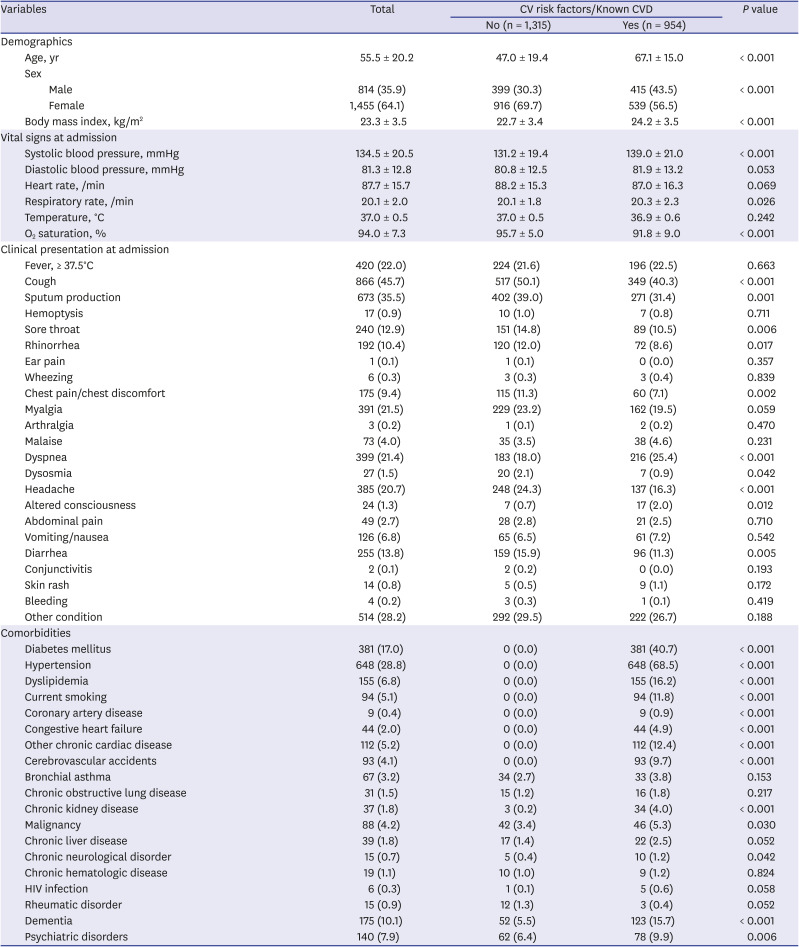
Table 1

Baseline characteristics of study subject

jkms-36-e15-i001
Variables Total CV risk factors/Known CVD P value
No (n = 1,315) Yes (n = 954)
Demographics
Age, yr 55.5 ± 20.2 47.0 ± 19.4 67.1 ± 15.0 < 0.001
Sex
Male 814 (35.9) 399 (30.3) 415 (43.5) < 0.001
Female 1,455 (64.1) 916 (69.7) 539 (56.5)
Body mass index, kg/m2 23.3 ± 3.5 22.7 ± 3.4 24.2 ± 3.5 < 0.001
Vital signs at admission
Systolic blood pressure, mmHg 134.5 ± 20.5 131.2 ± 19.4 139.0 ± 21.0 < 0.001
Diastolic blood pressure, mmHg 81.3 ± 12.8 80.8 ± 12.5 81.9 ± 13.2 0.053
Heart rate, /min 87.7 ± 15.7 88.2 ± 15.3 87.0 ± 16.3 0.069
Respiratory rate, /min 20.1 ± 2.0 20.1 ± 1.8 20.3 ± 2.3 0.026
Temperature, °C 37.0 ± 0.5 37.0 ± 0.5 36.9 ± 0.6 0.242
O2 saturation, % 94.0 ± 7.3 95.7 ± 5.0 91.8 ± 9.0 < 0.001
Clinical presentation at admission
Fever, ≥ 37.5°C 420 (22.0) 224 (21.6) 196 (22.5) 0.663
Cough 866 (45.7) 517 (50.1) 349 (40.3) < 0.001
Sputum production 673 (35.5) 402 (39.0) 271 (31.4) 0.001
Hemoptysis 17 (0.9) 10 (1.0) 7 (0.8) 0.711
Sore throat 240 (12.9) 151 (14.8) 89 (10.5) 0.006
Rhinorrhea 192 (10.4) 120 (12.0) 72 (8.6) 0.017
Ear pain 1 (0.1) 1 (0.1) 0 (0.0) 0.357
Wheezing 6 (0.3) 3 (0.3) 3 (0.4) 0.839
Chest pain/chest discomfort 175 (9.4) 115 (11.3) 60 (7.1) 0.002
Myalgia 391 (21.5) 229 (23.2) 162 (19.5) 0.059
Arthralgia 3 (0.2) 1 (0.1) 2 (0.2) 0.470
Malaise 73 (4.0) 35 (3.5) 38 (4.6) 0.231
Dyspnea 399 (21.4) 183 (18.0) 216 (25.4) < 0.001
Dysosmia 27 (1.5) 20 (2.1) 7 (0.9) 0.042
Headache 385 (20.7) 248 (24.3) 137 (16.3) < 0.001
Altered consciousness 24 (1.3) 7 (0.7) 17 (2.0) 0.012
Abdominal pain 49 (2.7) 28 (2.8) 21 (2.5) 0.710
Vomiting/nausea 126 (6.8) 65 (6.5) 61 (7.2) 0.542
Diarrhea 255 (13.8) 159 (15.9) 96 (11.3) 0.005
Conjunctivitis 2 (0.1) 2 (0.2) 0 (0.0) 0.193
Skin rash 14 (0.8) 5 (0.5) 9 (1.1) 0.172
Bleeding 4 (0.2) 3 (0.3) 1 (0.1) 0.419
Other condition 514 (28.2) 292 (29.5) 222 (26.7) 0.188
Comorbidities
Diabetes mellitus 381 (17.0) 0 (0.0) 381 (40.7) < 0.001
Hypertension 648 (28.8) 0 (0.0) 648 (68.5) < 0.001
Dyslipidemia 155 (6.8) 0 (0.0) 155 (16.2) < 0.001
Current smoking 94 (5.1) 0 (0.0) 94 (11.8) < 0.001
Coronary artery disease 9 (0.4) 0 (0.0) 9 (0.9) < 0.001
Congestive heart failure 44 (2.0) 0 (0.0) 44 (4.9) < 0.001
Other chronic cardiac disease 112 (5.2) 0 (0.0) 112 (12.4) < 0.001
Cerebrovascular accidents 93 (4.1) 0 (0.0) 93 (9.7) < 0.001
Bronchial asthma 67 (3.2) 34 (2.7) 33 (3.8) 0.153
Chronic obstructive lung disease 31 (1.5) 15 (1.2) 16 (1.8) 0.217
Chronic kidney disease 37 (1.8) 3 (0.2) 34 (4.0) < 0.001
Malignancy 88 (4.2) 42 (3.4) 46 (5.3) 0.030
Chronic liver disease 39 (1.8) 17 (1.4) 22 (2.5) 0.052
Chronic neurological disorder 15 (0.7) 5 (0.4) 10 (1.2) 0.042
Chronic hematologic disease 19 (1.1) 10 (1.0) 9 (1.2) 0.824
HIV infection 6 (0.3) 1 (0.1) 5 (0.6) 0.058
Rheumatic disorder 15 (0.9) 12 (1.3) 3 (0.4) 0.052
Dementia 175 (10.1) 52 (5.5) 123 (15.7) < 0.001
Psychiatric disorders 140 (7.9) 62 (6.4) 78 (9.9) 0.006
Data are presented as mean ± standard deviation or number (%).
CV = cardiovascular, CVD = cardiovascular disease, HIV = human immunodeficiency virus.
Table 2 presents the laboratory findings. White blood cell (WBC) count, C-reactive protein (CRP), high-sensitivity CRP (hs-CRP), lactate dehydrogenase, pro-calcitonin, blood urea nitrogen, and creatinine were significantly higher in patients with preexisting CVRFs or known CVDs than those without, whereas hemoglobin and lymphocyte counts at baseline were significantly lower. Moreover, these results were consistent with the laboratory findings during the follow-up.
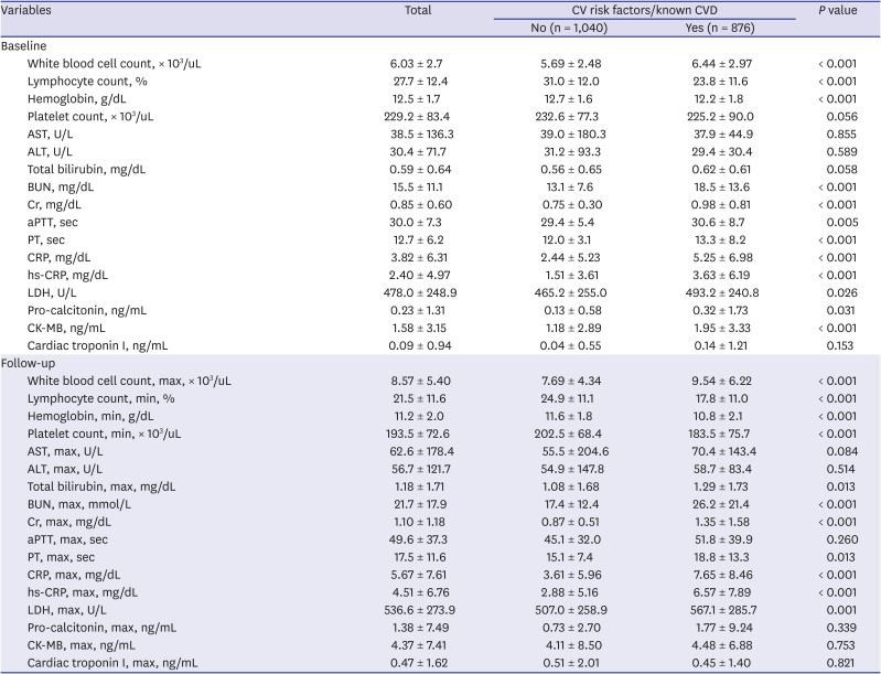
Table 2

Laboratory findings of study subject

jkms-36-e15-i002
Variables Total CV risk factors/known CVD P value
No (n = 1,040) Yes (n = 876)
Baseline
White blood cell count, × 103/uL 6.03 ± 2.7 5.69 ± 2.48 6.44 ± 2.97 < 0.001
Lymphocyte count, % 27.7 ± 12.4 31.0 ± 12.0 23.8 ± 11.6 < 0.001
Hemoglobin, g/dL 12.5 ± 1.7 12.7 ± 1.6 12.2 ± 1.8 < 0.001
Platelet count, × 103/uL 229.2 ± 83.4 232.6 ± 77.3 225.2 ± 90.0 0.056
AST, U/L 38.5 ± 136.3 39.0 ± 180.3 37.9 ± 44.9 0.855
ALT, U/L 30.4 ± 71.7 31.2 ± 93.3 29.4 ± 30.4 0.589
Total bilirubin, mg/dL 0.59 ± 0.64 0.56 ± 0.65 0.62 ± 0.61 0.058
BUN, mg/dL 15.5 ± 11.1 13.1 ± 7.6 18.5 ± 13.6 < 0.001
Cr, mg/dL 0.85 ± 0.60 0.75 ± 0.30 0.98 ± 0.81 < 0.001
aPTT, sec 30.0 ± 7.3 29.4 ± 5.4 30.6 ± 8.7 0.005
PT, sec 12.7 ± 6.2 12.0 ± 3.1 13.3 ± 8.2 < 0.001
CRP, mg/dL 3.82 ± 6.31 2.44 ± 5.23 5.25 ± 6.98 < 0.001
hs-CRP, mg/dL 2.40 ± 4.97 1.51 ± 3.61 3.63 ± 6.19 < 0.001
LDH, U/L 478.0 ± 248.9 465.2 ± 255.0 493.2 ± 240.8 0.026
Pro-calcitonin, ng/mL 0.23 ± 1.31 0.13 ± 0.58 0.32 ± 1.73 0.031
CK-MB, ng/mL 1.58 ± 3.15 1.18 ± 2.89 1.95 ± 3.33 < 0.001
Cardiac troponin I, ng/mL 0.09 ± 0.94 0.04 ± 0.55 0.14 ± 1.21 0.153
Follow-up
White blood cell count, max, × 103/uL 8.57 ± 5.40 7.69 ± 4.34 9.54 ± 6.22 < 0.001
Lymphocyte count, min, % 21.5 ± 11.6 24.9 ± 11.1 17.8 ± 11.0 < 0.001
Hemoglobin, min, g/dL 11.2 ± 2.0 11.6 ± 1.8 10.8 ± 2.1 < 0.001
Platelet count, min, × 103/uL 193.5 ± 72.6 202.5 ± 68.4 183.5 ± 75.7 < 0.001
AST, max, U/L 62.6 ± 178.4 55.5 ± 204.6 70.4 ± 143.4 0.084
ALT, max, U/L 56.7 ± 121.7 54.9 ± 147.8 58.7 ± 83.4 0.514
Total bilirubin, max, mg/dL 1.18 ± 1.71 1.08 ± 1.68 1.29 ± 1.73 0.013
BUN, max, mmol/L 21.7 ± 17.9 17.4 ± 12.4 26.2 ± 21.4 < 0.001
Cr, max, mg/dL 1.10 ± 1.18 0.87 ± 0.51 1.35 ± 1.58 < 0.001
aPTT, max, sec 49.6 ± 37.3 45.1 ± 32.0 51.8 ± 39.9 0.260
PT, max, sec 17.5 ± 11.6 15.1 ± 7.4 18.8 ± 13.3 0.013
CRP, max, mg/dL 5.67 ± 7.61 3.61 ± 5.96 7.65 ± 8.46 < 0.001
hs-CRP, max, mg/dL 4.51 ± 6.76 2.88 ± 5.16 6.57 ± 7.89 < 0.001
LDH, max, U/L 536.6 ± 273.9 507.0 ± 258.9 567.1 ± 285.7 0.001
Pro-calcitonin, max, ng/mL 1.38 ± 7.49 0.73 ± 2.70 1.77 ± 9.24 0.339
CK-MB, max, ng/mL 4.37 ± 7.41 4.11 ± 8.50 4.48 ± 6.88 0.753
Cardiac troponin I, max, ng/mL 0.47 ± 1.62 0.51 ± 2.01 0.45 ± 1.40 0.821
CV = cardiovascular, CVD = cardiovascular disease, AST = aspartate aminotransferase, ALT = alanine aminotransferase, BUN = blood urea nitrogen, Cr = creatinine, aPTT = activated partial thromboplastin time, PT = prothrombin time, CRP = C-reactive protein, hs-CRP = high-sensitivity CRP, LDH = lactate dehydrogenase, CK-MB = creatine kinase-MB.
Table 3 shows the univariate analysis for in-hospital death. In total, 164 (7.2%) patients died in the hospital. In the demographic findings, deceased patients were older and more likely to be male and obese. Regarding the vital signs at admission, the deceased patients had higher respiratory rates, lower O2 saturation, and lower diastolic blood pressures at baseline. Patients with fever upon admission (P < 0.001) and systemic symptoms such as body malaise (P < 0.001) and myalgia (P = 0.001) had higher in-hospital mortality. Among the respiratory symptoms, hemoptysis (P = 0.001) and dyspnea (P < 0.001) were lower in the survivors than in the deceased patients, whereas cough, sore throat, and rhinorrhea were higher. Altered consciousness was higher (P < 0.001) and headache was lower (P < 0.001) in the deceased patients than in the survivors. Among the comorbidities, patients with preexisting CVRFs or CVDs such as diabetes mellitus (P < 0.001), hypertension (P < 0.001), coronary artery disease (P = 0.002), congestive heart failure (P < 0.001), and chronic cardiac disease (P < 0.001) had a higher in-hospital mortality. Among the other comorbidities, patients with bronchial asthma (P = 0.018), chronic obstructive lung disease (P = 0.025), chronic kidney disease (P < 0.001), malignancy (P < 0.001), chronic neurologic disease (P < 0.001), dementia (P < 0.001), and psychiatric disorders (P = 0.038) had higher in-hospital mortality. Among the laboratory findings, the inflammatory markers such as WBC count, CRP, hs-CRP, and pro-calcitonin; hepatic function markers such as aspartate aminotransferase, total bilirubin, and lactate dehydrogenase; renal function markers such as blood urea nitrogen and creatinine; and creatine kinase myocardial band (CK-MB) were higher in patients with in-hospital death at baseline and during follow-up, whereas lymphocyte and platelet counts and hemoglobin were lower (Table 4). WBC count, CRP, hs-CRP, and pro-calcitonin were statistically significantly higher in patients requiring intensive care and invasive MV at baseline and during follow-up (Supplementary Fig. 2). Pulmonary infiltration on chest X-ray was presented in 44.3% (n = 989) and 55.6% (n = 1,240) of the patients at the time of admission and during hospitalization, respectively. The use of invasive MV was significantly greater in patients with pulmonary infiltration at the time of admission (0.9% vs. 5.2%, P < 0.001) and during hospitalization (0.5% vs. 4.6%, P < 0.001).
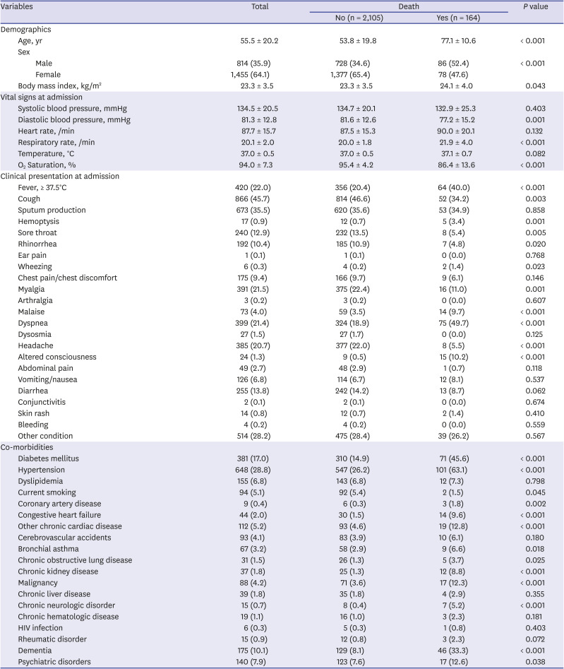
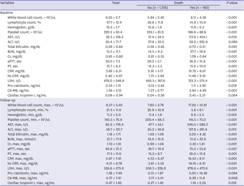
Table 3

Univariate analysis for death

jkms-36-e15-i003
Variables Total Death P value
No (n = 2,105) Yes (n = 164)
Demographics
Age, yr 55.5 ± 20.2 53.8 ± 19.8 77.1 ± 10.6 < 0.001
Sex
Male 814 (35.9) 728 (34.6) 86 (52.4) < 0.001
Female 1,455 (64.1) 1,377 (65.4) 78 (47.6)
Body mass index, kg/m2 23.3 ± 3.5 23.3 ± 3.5 24.1 ± 4.0 0.043
Vital signs at admission
Systolic blood pressure, mmHg 134.5 ± 20.5 134.7 ± 20.1 132.9 ± 25.3 0.403
Diastolic blood pressure, mmHg 81.3 ± 12.8 81.6 ± 12.6 77.2 ± 15.2 0.001
Heart rate, /min 87.7 ± 15.7 87.5 ± 15.3 90.0 ± 20.1 0.132
Respiratory rate, /min 20.1 ± 2.0 20.0 ± 1.8 21.9 ± 4.0 < 0.001
Temperature, °C 37.0 ± 0.5 37.0 ± 0.5 37.1 ± 0.7 0.082
O2 Saturation, % 94.0 ± 7.3 95.4 ± 4.2 86.4 ± 13.6 < 0.001
Clinical presentation at admission
Fever, ≥ 37.5°C 420 (22.0) 356 (20.4) 64 (40.0) < 0.001
Cough 866 (45.7) 814 (46.6) 52 (34.2) 0.003
Sputum production 673 (35.5) 620 (35.6) 53 (34.9) 0.858
Hemoptysis 17 (0.9) 12 (0.7) 5 (3.4) 0.001
Sore throat 240 (12.9) 232 (13.5) 8 (5.4) 0.005
Rhinorrhea 192 (10.4) 185 (10.9) 7 (4.8) 0.020
Ear pain 1 (0.1) 1 (0.1) 0 (0.0) 0.768
Wheezing 6 (0.3) 4 (0.2) 2 (1.4) 0.023
Chest pain/chest discomfort 175 (9.4) 166 (9.7) 9 (6.1) 0.146
Myalgia 391 (21.5) 375 (22.4) 16 (11.0) 0.001
Arthralgia 3 (0.2) 3 (0.2) 0 (0.0) 0.607
Malaise 73 (4.0) 59 (3.5) 14 (9.7) < 0.001
Dyspnea 399 (21.4) 324 (18.9) 75 (49.7) < 0.001
Dysosmia 27 (1.5) 27 (1.7) 0 (0.0) 0.125
Headache 385 (20.7) 377 (22.0) 8 (5.5) < 0.001
Altered consciousness 24 (1.3) 9 (0.5) 15 (10.2) < 0.001
Abdominal pain 49 (2.7) 48 (2.9) 1 (0.7) 0.118
Vomiting/nausea 126 (6.8) 114 (6.7) 12 (8.1) 0.537
Diarrhea 255 (13.8) 242 (14.2) 13 (8.7) 0.062
Conjunctivitis 2 (0.1) 2 (0.1) 0 (0.0) 0.674
Skin rash 14 (0.8) 12 (0.7) 2 (1.4) 0.410
Bleeding 4 (0.2) 4 (0.2) 0 (0.0) 0.559
Other condition 514 (28.2) 475 (28.4) 39 (26.2) 0.567
Co-morbidities
Diabetes mellitus 381 (17.0) 310 (14.9) 71 (45.6) < 0.001
Hypertension 648 (28.8) 547 (26.2) 101 (63.1) < 0.001
Dyslipidemia 155 (6.8) 143 (6.8) 12 (7.3) 0.798
Current smoking 94 (5.1) 92 (5.4) 2 (1.5) 0.045
Coronary artery disease 9 (0.4) 6 (0.3) 3 (1.8) 0.002
Congestive heart failure 44 (2.0) 30 (1.5) 14 (9.6) < 0.001
Other chronic cardiac disease 112 (5.2) 93 (4.6) 19 (12.8) < 0.001
Cerebrovascular accidents 93 (4.1) 83 (3.9) 10 (6.1) 0.180
Bronchial asthma 67 (3.2) 58 (2.9) 9 (6.6) 0.018
Chronic obstructive lung disease 31 (1.5) 26 (1.3) 5 (3.7) 0.025
Chronic kidney disease 37 (1.8) 25 (1.3) 12 (8.8) < 0.001
Malignancy 88 (4.2) 71 (3.6) 17 (12.3) < 0.001
Chronic liver disease 39 (1.8) 35 (1.8) 4 (2.9) 0.355
Chronic neurologic disorder 15 (0.7) 8 (0.4) 7 (5.2) < 0.001
Chronic hematologic disease 19 (1.1) 16 (1.0) 3 (2.3) 0.181
HIV infection 6 (0.3) 5 (0.3) 1 (0.8) 0.403
Rheumatic disorder 15 (0.9) 12 (0.8) 3 (2.3) 0.072
Dementia 175 (10.1) 129 (8.1) 46 (33.3) < 0.001
Psychiatric disorders 140 (7.9) 123 (7.6) 17 (12.6) 0.038
Data are presented as mean ± standard deviation or number (%).
HIV = human immunodeficiency virus.
Table 4

Laboratory findings of study subjects

jkms-36-e15-i004
Variables Total Death P value
No (n = 1,756) Yes (n = 160)
Baseline
White blood cell count, × 103/uL 6.03 ± 2.7 5.84 ± 2.42 8.11 ± 4.58 < 0.001
Lymphocyte count, % 27.7 ± 12.4 28.8 ± 11.8 14.9 ± 10.9 < 0.001
Hemoglobin, g/dL 12.5 ± 1.7 12.6 ± 1.6 11.8 ± 2.3 < 0.001
Platelet count, × 103/uL 229.2 ± 83.4 233.1 ± 81.8 186.6 ± 88.8 < 0.001
AST, U/L 38.5 ± 136.3 31.4 ± 34.5 117.0 ± 454.1 0.019
ALT, U/L 30.4 ± 71.7 27.8 ± 33.2 59.2 ± 222.9 0.080
Total bilirubin, mg/dL 0.59 ± 0.64 0.58 ± 0.65 0.70 ± 0.51 0.023
BUN, mg/dL 15.5 ± 11.1 14.5 ± 9.2 27.7 ± 19.6 < 0.001
Cr, mg/dL 0.85 ± 0.60 0.81 ± 0.55 1.29 ± 0.84 < 0.001
aPTT, sec 30.0 ± 7.3 29.2 ± 5.1 36.8 ± 15.6 < 0.001
PT, sec 12.7 ± 6.2 12.3 ± 5.5 15.6 ± 10.0 0.001
CRP, mg/dL 3.82 ± 6.31 2.91 ± 5.17 12.72 ± 9.01 < 0.001
hs-CRP, mg/dL 2.40 ± 4.97 1.75 ± 3.64 11.46 ± 9.91 < 0.001
LDH, U/L 478.0 ± 248.9 458.3 ± 187.8 723.3 ± 575.4 < 0.001
Pro-calcitonin, ng/mL 0.23 ± 1.31 0.13 ± 0.65 1.11 ± 3.58 0.018
CK-MB, ng/mL 1.58 ± 3.15 1.27 ± 2.77 3.64 ± 4.49 < 0.001
Cardiac troponin I, ng/mL 0.09 ± 0.94 0.04 ± 0.56 0.45 ± 2.21 0.064
Follow-up
White blood cell count, max, × 103/uL 8.57 ± 5.40 7.80 ± 3.78 17.20 ± 10.81 < 0.001
Lymphocyte count, min, % 21.5 ± 11.6 22.9 ± 10.9 5.8 ± 6.1 < 0.001
Hemoglobin, min, g/dL 11.2 ± 2.0 11.4 ± 1.9 9.6 ± 2.5 < 0.001
Platelet count, min, × 103/uL 193.5 ± 72.6 200.4 ± 68.5 116.5 ± 73.0 < 0.001
AST, max, U/L 62.6 ± 178.4 47.7 ± 54.1 248.0 ± 598.3 < 0.001
ALT, max, U/L 56.7 ± 121.7 50.2 ± 56.8 137.6 ± 391.6 0.013
Total bilirubin, max, mg/dL 1.18 ± 1.71 1.02 ± 1.09 3.00 ± 4.32 < 0.001
BUN, max, mmol/L 21.7 ± 17.9 19.0 ± 12.8 51.6 ± 32.9 < 0.001
Cr, max, mg/dL 1.10 ± 1.18 0.99 ± 1.04 2.32 ± 1.81 < 0.001
aPTT, max, sec 49.6 ± 37.3 38.7 ± 19.9 75.2 ± 53.8 < 0.001
PT, max, sec 17.5 ± 11.6 15.3 ± 8.7 22.6 ± 15.6 0.001
CRP, max, mg/dL 5.67 ± 7.61 4.55 ± 6.47 18.43 ± 8.11 < 0.001
hs-CRP, max, mg/dL 4.51 ± 6.76 3.61 ± 5.52 18.16 ± 8.91 < 0.001
LDH, max, U/L 536.6 ± 273.9 508.3 ± 228.9 878.6 ± 473.6 < 0.001
Pro-calcitonin, max, ng/mL 1.38 ± 7.49 0.51 ± 1.87 5.00 ± 16.28 0.094
CK-MB, max, ng/mL 4.37 ± 7.41 3.17 ± 5.01 8.39 ± 11.6 0.006
Cardiac troponin I, max, ng/mL 0.47 ± 1.62 0.27 ± 1.42 1.16 ± 2.05 0.012
Data are presented as mean ± standard deviation.
AST = aspartate aminotransferase, ALT = alanine aminotransferase, BUN = blood urea nitrogen, Cr = creatinine, aPTT = activated partial thromboplastin time, PT = prothrombin time, CRP = C-reactive protein, hs-CRP = high-sensitivity CRP, LDH = lactate dehydrogenase, CK-MB = creatine kinase-MB.
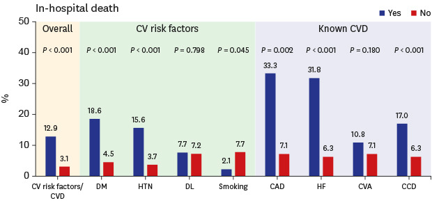
The prevalence rates of preexisting CVRFs or CVDs increased with age (P < 0.001) (Table 5). Moreover, the number of patients requiring intensive care (P < 0.001) and invasive MV (P < 0.001) increased with age. In-hospital death was significantly higher in the elderly (P < 0.001). During hospitalization, the need for intensive care (5.3% vs. 1.6%; P < 0.001) and invasive MV (4.3% vs. 1.7%; P < 0.001) was significantly greater in patients with preexisting CVRFs or CVDs than those without. Moreover, in-hospital mortality (12.9% vs. 3.1%; P < 0.001) was significantly higher in patients with preexisting CVRFs or CVDs. Among the CVRFs, diabetes mellitus (P < 0.001) and hypertension (P < 0.001) were associated with increased requirement of intensive care and invasive MV and in-hospital death (Fig. 1). Among the CVDs, coronary artery disease (22.2% vs. 2.7%; P < 0.001) was associated with invasive MV. Coronary artery disease (33.3% vs. 7.1%; P = 0.002) and congestive heart failure (31.8% vs. 6.3%; P < 0.001) were associated with in-hospital death. Based on the multivariate analysis, preexisting CVRFs or CVDs (OR, 1.79; 95% CI, 1.07–3.01; P = 0.027) were independent predictors of in-hospital death after adjusting for confounding variables. Among the individual CVRF or CVD components, diabetes mellitus (OR, 2.43; 95% CI, 1.51–3.90; P < 0.001) and congestive heart failure (OR, 2.43; 95% CI, 1.06–5.87; P = 0.049) were independent predictors of in-hospital death (Table 6). Moreover, advanced age, male gender, respiratory rate > 20/min, fever ≥ 37.5°C, altered consciousness, hemoptysis, and chronic neurologic disorders were independent predictors of in-hospital death.
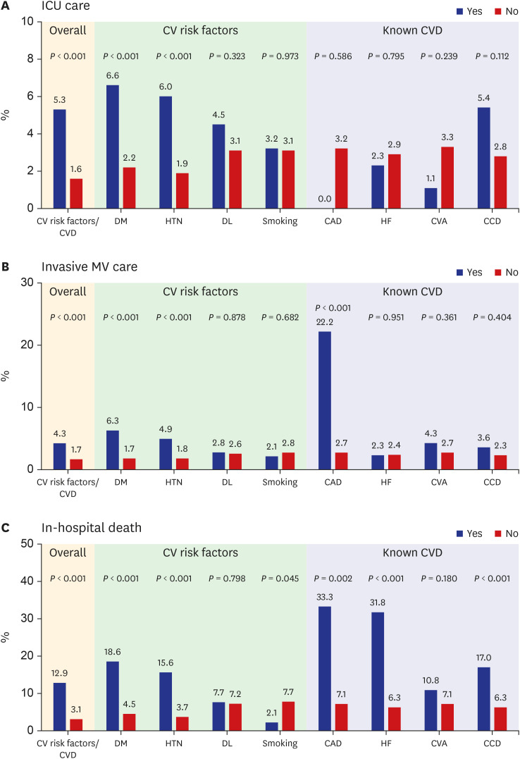
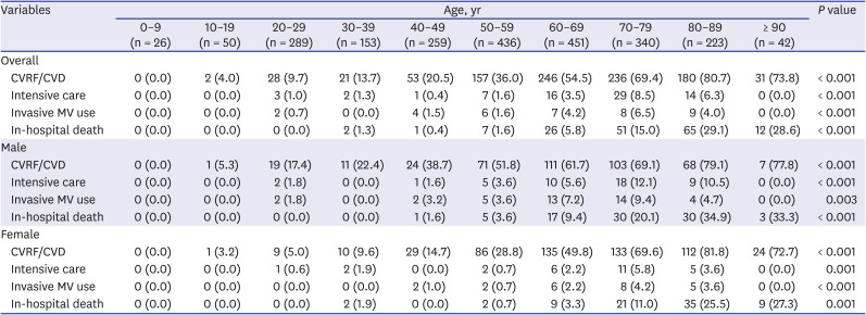
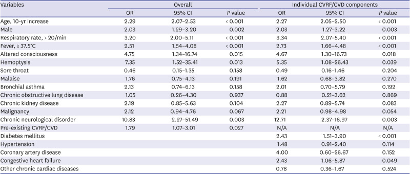
Table 5

Clinical measures and outcomes by 10-year intervals of patients hospitalized with coronavirus disease 2019

jkms-36-e15-i005
Variables Age, yr P value
0–9 (n = 26) 10–19 (n = 50) 20–29 (n = 289) 30–39 (n = 153) 40–49 (n = 259) 50–59 (n = 436) 60–69 (n = 451) 70–79 (n = 340) 80–89 (n = 223) ≥ 90 (n = 42)
Overall
CVRF/CVD 0 (0.0) 2 (4.0) 28 (9.7) 21 (13.7) 53 (20.5) 157 (36.0) 246 (54.5) 236 (69.4) 180 (80.7) 31 (73.8) < 0.001
Intensive care 0 (0.0) 0 (0.0) 3 (1.0) 2 (1.3) 1 (0.4) 7 (1.6) 16 (3.5) 29 (8.5) 14 (6.3) 0 (0.0) < 0.001
Invasive MV use 0 (0.0) 0 (0.0) 2 (0.7) 0 (0.0) 4 (1.5) 6 (1.6) 7 (4.2) 8 (6.5) 9 (4.0) 0 (0.0) < 0.001
In-hospital death 0 (0.0) 0 (0.0) 0 (0.0) 2 (1.3) 1 (0.4) 7 (1.6) 26 (5.8) 51 (15.0) 65 (29.1) 12 (28.6) < 0.001
Male
CVRF/CVD 0 (0.0) 1 (5.3) 19 (17.4) 11 (22.4) 24 (38.7) 71 (51.8) 111 (61.7) 103 (69.1) 68 (79.1) 7 (77.8) < 0.001
Intensive care 0 (0.0) 0 (0.0) 2 (1.8) 0 (0.0) 1 (1.6) 5 (3.6) 10 (5.6) 18 (12.1) 9 (10.5) 0 (0.0) < 0.001
Invasive MV use 0 (0.0) 0 (0.0) 2 (1.8) 0 (0.0) 2 (3.2) 5 (3.6) 13 (7.2) 14 (9.4) 4 (4.7) 0 (0.0) 0.003
In-hospital death 0 (0.0) 0 (0.0) 0 (0.0) 0 (0.0) 1 (1.6) 5 (3.6) 17 (9.4) 30 (20.1) 30 (34.9) 3 (33.3) < 0.001
Female
CVRF/CVD 0 (0.0) 1 (3.2) 9 (5.0) 10 (9.6) 29 (14.7) 86 (28.8) 135 (49.8) 133 (69.6) 112 (81.8) 24 (72.7) < 0.001
Intensive care 0 (0.0) 0 (0.0) 1 (0.6) 2 (1.9) 0 (0.0) 2 (0.7) 6 (2.2) 11 (5.8) 5 (3.6) 0 (0.0) 0.001
Invasive MV use 0 (0.0) 0 (0.0) 0 (0.0) 0 (0.0) 2 (1.0) 2 (0.7) 6 (2.2) 8 (4.2) 5 (3.6) 0 (0.0) < 0.001
In-hospital death 0 (0.0) 0 (0.0) 0 (0.0) 2 (1.9) 0 (0.0) 2 (0.7) 9 (3.3) 21 (11.0) 35 (25.5) 9 (27.3) 0.001
Data are presented as number (%).
CVRF = cardiovascular risk factor, CVD = cardiovascular disease, MV = mechanical ventilator.
Fig. 1

Frequency according to the presence (“yes”) or absence (“no”) of preexisting cardiovascular risk factors or cardiovascular disease. Frequency of intensive care unit utilization (A), invasive mechanical ventilator utilization (B), and in-hospital death (C) according to the presence (“yes”) or absence (“no”) of preexisting cardiovascular risk factors or cardiovascular disease.

ICU = intensive care unit, CV = cardiovascular, CVD = cardiovascular disease, DM = diabetes mellitus, HTN = hypertension, DL = dyslipidemia, CAD = coronary artery disease, HF = heart failure, CVA = cerebrovascular accidents, CCD = chronic cardiac disease, MV = mechanical ventilator.
jkms-36-e15-g001
Table 6

Multivariate analysis for in-hospital death

jkms-36-e15-i006
Variables Overall Individual CVRF/CVD components
OR 95% CI P value OR 95% CI P value
Age, 10-yr increase 2.29 2.07–2.53 < 0.001 2.27 2.05–2.50 < 0.001
Male 2.03 1.29–3.20 0.002 2.03 1.27–3.22 0.003
Respiratory rate, > 20/min 3.20 2.00–5.11 < 0.001 3.34 2.07–5.40 < 0.001
Fever, ≥ 37.5°C 2.51 1.54–4.08 < 0.001 2.73 1.66–4.48 < 0.001
Altered consciousness 4.75 1.34–16.74 0.015 4.67 1.30–16.73 0.018
Hemoptysis 7.35 1.52–35.41 0.013 5.35 1.08–26.43 0.039
Sore throat 0.46 0.15–1.35 0.158 0.49 0.16–1.46 0.204
Malaise 1.76 0.75–4.13 0.191 1.62 0.68–3.82 0.270
Bronchial asthma 2.13 0.74–6.13 0.158 2.01 0.70–5.79 0.192
Chronic obstructive lung disease 1.05 0.26–4.30 0.937 0.88 0.21–3.62 0.869
Chronic kidney disease 2.19 0.85–5.63 0.104 2.27 0.89–5.74 0.083
Malignancy 2.12 0.94–4.76 0.067 2.21 0.98–4.98 0.054
Chronic neurological disorder 10.83 2.27–51.49 0.003 12.71 2.37–16.97 0.003
Pre-existing CVRF/CVD 1.79 1.07–3.01 0.027 N/A N/A N/A
Diabetes mellitus 2.43 1.51–3.90 < 0.001
Hypertension 1.48 0.91–2.40 0.114
Coronary artery disease 4.00 0.60–26.67 0.152
Congestive heart failure 2.43 1.06–5.87 0.049
Other chronic cardiac diseases 0.78 0.36–1.67 0.524
CVRF = cardiovascular risk factor, CVD = cardiovascular disease, OR = odds ratio, CI = confidence interval.

DISCUSSION

The principal findings of this large observational study are as follows. First, it is uncertain whether diabetes mellitus and hypertension increase the risk of COVID-19 infection. Second, patients with COVID-19 with preexisting CVRFs or CVDs were more likely to have severe disease progression. Third, preexisting CVRFs or CVDs increased the mortality of patients with COVID-19.
Furthermore, three important clinical questions regarding the association between preexisting CVRFs or CVDs and COVID-19 infection should be considered. Our study provided important findings to address these important questions through a comprehensive analysis.
The first clinical question is whether diabetes mellitus and hypertension increase the risk of COVID-19 infection. SARS-CoV-2 binds to the angiotensin-converting enzyme 2 (ACE2) receptors in the lungs which are also associated with heart function, high blood pressure, and diabetes mellitus.13 In animal studies, the expression of ACE2 is markedly increased in patients with diabetes mellitus, hypertension, and failing heart as an adaptive response to counteract the elevated level of angiotensin II.14 Therefore, theoretically, diabetes mellitus and hypertension could increase the risk of COVID-19 infection. In studies from China that enrolled a small sample of patients with COVID-19 (200 or less), the prevalence rates of hypertension, diabetes, and CVD were comparable to those of the general population in 2018.23456 However, the prevalence rate of CVRFs in patients with COVID-19 was less than that in the general population based on the findings of studies with a large sample of patients with COVID-19 (more than 1,000).789 However, these results are inconsistent with those in western countries. Based on the data from New York City, patients with COVID-19 had higher prevalence rates of hypertension (56.6% vs. 45%) and diabetes (33.8% vs. 10.5%) than the general population.15 In the present study, the prevalence rates of hypertension and diabetes mellitus in patients with COVID-19 were comparable to those in the general population in the KNHANES 2018. However, the prevalence rate of diabetes mellitus in patients with COVID-19 showed numerically higher trend compared with those in the general population of Daegu Metropolitan City in the CHS 2019. Therefore, it is still uncertain whether diabetes mellitus and hypertension increase the risk of COVID-19 infection.
The second clinical question is whether patients with COVID-19 with preexisting CVRFs or CVDs are more likely to have severe disease. SARS-CoV-2 infection is a mild disease in most patients, but in some patients, it progresses to a serious respiratory disease causing hyper-inflammation, multi-organ failure, and death.16 Although a study from China reported that in patients with COVID-19, the requirement of invasive MV was greater in those with preexisting CVRFs or CVDs, this result was inconsistent with another study from China.23 However, in the latest study, patients with COVID-19 with preexisting CVRFs or CVDs were more likely to progress to severe disease.7 In our study, in patients with COVID-19, the requirement for intensive care and invasive MV was significantly greater in those with preexisting CVRFs or CVDs. In particular, preexisting coronary artery disease was associated with the use of invasive MV. Although the effect of preexisting coronary artery disease on the clinical course of COVID-19 has not been fully investigated, it has been known that severe respiratory failure and multi-organ failure might be directly or indirectly related with coronary artery disease.17 COVID-19 is an infectious disease. Therefore, severe inflammatory response is associated with disease progression and poor prognosis. In this study, surrogate markers of severe inflammatory reaction such as WBC count, CRP, hs-CRP, and pro-calcitonin were greater in patients with preexisting CVRFs or CVDs at baseline and during the follow-up. Moreover, these surrogate markers were statistically significantly higher in patients requiring intensive care and invasive MV and in deceased patients. Although multiple possible explanations regarding the clear association between preexisting CVRFs or CVDs and COVID-19 severity exist, the greater inflammatory response in patients with preexisting CVRF or CVD is an important reason why patients with preexisting CVRFs or CVDs are more likely to have severe COVID-19.
The third clinical question is whether preexisting CVRFs or CVDs increase the mortality of patients with COVID-19. Studies from China reported that CVRFs or known CVDs are associated with increased in-hospital mortality.456 However, a study from a Western country reported that diabetes mellitus was associated with in-hospital death in patients with COVID-19 requiring intensive care and invasive MV, but hypertension was not.15 It can be assumed that patients with preexisting CVRFs or CVDs are more likely to be older than those without. Advanced age has consistently been shown to be associated with poor prognosis in patients with COVID-19. However, most of the aforementioned studies did not adjust for age. Moreover, some studies showed that female were more resistant to viral infections than male, which is consistent with the results of this study. In animal models, male mice showed a higher susceptibility to SARS-CoV-1 infection.18 Moreover, initial vital signs and presenting characteristics reflected the severity of COVID-19 infection. Furthermore, patients with multiple comorbidities were more likely to have severe disease and subsequent mortality. Nonetheless, a comprehensive analysis considering age, sex, vital signs at admission, presenting characteristics, and comorbidities has not yet been conducted. The present study clearly demonstrated that preexisting CVRFs or CVDs were an independent predictor of in-hospital mortality after adjusting for the confounding variables. In particular, diabetes mellitus, among the CVRFs, and congestive heart failure, among the CVDs, were independent predictors of in-hospital morality after adjusting for all variables.
However, this study has several limitations to consider. First, since the Daegu COVID-19 Research Project was an observational study, we cannot exclude the possibility of having residual confounding factors. Therefore, our results should only be regarded as hypothesis generating. Second, the study population only included patients with COVID-19 hospitalized in a Korean healthcare system in Daegu Metropolitan City. Third, history taking, and laboratory findings were not available in some patients. Moreover, some of the patients had missing laboratory data. Fourth, since this analysis was performed based on chart review without external prospective ascertainment, the results need to be interpreted with caution. Fifth, although the use of invasive MV might be related with the severity of pneumonia, the status of pneumonia was not obtained in our registry. Sixth, the frequency of ICU utilization in patients with coronary artery disease or cerebrovascular accidents was too small to explain the differences between patients with coronary artery disease or cerebrovascular accidents and those without. Further studies are required to clarify these associations. However, the limitations should not undermine the strength of this study including the overall consecutive patients with COVID-19 encountered in day-to-day clinical practice during the COVID-19 pandemic.
In conclusion, it remains uncertain whether patients with diabetes mellitus and hypertension are more causally vulnerable to COVID-19 infection. However, the patients with preexisting CVRFs or CVDs had worse clinical COVID-19 outcomes, mainly driven by a severe inflammatory reaction. Therefore, we should emphasize that appropriate risk stratification at triage for patients with preexisting CVRFs or CVDs is necessary for the patients' survival especially during this COVID-19 pandemic.

Notes

Funding: This work was supported by the Research Program of Medicity Daegu Council funded by Daegu Metropolitan City (fund code COVID19_DM03).

Disclosure: The authors have no potential conflicts of interest to disclose.

Author Contributions:

  • Conceptualization: Park BE, Lee JH, Park HS, Nam CW.

  • Data curation: Lee JH, Lee BY, Nam CW.

  • Formal analysis: Park BE, Lee BY, Lee JB.

  • Funding acquisition: Park BE.

  • Investigation: Kim HN, Kim U.

  • Methodology: Park HK, Jang SY.

  • Project administration: Park HK, Jang SY, Kim U.

  • Resources: Lee JH, Bae MH, Lee JB.

  • Software: Park HK, Bae MH.

  • Supervision: Yang DH, Chae SC.

  • Validation: Park BE, Yang DH, Park HS, Cho Y, Chae SC.

  • Visualization: Park BE.

  • Writing - original draft: Park BE, Lee JH.

  • Writing - review & editing: Park BE, Lee JH.

References

1. World Health Organization. WHO Director- General's opening remarks at the media briefing on COVID-19 - 11 March 2020. Updated 2020. Accessed March 12, 2020. https://www.who.int/director-general/speeches/detail/who-director-general-s-opening-remarks-at-the-media-briefing-on-covid-19---11-march-2020.
2. Huang C, Wang Y, Li X, Ren L, Zhao J, Hu Y, et al. Clinical features of patients infected with 2019 novel coronavirus in Wuhan, China. Lancet. 2020; 395(10223):497–506. PMID: 31986264.
crossref
3. Wang D, Hu B, Hu C, Zhu F, Liu X, Zhang J, et al. Clinical characteristics of 138 hospitalized patients with 2019 novel coronavirus-infected pneumonia in Wuhan, China. JAMA. 2020; 323(11):1061–1069. PMID: 32031570.
crossref
4. Ruan Q, Yang K, Wang W, Jiang L, Song J. Clinical predictors of mortality due to COVID-19 based on an analysis of data of 150 patients from Wuhan, China. Intensive Care Med. 2020; 46(5):846–848. PMID: 32125452.
crossref
5. Zhou F, Yu T, Du R, Fan G, Liu Y, Liu Z, et al. Clinical course and risk factors for mortality of adult inpatients with COVID-19 in Wuhan, China: a retrospective cohort study. Lancet. 2020; 395(10229):1054–1062. PMID: 32171076.
crossref
6. Wu C, Chen X, Cai Y, Xia J, Zhou X, Xu S, et al. Risk factors associated with acute respiratory distress syndrome and death in patients with coronavirus disease 2019 pneumonia in Wuhan, China. JAMA Intern Med. 2020; 180(7):934–943. PMID: 32167524.
crossref
7. Guan WJ, Ni ZY, Hu Y, Liang WH, Ou CQ, He JX, et al. Clinical characteristics of coronavirus disease 2019 in China. N Engl J Med. 2020; 382(18):1708–1720. PMID: 32109013.
8. Fang Z, Yi F, Wu K, Kang Wu, Lai K, Sun X, et al. Clinical characteristics of coronavirus pneumonia 2019 (COVID-19): an updated systematic review. MedRxiv;Updated 2020. Accessed August 7, 2020. https://www.medrxiv.org/content/10.1101/2020.03.07.20032573v2.
9. Wu Z, McGoogan JM. Characteristics of and important lessons from the coronavirus disease 2019 (COVID-19) outbreak in China: summary of a report of 72 314 cases from the Chinese Center for Disease Control and Prevention. JAMA. 2020; 323(13):1239–1242. PMID: 32091533.
10. Li B, Yang J, Zhao F, Zhi L, Wang X, Liu L, et al. Prevalence and impact of cardiovascular metabolic diseases on COVID-19 in China. Clin Res Cardiol. 2020; 109(5):531–538. PMID: 32161990.
crossref
11. Xiong TY, Redwood S, Prendergast B, Chen M. Coronaviruses and the cardiovascular system: acute and long-term implications. Eur Heart J. 2020; 41(19):1798–1800. PMID: 32186331.
crossref
12. Zheng YY, Ma YT, Zhang JY, Xie X. COVID-19 and the cardiovascular system. Nat Rev Cardiol. 2020; 17(5):259–260. PMID: 32139904.
crossref
13. Walls AC, Park YJ, Tortorici MA, Wall A, McGuire AT, Veesler D. Structure, function, and antigenicity of the SARS-CoV-2 spike glycoprotein. Cell. 2020; 181(2):281–292.e6. PMID: 32155444.
crossref
14. Turner AJ, Hiscox JA, Hooper NM. ACE2: from vasopeptidase to SARS virus receptor. Trends Pharmacol Sci. 2004; 25(6):291–294. PMID: 15165741.
crossref
15. Richardson S, Hirsch JS, Narasimhan M, Crawford JM, McGinn T, Davidson KW, et al. Presenting characteristics, comorbidities, and outcomes among 5700 patients hospitalized with COVID-19 in the New York City area. JAMA. 2020; 323(20):2052–2059. PMID: 32320003.
crossref
16. Siddiqi HK, Mehra MR. COVID-19 illness in native and immunosuppressed states: A clinical-therapeutic staging proposal. J Heart Lung Transplant. 2020; 39(5):405–407. PMID: 32362390.
crossref
17. Libby P, Loscalzo J, Ridker PM, Farkouh ME, Hsue PY, Fuster V, et al. Inflammation, immunity, and infection in atherothrombosis: JACC review topic of the week. J Am Coll Cardiol. 2018; 72(17):2071–2081. PMID: 30336831.
18. Channappanavar R, Fett C, Mack M, Ten Eyck PP, Meyerholz DK, Perlman S. Sex-based differences in susceptibility to severe acute respiratory syndrome coronavirus infection. J Immunol. 2017; 198(10):4046–4053. PMID: 28373583.
crossref

SUPPLEMENTARY MATERIALS

Supplementary Fig. 1

Prevalence rates of diabetes mellitus and hypertension in patients with COVID-19 and the general population. Prevalence rates of diabetes mellitus (A) and hypertension (B) in patients with COVID-19 and the general population in Korea National Health And Nutrition Examination Survey 2018. Prevalence rates of diabetes mellitus (C) and hypertension (D) in patients with COVID-19 and the general population of Daegu Metropolitan City from Community Health Survey 2019.
jkms-36-e15-s001.doc

Supplementary Fig. 2

Comparison of laboratory findings between patients admitted to intensive care units and those not admitted and between patients on invasive mechanical ventilation and those who are not at baseline and during the follow-up. Comparison of white blood cell count, C-reactive protein, high-sensitivity C-reactive protein, and pro-calcitonin between patients admitted to intensive care units and those not admitted (A). Comparison of white blood cell count, C-reactive protein, high-sensitivity C-reactive protein, and pro-calcitonin between patients on invasive mechanical ventilation and those who are not at baseline and during the follow-up (B).
jkms-36-e15-s002.doc
TOOLS
Similar articles