

 PDF
PDF Citation
 Print
Print


INTRODUCTION
Treatment of malignant brain tumors in very young children is challenging because radiotherapy (RT), and particularly craniospinal radiotherapy (CSRT), should be used minimally due to the risk of functional impairment of the developing brain and late adverse effects, which include global reduction in intelligence quotient, cognitive deficits, and neuroendocrine dysfunction.1 In an effort to minimize the adverse effects of RT, most institutions and national groups have adopted chemotherapy-based strategies to avoid or delay RT; however, the outcomes of these approaches have been largely unsatisfactory.234
A strategy using high-dose chemotherapy and autologous stem cell transplantation (HDCT/auto-SCT) to improve the prognosis of patients with high-risk solid tumors has been explored.567 Following success in high-risk neuroblastoma, studies have investigated the effectiveness of HDCT/auto-SCT in improving the prognosis of high-risk brain tumors and/or to avoid or minimize RT.89101112 Recently, it has been demonstrated that further dose intensification by tandem HDCT/auto-SCT may further improve outcomes in patients with high-risk brain tumors.1314
Previously, we reported the early results of a single-arm pilot trial using tandem HDCT/auto-SCT to minimize RT dose and volume in very young children with malignant brain tumors.1 The results suggested that it was possible to avoid or defer RT until 3 years of age while improving survival rates. However, the results were not confirmative because the number of patients was small, and the short follow-up prevented determination of long-term effects. Therefore, the study has been extended. Here, we report the results of the study with a larger cohort of patients and a longer follow-up duration. In the present study, we focused on very young children with non-metastatic malignant brain tumors.
METHODS
Patients
Children younger than 3 years who were diagnosed with non-metastatic malignant brain tumors between September 2004 and November 2017 were eligible for the study. A pediatric neuropathologist reviewed all cases according to the WHO criteria. Past diagnosis based on the 2007 WHO classification was revised using the 2016 WHO classification if a tumor sample was available. Immunohistochemistry and/or next-generation sequencing using a targeted panel (PedSCAN™) were performed. For molecular subgrouping of medulloblastoma, nanoString-based RNA assays were also performed.15 The extent of disease at the time of diagnosis was assessed using brain and spinal magnetic resonance imaging (MRI) and cerebrospinal fluid (CSF) cytology.
Induction chemotherapy
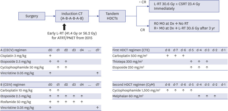
Fig. 1 shows the treatment scheme used in this study. Surgery was the primary treatment for all resectable tumors. Second-look surgery was performed whenever possible. Prior to HDCT/auto-SCT, six cycles of induction chemotherapy were administered consisting of alternating CECV (cisplatin + etoposide + cyclophosphamide + vincristine) and CEIV (carboplatin + etoposide + ifosfamide + vincristine) regimens. Induction chemotherapy cycles were scheduled 4 weeks apart, but some delays were permitted to allow recovery of absolute neutrophil count (ANC) and platelet count to 1,000/µL and 100,000/µL, respectively. Peripheral blood stem cells (PBSCs) were collected during the recovery phase of the first chemotherapy cycle. For patients with atypical teratoid/rhabdoid tumor (ATRT) or primitive neuroectodermal tumor (PNET), intrathecal methotrexate (6 mg for age < 1 year, 8 mg for age 1–2 years, 10 mg for age 2–3 years) was administered on day 0 of each induction chemotherapy starting in September 2015.
Tandem HDCT/auto-SCT
CTE (carboplatin + thiotepa + etoposide) and CyM (cyclophosphamide + melphalan) regimens were used for the first and second HDCT/auto-SCT, respectively (Fig. 1). We allowed an approximately 12-week interval between the first and second HDCT/auto-SCT to minimize toxicity. Approximately half of the PBSCs collected during a single round of leukapheresis was infused for bone marrow rescue during each HDCT/auto-SCT session.
RT
RT was administered with 3-dimensional photon beams until December 2015, after which patients were treated with proton beams. Proton beam doses were same as photon therapy with a relative biologic effectiveness of 1.1. Local RT (L-RT, 30.6 Gy) with 1–2 cm margins from surgical defects was administered after the patient reached 3 years of age only if gross residual tumor (> 1.5 cm2) remained after initial or second-look surgery. Otherwise, no RT was given if the patient remained progression-free. However, immediate RT was administered if the patient experienced relapse or progression. For patients who experienced metastatic relapse, both CSRT (23.4 Gy) and L-RT (30.6 Gy) were administered. The daily fraction size was 1.8 Gy. Because the outcome of patients with ATRT or PNET was disappointing due to early progression in the early phase of the study,1 early post-operative L-RT was administered starting in September 2015; patients without residual tumors received 41.4 Gy, and patients with residual tumors received 56.2 Gy. During early L-RT, the dose of concomitant chemotherapy was reduced to 75% of the full dose.
Response and toxicity criteria
Treatment response was evaluated by brain and spinal MRI and CSF cytology after every 2 cycles of induction chemotherapy, between the first and second HDCT/auto-SCT, every 3 months for the first year after the second HDCT/auto-SCT, every 4 months for the second year, every 6 months for the third year, and every 12 months thereafter. Tumor size was estimated by MRI as the product of the greatest diameter and the longest perpendicular diameter. Treatment responses were categorized as follows: complete response (CR), complete disappearance of all previously measurable tumor; partial response (PR), greater than 50% decrease in tumor size; minor response (MR), 25% to 50% decrease in tumor size; stable disease (SD), less than 25% change in tumor size; and progressive disease (PD), greater than 25% increase in tumor size or appearance of a new area of tumor. Toxicities were graded using the National Cancer Institute's Common Terminology Criteria (version 4.0).
Evaluation of late adverse effects
Late adverse effects were evaluated yearly after completion of the second HDCT/auto-SCT. Neuroendocrine, cardiac, pulmonary, renal, auditory, ophthalmologic, and cognitive functions were evaluated. Thyroid hormones (T3 and free T4), thyroid stimulating hormone, morning cortisol and ACTH, insulin like growth factor-1, bone age, gonadotropin (FSH, LH), sex hormones (testosterone or estradiol), blood lipid profile, hemoglobin A1c, insulin and C-peptide were checked to screen for neuroendocrine dysfunction. Cardiac evaluation was performed by two-dimensional echocardiography and electrocardiography. The respiratory system was evaluated using pulmonary function test and computed tomography scan. Renal function was evaluated using the serum and urine biomarkers. Auditory evaluation including pure tone and speech audiometry was performed by the otolaryngologist. An ophthalmic examination by an ophthalmologist was also performed. Cognitive function was evaluated using the Korean-Wechsler Preschool and Primary Scale of Intelligence and the Korean-Wechsler Intelligence Scale for Children-IV.
Statistics
Event-free survival (EFS) was calculated from the date of diagnosis until the date of relapse, progression, secondary malignancy or death from any cause, whichever occurred first. Overall survival (OS) was calculated from the date of diagnosis until death from any cause. CSRT-free survival was calculated from the date of diagnosis until initiation of CSRT or death from any cause. Survival rates and standard errors were estimated using the Kaplan–Meier method. Differences in survival rates between the two groups were compared using the log-rank test. P values less than 0.05 were considered significant.
RESULTS
Patient characteristics
Patient characteristics are listed in Table 1. During the study period, 20 consecutive patients (15 boys and 5 girls) with various malignant brain tumors (anaplastic ependymoma in four, medulloblastoma in four, choroid plexus carcinoma in three, high-grade glioma in two, PNET in four, immature teratoma in one, malignant fibrous histiocytoma in one, and ATRT in one) were enrolled. The median age at diagnosis was 1.5 (range, 0.2−3.0) years, and 17 patients were younger than 2 years at diagnosis. The tumor was located at the posterior fossa in six patients and supratentorially in 14 patients. Gross residual tumor (> 1.5 cm2) remained after surgery in eight patients. After revision of the diagnoses according to the WHO 2016 classification, the diagnosis of PNET was changed to anaplastic ependymoma in two patients, embryonal tumor with multilayered rosette C19MC-altered in one patient, and pineoblastoma in one patient. One anaplastic glioneuronal tumor was also reclassified as embryonal tumor, NOS. Among four medulloblastoma cases, we could identify a molecular subgroup in three: all were SHH-activated and TP53-wild type medulloblastoma.
Table 1
Patient characteristics
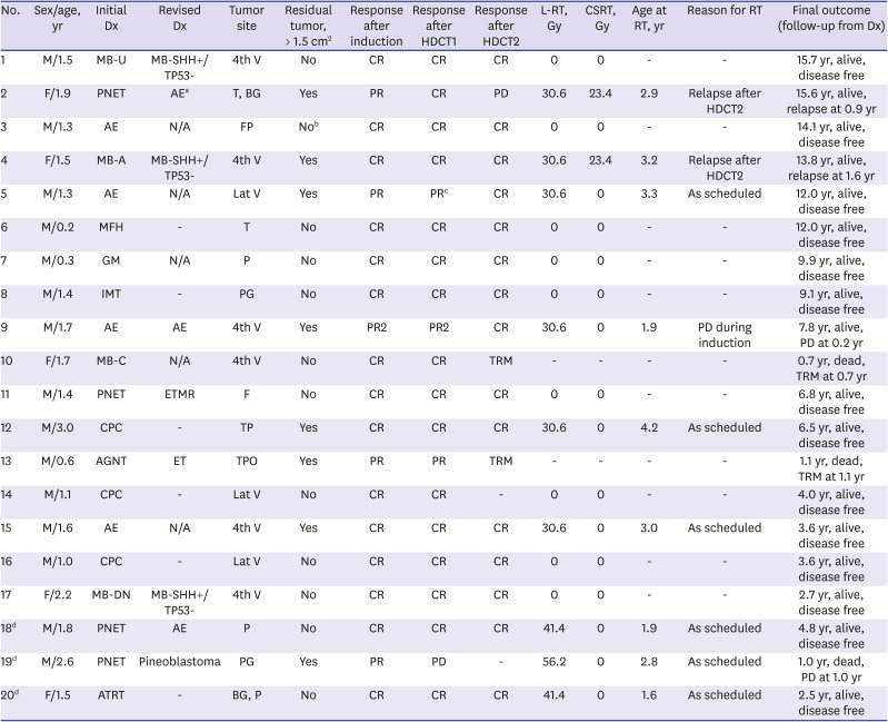
Dx = diagnosis, HDCT1 = first high-dose chemotherapy, HDCT2 = second HDCT, L-RT = local radiotherapy, CSRT = craniospinal RT, N/A = not available, MB-U = medulloblastoma of unknown pathology, MB-SHH+/TP53- = medulloblastoma, SHH-activated and TP53-wildtype, PNET = primitive neuroectodermal tumor, AE = anaplastic ependymoma, MB-A = anaplastic medulloblastoma, MFH = malignant fibrous histiocytoma, GM = glioblastoma multiforme, IMT = immature teratoma, MB-C = classic medulloblastoma, CPC = Choroid plexus carcinoma, AGNT = anaplastic glioneuronal tumor, MB-DN = desmoplastic/nodular medulloblastoma, ATRT = atypical teratoid/rhabdoid tumor, ETMR = embryonal tumor with multilayered rosette, C19MC-altered, ET = embryonal tumor, NOS, 4th V = 4th ventricle, T = temporal lobe, BG = basal ganglia, FP = fronto-parietal lobe, Lat V = lateral ventricle, P = parietal lobe, PG = pineal gland, F = frontal lobe, TP = temporo-parietal lobe, TPO = temporo-parieto-occipital lobe, CR = complete response, PR = partial response, PR2 = second PR, PD = progressive disease, TRM = treatment-related mortality, VOD = hepatic veno-occlusive disease.
aL1CAM-positive tumor, however, we could not check RELA fusion because of the insufficient tumor sample; bNo residual tumor remained after second-look surgery; cNear total resection was possible after first HDCT/auto-SCT; dBecause these patients were diagnosed with PNET or ATRT after September 2015, early local RT before 3 years old were administered.
Induction treatment
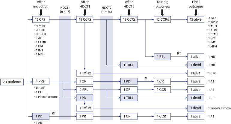
Fig. 2 shows the flow of patients. All patients experienced neutropenic fever during induction chemotherapy; however, non-hematologic toxicity was not common. In one patient (No. 3), gross total resection was possible during second-look surgery after six cycles of induction chemotherapy. Another patient (No. 9) experienced local progression at the primary tumor site and received L-RT with concomitant chemotherapy. Otherwise, all patients remained progression-free during induction chemotherapy. Tumor status at the end of induction treatment was CR in 15 patients, PR in four, and second PR in one. The median number of CD34+ cells collected during a median of 3 (range, 2−10) leukapheresis events was 68.6 (range, 3.8−223.0) × 106 cells/kg.
Fig. 2
Flow of patients.
HDCT1= first high-dose chemotherapy, HDCT2 = second HDCT, CR = complete response, CCR = continuous CR, PR = partial response, PD = progressive disease, REL = relapse, TRM = treatment-related mortality, off-Tx = off treatment, RT = radiotherapy, MB = medulloblastoma, AE = anaplastic ependymoma, CPC = Choroid plexus carcinoma, ATRT = atypical teratoid/rhabdoid tumor, ETMR = embryonal tumor with multilayered rosette, C19MC-altered, GM = glioblastoma multiforme, IMT = immature teratoma, MFH = malignant fibrous histiocytoma, ET = embryonal tumor, NOS.
Tandem HDCT/auto-SCT
All 20 patients, including a patient (No. 9) who experienced progression during induction chemotherapy, underwent the first HDCT/auto-SCT. The median age at the time of the first HDCT/auto-SCT was 2.1 (range, 0.8−3.6) years. In one patient (No. 5) who was still in PR after the first HDCT/auto-SCT, near total resection of the tumor was performed prior to the second HDCT/auto-SCT. In another patient (No. 14), who was in CR after the first HDCT/auto-SCT, parents refused the second HDCT/auto-SCT. In another patient (No.19), who experienced progression after the first HDCT/auto-SCT, parents refused further treatment. Consequently, a total of 18 patients proceeded to the second HDCT/auto-SCT. The median interval between the first auto-SCT and initiation of the second HDCT was 83 (range, 75−122) days. Table 2 shows the toxicities observed during tandem HDCT/auto-SCT. Hematologic recovery was rapid in both the first and second HDCT/auto-SCT. Frequent grade 3/4 toxicity included fever, stomatitis, diarrhea, elevation of liver enzymes, and hypokalemia. No treatment-related mortality (TRM) occurred during the first HDCT/auto-SCT; however, two patients died from toxicities (asphyxia and hepatic veno-occlusive disease) during the second HDCT/auto-SCT.
Table 2
Grade 3/4 toxicities during tandem HDCT/auto-SCT
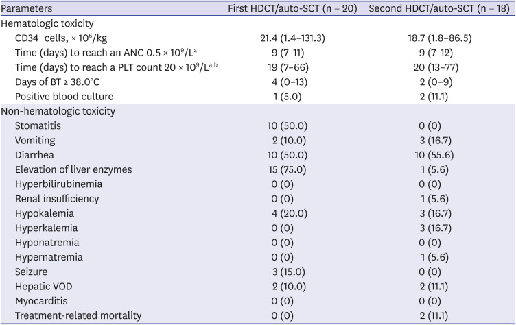
Data are presented as median (range) or number (%).
ANC = absolute neutrophil count, PLT = platelet, BT = body temperature, VOD = hepatic veno-occlusive disease.
aThe first day when ANC exceeded 0.5 × 10⁹/L for 3 consecutive days; bThe first day when PLT count exceeded 20 × 109/L without transfusion for 7 days.
RT
Of 12 patients without gross residual tumor after surgery, all but one (No. 10, who died from asphyxia during the second HDCT/auto-SCT) remained event-free and did not receive RT except two patients who received early L-RT. Of eight patients with gross residual tumor, three remained event-free and received L-RT alone as scheduled. One patient died from hepatic veno-occlusive disease during the second HDCT/auto-SCT. The remaining four patients experienced relapse/progression. A patient (No. 9) with anaplastic ependymoma who experienced local progression during induction treatment received L-RT (30.6 Gy) after progression. Two patients with anaplastic ependymoma (No. 2) and medulloblastoma (No. 4) who experienced metastatic relapse after tandem HDCT/auto-SCT received both CSRT (23.4 Gy) and L-RT (30.6 Gy). The remaining patient (No. 19) with pineoblastoma received early L-RT during induction chemotherapy; however, metastatic relapse occurred after the first HDCT/auto-SCT, and the patient died without further treatment. Proton beam was used in three patients. Collectively, among 17 survivors, nine received no RT, six received L-RT alone, and two received both CSRT and L-RT. The median age at initiation of RT was 2.9 (range, 1.6–4.2) years, and five patients received RT at younger than 3 years.
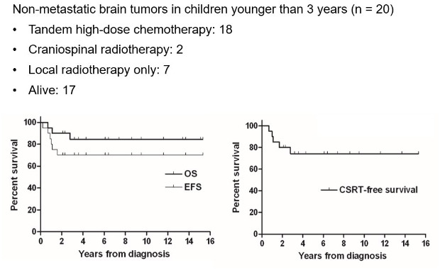
Survival
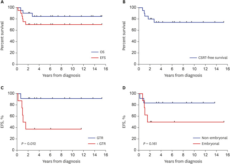
Of four patients who experienced relapse/progression, three remain alive after salvage treatment including RT. The remaining patient (No. 19) died without salvage treatment. Two patients died from toxicities during the second HDCT/auto-SCT. Therefore, a total of 17 patients remain alive at a median follow-up of 7.8 (range, 2.5−15.7) years from diagnosis. The 5-year OS and EFS rates were 85.0% ± 8.0% and 70.0% ± 10.2%, respectively (Fig. 3A). The 5-year CSRT-free survival rate was 75.0% ± 9.7% (Fig. 3B). The EFS was lower in patients with gross residual tumor than in those without (37.5% ± 17.1% versus 91.7% ± 8.0%, P = 0.012) (Fig. 3C). There was no difference in EFS between embryonal and non-embryonal tumors (Fig. 3D). All six patients with anaplastic ependymoma remain alive although two of them experienced relapse/progression. In addition, there were no differences in survival rates according to other clinical factors such as primary tumor site or age at diagnosis.
Fig. 3
Survival rates. (A) The 5-year OS and EFS rates were 85.0% ± 8.0% and 70.0% ± 10.2%, respectively. (B) The 5-year CSRT-free survival was 75.0% ± 9.7%. (C) The EFS was lower in patients with gross residual tumor than in those without (37.5% ± 17.1% versus 91.7% ± 8.0%, P = 0.012). (D) There was no difference in EFS between embryonal and non-embryonal tumors.
OS = overall survival, EFS = event-free survival, CSRT = craniospinal radiotherapy, GTR = gross total resection.
Late adverse effects
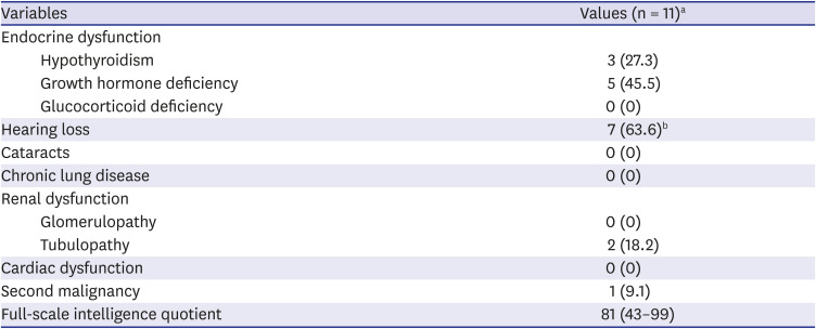
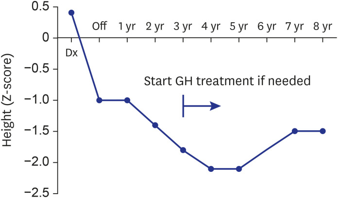
Table 3 shows late adverse effects observed 5 years after the second HDCT/auto-SCT in 11 patients, two of whom received both CSRT and L-RT. Frequent late adverse effects included hypothyroidism, growth hormone deficiency, sensory neural hearing loss, and renal tubulopathy; however, except in one patient with grade 4 hearing loss, all adverse effects were grade 1 or 2. Until 4 years after tandem HDCT/auto-SCT, continuous deceleration in vertical growth was observed (Fig. 4). The median height at 5 years after the second HDCT/auto-SCT was –2.2 (range, 0.2 to –3.2) standard deviations from mean for patient age. The median value for full-scale intelligence quotient evaluated at 5 years after the second HDCT/auto-SCT was 81 (range, 43–99).
Table 3
Late adverse effects 5 years after tandem HDCT/auto-SCT
DISCUSSION
The adverse effects of RT vary according to dose/volume and age at the time of RT, with late neurocognitive deficits being more pronounced in children treated before 3 years of age.16 Several studies using HDCT/auto-SCT to minimize RT, including Head Start I-III and CCG 99703, have reported encouraging outcomes in very young patients with malignant brain tumors.891011121314 Previously, we also reported encouraging preliminary results of a single-arm trial using tandem HDCT/auto-SCT in very young children with malignant brain tumors. However, the number of enrolled patients was small, and the short follow-up duration prevented analysis of long-term late effects. In the present study, we report the long-term follow-up results of an extended study, focusing on very young children with non-metastatic malignant brain tumors. Fifteen of 17 survivors did not receive CSRT, and nine of them did not receive RT. The results suggest that tandem HDCT/auto-SCT may improve the survival rates of patients with non-metastatic brain tumors while minimizing RT, particularly CSRT, and that late adverse effects were generally acceptable.
One of the most interesting observations in the present study is that all 12 patients without gross residual tumor remain progression-free, although one died from toxicity. In contrast, four of eight patients with gross residual tumor experienced relapse/progression (metastatic relapse in three). Tumors relapsed/progressed before scheduled RT in three of the patients, although they remain alive after salvage treatment including RT. These findings suggest that early L-RT might improve the outcome in patients with gross residual tumor. In the results of the Children's Oncology Group study P9934 by Ashley et al.,17 addition of conformal RT to the posterior fossa and tumor bed after induction chemotherapy increased EFS compared with use of chemotherapy alone, and neurodevelopmental assessments did not show a decline in cognitive or motor function. Another therapeutic option to prevent relapse/progression in very young children might be use of intrathecal or intraventricular chemotherapy. A previous report of the HIT 2000 trial demonstrated the efficacy of intraventricular methotrexate with acceptable toxicity.18 Recently, researchers have reported that administration of intrathecal topotecan is an effective strategy for treatment of various relapsed tumors.1920 During the late period of this study, early L-RT and intrathecal chemotherapy were administered to three patients with ATRT or PNET. Although one patient with residual tumor experienced metastatic relapse, two patients remain event-free. To evaluate the effectiveness of this treatment strategy, further study with a longer follow-up in a larger cohort is needed.
Tandem HDCT/auto-SCT is associated with greater toxicity and a higher TRM rate than single HDCT/auto-SCT, particularly during the second HDCT/auto-SCT.21 In the present study, two patients younger than 2 years of age died from TRM in the second HDCT/auto-SCT. This finding is consistent with the results of our previous report evaluating toxicities during tandem HDCT/auto-SCT in patients with brain tumors.21 The study suggests that dose intensity during the second HDCT/auto-SCT might be relatively higher in younger children than in older children, and dose reduction might reduce the TRM rate during the second HDCT/auto-SCT in younger children. However, dose reduction should be weighed against a probable increase in relapse rate. Therefore, further study is needed to determine the optimal dose in very young children with malignant brain tumors.
Late adverse effects were generally acceptable, and all but one of the adverse late effects observed at 5 years after the second HDCT/auto-SCT were grade 1 or 2. However, patients who did not receive CSRT also showed substantial late adverse effects. These late effects might be associated with dose-intense tandem HDCT/auto-SCT. Further dose escalation and addition of more drugs during tandem HDCT/auto-SCT might be associated with more significant late adverse effects compared with single HDCT/auto-SCT. Therefore, longer follow-up is needed to assess whether the probable survival benefits from tandem HDCT/auto-SCT will ultimately outweigh the overall late adverse effects.
Risk stratification and choice of RT in the present study were based on conventional clinical parameters (age, presence/absence of metastasis, and significant post-operative residual tumor). Tumor type might be important when determining treatment strategies including RT in very young children with malignant brain tumors. Other than early L-RT given to ATRT/PNET patients starting in September 2015, all patients in the present study received uniform treatment regardless of tumor type. In addition, recent efforts at stratifying brain tumors on the basis of their molecular/genetic features have subdivided brain tumors into various distinct subgroups characterized by disparate genetic and clinical features. For example, prognosis is quite variable with medulloblastomas according to molecular/genetic subtype.2223 Moreover, with the emergence of molecular/genetic characteristics as an important factor, molecular/genetic parameters are now used to establish brain tumor diagnoses in the revised 2016 WHO classification.24 In three of our patients, diagnosis according to the 2016 WHO classification was SHH-activated and TP53-wild type medulloblastoma. It is not clear whether our treatment strategy is appropriate for these patients. In the next phase of clinical trials using HDCT/auto-SCT, selection of patients will also be based on these molecular/genetic stratification data, potentially avoiding an intensive treatment regimen in some patients previously considered high risk.
One of the important limitations of this study is that it included patients with heterogeneous diagnoses. The role of chemotherapy is well established in embryonal tumors, however, it is controversial in other tumors such as immature teratomas and ependymomas. Due to the rarity of immature teratomas, standard treatment have not been established. In several research groups, patients with immature teratomas were enrolled in nongerminomatous germ cell tumor trials and treated with protocols including chemotherapy.25 While there have been some reports that chemotherapy may benefit patients with immature teratomas, several reports have shown that chemotherapy does not significantly affect survival.2526272829 Therefore, the efficacy of tandem HDCT/auto-SCT in patients with immature teratomas could be highly controversial. Despite controversies about the effectiveness of chemotherapy in ependymomas,3031 some studies have suggested that chemotherapy may be beneficial for ependymomas that are not completely resected,32 making it possible to either delay RT without compromising survival3133 or facilitate resection of residual tumors.34 This potential beneficial effects of chemotherapy are expected to be more prominent in anaplastic ependymomas. Our previous reports suggests that multimodal treatment including tandem HDCT/auto-SCT could be a feasible option for improving survival in children, particularly very young children, with anaplastic ependymomas.13536 However, Rajagopal et al.37 suggested that treatment with induction chemotherapy and HDCT/auto-SCT is less likely to hold the remission status without RT. And recent Children’s Oncology Group ACNS0121 trial showed that conformal RT without chemotherapy can be an alternative option for very young pediatric patients with completely resected anaplastic ependymoma.38 In this study, all six patients with anaplastic ependymoma remain alive although two of them experienced relapse/progression. In the future, optimal treatment based on the molecular classification of ependymomas will be investigated.
The limitations of small patient number (various pathologies in a few patients) and study design (single-arm study) due to the rarity of malignant brain tumors in very young children at a single center make it difficult to draw a robust conclusion regarding the applicability of our treatment strategy in very young children with malignant brain tumors. Therefore, multi-center, prospective, randomized, controlled studies with a larger cohort of patients are needed to compare the efficacy and toxicity among treatment strategies, including ours.
In conclusion, the results of our single-arm study suggest that non-metastatic malignant brain tumors in very young children could be treated with multimodal treatment including tandem HDCT/auto-SCT while minimizing RT, particularly CSRT. However, further study with a larger cohort of patients is needed to confirm these findings.
 XML Download