

 PDF
PDF Citation
 Print
Print


INTRODUCTION
A book entitled “Visually Memorable Regional Anatomy (VMRA)” was elaborated by the first and second authors. The book, dealing with concise anatomic knowledge, consists of extremely schematic figures and simple explanations to help memorization. This electronic book is available either off-line using a portable document format (PDF) file or on-line.1
On the other hand, the Visible Korean (VK) project, done by all the authors, yielded the sectioned images of a male cadaver head.2 From the sectioned images, three-dimensional (3D) surface models were reconstructed. Using another PDF file, the surface models can be selected in any combinations and freely rotated off-line.3
Both the PDF file of VMRA and the PDF file of VK are downloadable from the homepage (www.anatomy.co.kr) without charge or registration.4 The schematics of head structures and their surface models complement one another in the way that simple maps and real maps do. Therefore, students were suggested to learn the head anatomy using the two PDF files alternately. However, it was inconvenient to find the corresponding contents in two sources; in order to solve the problem, we decided to combine the two PDF files.
Purpose of the present study was to verify the effect of the combination of schematics and surface models of the head structures on anatomy learning and cadaver dissection. For achieving the purpose, the surface models corresponding to the schematics were extracted from the PDF file of VK and embedded in the PDF file of VMRA. The responses of the medical students who used the PDF file were gathered. The relationship between the usage of the PDF file and the anatomy examination scores were assessed. The method of using schematics and realistic models together was discussed in comparison with other teaching methods.
METHODS
Production of the PDF file
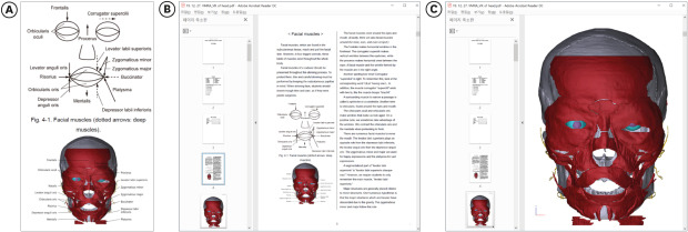
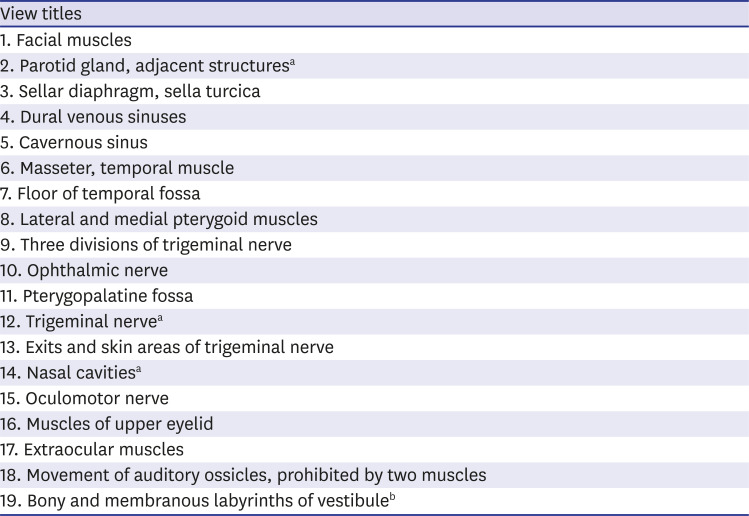
In the PDF file of VMRA, only the head chapter remained. Schematics in the PDF file of VMRA and surface models in the PDF file of VK were reviewed to find the common features. In the PDF file of VK, the surface models of the appropriate ones among the 297 head structures were selected and rotated to capture anterior, posterior, superior, inferior, or lateral views. This trial was repeated to find 19 views of the surface models. Among the 19 views, 15 views corresponded well with the schematics (Fig. 1A); 3 views did not correspond well, and 1 view corresponded very poorly (Table 1).
Fig. 1
Composition of the PDF file of Visually Memorable Regional Anatomy and Visible Korean. (A) A schematic figure and a captured view of surface models with annotations of the facial muscles. (B) PDF file containing the schematic figure, captured view, and explanations. (C) PDF file containing a page of the surface models.
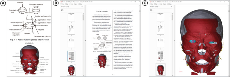
Table 1
Nineteen view titles of the surface models that are corresponding with the schematics
On the captured views of the surface models, the structures were manually annotated, whereas the schematics already had the annotations. Each captured view with the annotations was inserted into the PDF file of VMRA to be placed next to the equivalent schematic figure. For example, the surface models (anterior view) of facial muscles were paired with the schematic figures (Table 1). The common labels were frontalis, orbicularis oculi, nasalis, levator anguli oris, orbicularis oris, risorius, depressor anguli oris, mentalis, procerus, levator labii superioris, zygomaticus minor, zygomaticus major, buccinator, depressor labii inferioris, and platysma (Fig. 1A).
From the PDF file of VK, a page of the surface models of facial muscles and adjacent structures (cranium, facial nerve, facial artery) was embedded in the PDF file of VMRA as the page next to the schematic figure and captured view (Fig. 1B and C). In the same manner, the other 18 pages were embedded to yield the PDF file of VMRA & VK (Table 1).
Evaluation of the PDF file
The resultant PDF file of VMRA & VK was loaded on the two websites (www.anatomy.co.kr; www.neuroanatomy.kr). The PDF file was downloadable without payment or registration (menu: Visually Memorable Regional Anatomy [head chapter with surface models]).
In October 2018, Flag Counter (www.flagcounter.com) was installed on the websites to count visitors from different nations, and the number of downloads of the PDF file was counted.
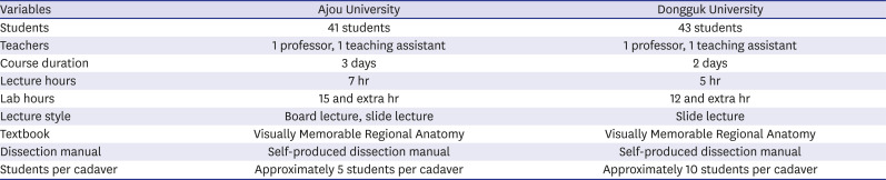
To assess the PDF file's learning effect, Korean medical students in two universities (Ajou University and Dongguk University), where the authors are affiliated, were asked to volunteer. The authors introduced the PDF file as a tool for their learning of head anatomy. The learning situations in the two universities were summarized in Table 2.
Table 2
Learning situations of head anatomy in the two universities
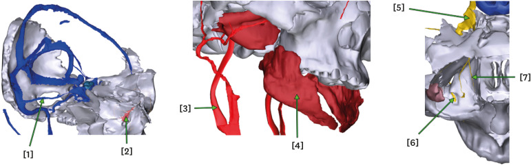
Written examination on head anatomy was partly made with the captured views of the surface models that were freely rotated by the authors (Fig. 2). These views differed from the orthogonal views in the PDF file (Fig. 1).
Fig. 2
Written examination made with the captured views of rotated surface models. The answers are [1] superior petrosal sinus, [2] lacrimal bone, [3] internal carotid artery, [4] buccinator, [5] trigeminal ganglion, [6] maxillary nerve, and [7] nasociliary nerve.
After the course, the students were asked to answer how many hours they had used the surface models in the PDF file. The usage hours and the scores on the examination on the captured views (Fig. 2) were statistically analyzed by calculating the Pearson's correlation coefficient and P value. Statistical Package for the Social Sciences (SPSS), version 20 (IBM Corp., Armonk, NY, USA) was employed for the statistical analysis.
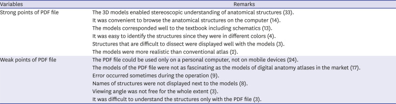
The participants were asked to answer how much the PDF file was helpful for understanding anatomy and for doing cadaver dissection (four degrees: not, hardly, slightly, and greatly). Furthermore, they were asked to provide their positive and negative remarks on the PDF file. During arrangement, the repeated numbers of the remarks were indicated (Table 3).
Table 3
Narrative remarks by the students on the PDF file of Visually Memorable Regional Anatomy & Visible Korean
RESULTS
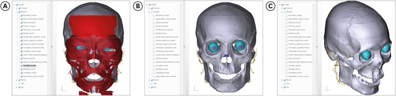
The PDF file of VMRA & VK could be opened using Adobe Reader, version 9 (Adobe Systems, Inc., San Jose, CA, USA). After one of the 19 pages of the surface models was activated, a surface model could be clicked to highlight its anatomical name in the left window, or an anatomical name could be clicked in the left window to highlight its surface model (Fig. 3A). Each surface model could be concealed by selection in the left window (Fig. 3B) or made semitransparent. The surface models could be magnified or rotated at arbitrary angles by manipulation of the computer mouse (Fig. 3C).
Fig. 3
Manipulation of the surface models in the PDF file of Visually Memorable Regional Anatomy & Visible Korean. (A) Simultaneous highlight of the anatomical name and surface model of the frontalis. (B) Concealing of the surface models of facial muscles to reveal those of the cranium; (C) rotation of the surface models.
During 16 months, most of the visitors to the website of the PDF file of VMRA & VK were Koreans (429 visitors), some of whom must have been students learning head anatomy from the authors. The visitors included Americans (19 visitors) and Hungarians (13 visitors), and the PDF file had been downloaded 602 times. The number of visitors did not include the repeated IP addresses, while the number of downloads did include.
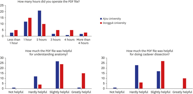
In both universities, the students operated the PDF file for two hours on average. In Ajou University, 68.3% of the students answered that the PDF file was helpful for understanding anatomy, and 41.5% of the students answered that the PDF file was helpful for doing cadaver dissection. In Dongguk University, 90.7% of the students answered that the PDF file was helpful for understanding anatomy, and 86.0% of the students answered that the PDF file was helpful for doing cadaver dissection (Fig. 4).
Fig. 4
Result of questionnaire survey given to the medical students who used the PDF file of Visually Memorable Regional Anatomy & Visible Korean.
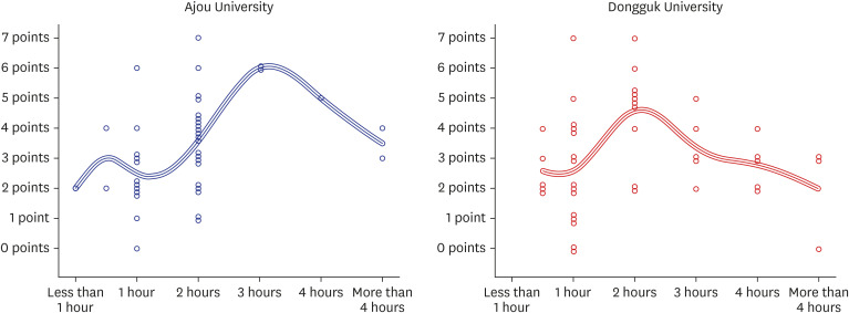
The number of hours that the PDF file was used and the scores on the written examination on the captured views (Fig. 2) showed a low positive correlation in Ajou University (Pearson correlation coefficient 0.353, P value = 0.023), but they showed no correlation in Dongguk University (Fig. 5).
Fig. 5
Scatter plot of the operating hours of the PDF file of Visually Memorable Regional Anatomy & Visible Korean and the scores of written examination on the captured views.
As for the strong points of the PDF file, the students answered that the surface models enabled stereoscopic understanding of anatomical structures. They were satisfied with the correspondence between the surface models and schematics as well as with the convenience of the PDF file. As the weak points, the PDF file with the surface models was not available on mobile devices, such as a tablet or smartphone. Some students claimed that the PDF file had a less attractive appearance and function than that of the commercial software (Table 3).
DISCUSSION
The PDF file of VMRA & VK, which includes both the schematics and operable surface models, is expected to contribute to anatomy learning for two reasons.
First, the schematics and surface models complement one another. The schematics have the problem of being quite different from the real feature of the human body. As a solution, the schematics should be compared to the realistic pictures in an anatomy atlas1 or the accurate surface models on a computer (Fig. 1).
There are many software packages to deal with the surface models of the human body.5-10 The surface models have problems in that they are sometimes difficult for novice students to understand. As a solution, the schematics can be used by the newcomers, just as simple maps are used by the first visitors.11,12 For the same reason, some anatomists still draw the schematics on the black or white board during lectures.13-15
As far as the authors know, the PDF file of VMRA & VK was the first attempt to show both the schematics and the surface models with the same annotations. Moreover, the PDF file provided explanations of the figures, including mnemonics (Fig. 1).1
The students who has experienced the PDF file answered that it was helpful for understanding anatomy and doing cadaver dissection. The frequency of this positive result differed according to the universities (Fig. 4), perhaps because of different lecture styles or a different number of cadavers (Table 2). The students were satisfied with the correspondence between the schematics and surface models (Table 3).
As a negative result, the hours that the PDF file was used and the scores on the written examinations on the captured views did not show a definite correlation (Fig. 5). Seemingly, the scores depended on the hours of reading the textbook or other books, rather than on the hours of using the PDF file. In other words, such multimedia contents as the PDF file can serve as just a supportive tool.
Second, the schematics and surface models were approachable. Unlike the usual anatomy books and software packages, the PDF file of VMRA & VK is free of charge and runs off-line without time delay. Therefore, all the students in a class can observe the schematics and work with the surface models regardless of economic state, place, or time. It is also easy for teachers to make the figure examination for evaluating their students (Fig. 2). The students preferred the convenience of learning on the computer (Table 3).
The PDF file can be improved in quantity as follows. The PDF file of VMRA includes the other regions (trunk, upper limb, and low limb); it can be combined with other PDF files of VK dealing with the male whole body,16 the female whole body,17 or the female pelvis.18
The PDF file can be improved in quality as follows. Unlike surface models that are stereoscopically drawn, the surface models in VK are reconstructed from the sectioned images of cadavers. Hence the surface models can be replaced with the volume models that can be sectioned to show the sectional planes with real body color.19,20 The expandability and its learning effect should be considered together.
The surface models can be 3D printed out to be touched. The resultant printed models, equivalent to the surface models, would be more educational than are the regular plastic models. The printed models and the surface models could be the material for augmented reality in the digital anatomy field.21,22
The students wanted to work with the surface models on the mobile devices (Table 3). It would be worthwhile to develop applications of the smartphone as the secondary learning tool.23 The students also wanted to have the upgraded software that is convenient and free of error (Table 3). The new version of the software that is as excellent as the commercial one will be forthcoming. The improvements cannot be done only by the authors; so the authors are willing to provide interested investigators with the source files containing the 3D models, which are easily modified.
Overall, this study employs different educational methods from other recently introduced methods. This study is like a convergence between the affordable digital technology and classical board lecture, whereas other studies are usually interested only in the virtual reality. This study utilizes typical personal computer that any student can prepare, while other studies utilize cutting-edge tools such as virtual dissection table24,25 and head-mounted display.26,27 This study emphasizes the schematic drawings that have long-lasting learning effect by students' replication.28,29
The PDF file of VMRA & VK and its upgraded software packages would be the tool to help students learn head anatomy easily, interactively, and accurately by themselves.
 XML Download