

 PDF
PDF Citation
 Print
Print


INTRODUCTION
From veterinary medicine and evolutionary biology to biophysics, monkeys are useful animal models because of their similarities to humans.123 Monkey researchers and students require a basic understanding of veterinary anatomy and radiology. While it is typically necessary to dissect a monkey for veterinary anatomy research, it is very difficult to obtain monkeys for dissection. Knowledge of sectional anatomy is required to interpret computed tomographs (CTs) and magnetic resonance images (MRIs) of monkeys,4567 but there is no sectional anatomy atlas of monkey which can show vividly normal structures on sectional planes in detail.
To overcome the difficulties of dissection and the lack of appropriate atlases of a monkey, computer software to deal with two-dimensional (2D) and three-dimensional (3D) images of monkey is need. Typically, 2D and 3D software packages rely upon CTs and MRIs. However, in the software based on the CTs and MRIs, anatomical structures cannot be shown in detail, because of the 8-bit gray scale and low resolution (pixel size and intervals, > 0.1 mm) of the raw data. Therefore, it is ideal for the 2D and 3D software packages to use high-resolution sectioned images in real color.89101112
We previously produced the sectioned images of a rhesus monkey (color depth, 48-bit color; intervals, > 0.05 mm; pixel size, 0.024 mm) in the Visible Monkey project.13 Segmentation of the sectioned images is necessary to allow anatomical structures to be distinguished in the 2D and 3D software packages.141516
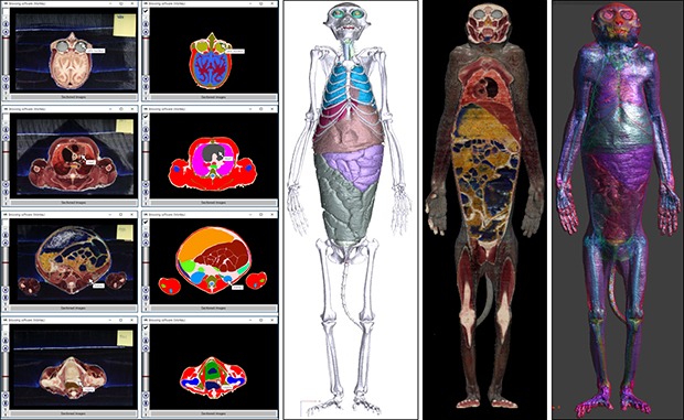
The purpose of this study is to present the utility of the segmented images, made from the sectioned images of Visible Monkey, with regard to the learning of monkey anatomy and radiology. To achieve this objective, sectioned images of a rhesus monkey were segmented; these segmented images were used to create 2D software (browsing software) and 3D software (PDF file).
Go to : 
METHODS
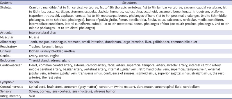
In our previous study, sectioned images of an entire monkey body were created (intervals of head, 0.05 mm; intervals of the rest regions, 0.5 mm; pixel size, 0.024 mm; resolution, 8,688 × 5,792; color depth, 48-bit color; BMP format) (Fig. 1A).13 One hundred sixty-seven structures to segment were selected (Table 1). Most structures were selected for the purposes of anatomy education. The cerebrum, cerebrospinal fluid, and eye structures were also selected for biophysical experiments.17 For segmentation, sectioned images were chosen at 0.5-mm intervals.
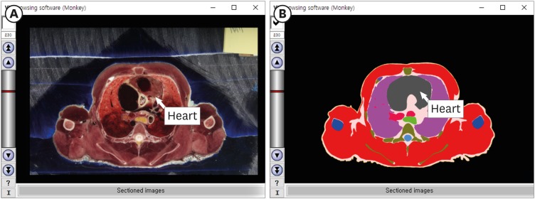 | Fig. 1Browsing software for a rhesus monkey. In the software, (A) the sectioned images and (B) segmented images of the thorax are able to be browsed serially. The name of the structure appears when a mouse pointer is placed over the image.
|
Table 1
One-hundred sixty-seven structures used for segmentation and surface modeling
![]()
Segmentation was performed using Photoshop CC 2015 (Adobe Systems, Inc., San Jose, CA, USA). The boundaries of structures in the sectioned images were chosen semi-automatically using the quick selection tool and magnetic lasso tool, or manually using the lasso tool.14 The boundaries were each filled automatically with a specific color using action and batch tools, then saved in BMP format to produce segmented images of an entire monkey body (Fig. 1B). To easily browse the sectioned images and segmented images, these images were converted from BMP format to JPG format and packaged into the browsing software (Fig. 1) created in our previous study.10
Surface modeling was performed using Mimics version 17.01 (Materialise, Leuven, Belgium). Each structure of the segmented images was remained using the threshold tool and reconstructed by surface modeling and saved in STL format. On Deep Exploration Standard (SAP America, Inc., Newtown Square, PA, USA), the STL files were assembled into a PDF file,18 which allowed users to manipulate the surface models freely using Adobe Reader version 9 or higher (Adobe Systems, Inc.) (Fig. 2).
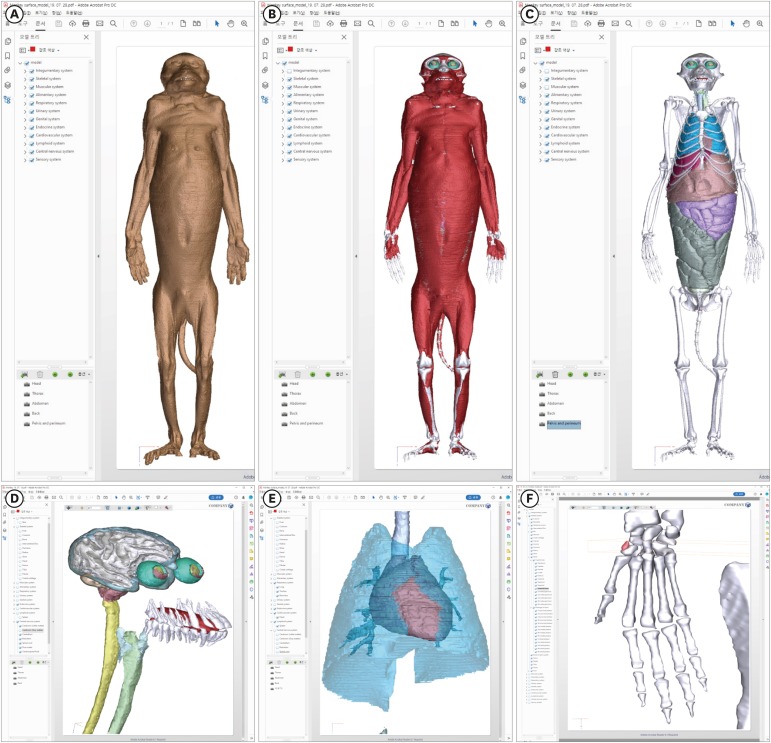 | Fig. 2Surface models of a rhesus monkey in Adobe Reader. The surface models of (A) the skin, (B) muscle, and (C) bones and organs are able to be displayed. (D, E, F) After the surface models are expanded, detailed shapes of segmented structures can be observed. (F) In front of the scaphoid, a sesamoid bone (red color) is visible.
|
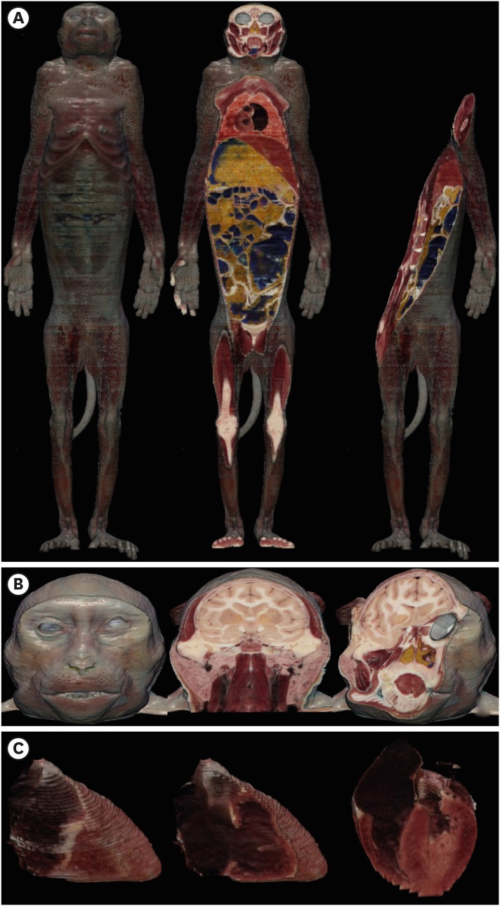
Volume modeling was carried out in MRIcroGL v1.0 (http://www.mccauslandcenter.sc.edu/mricrogl/) after converting sectioned images from BMP format to DICOM format using Photoshop. The DICOM files were imported and reconstructed automatically by volume modeling using the dcm2nii tool in MRIcroGL on a personal computer, thereby producing the volume model of the monkey body (Fig. 3A and B). In addition, based on the heart boundaries of segmented images, the heart alone was extracted from the images and saved in DICOM format using Photoshop. After the heart DICOM files had been reconstructed, a volume model of the heart was made using MRIcroGL (Fig. 3C).19
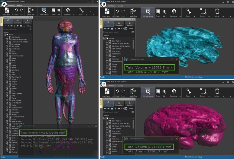
Spatial information of each segmented structure was checked by importing the STL files into Sim4Life (Zurich MedTech AG, Zurich, Switzerland) using a personal computer (operating system: Windows 10; Microsoft Corp., Redmond, WA, USA) (Fig. 4). Spatial volumes of segmented structures were multiplied by densities based on the Gabrial list and IT'IS foundation.2021 This enabled quantification of the weights of segmented structures.
Go to :
RESULTS
This study produced 839 segmented images of 167 structures throughout a monkey (intervals, 0.5 mm; pixel size, 0.024 mm; resolution, 8,688 × 5,792; color depth, 24-bit color; BMP format; individual image size, 157 MB; total image size, 128 GB) (Fig. 1). Sectioned and segmented images could be observed continuously and magnified using the browsing software. The names of structures could be visualized using a mouse pointer (Fig. 1). The surface models of 167 structures could be displayed and handled freely in a PDF file using Adobe Reader (file size, 52 MB) (Fig. 2). Using MRIcroGL, the real-color volume model of the whole body could be browsed and sectioned at any angle, similar to dissection of a real cadaver. Any region could be displayed in detail by magnification of the model (Fig. 3B). In addition, a volume model of the heart alone was able to be virtually dissected using MRIcroGL (Fig. 3C).
Some structures in monkey that definitely differ from structures in human were found using the browsing software, surface models, and volume models (Fig. 5). In the head, the eyeball, optic nerve, lateral geniculate nucleus, and hippocampus were relatively large (Fig. 5A and C) because of the specialized primate visual system.22 The salivary glands were also relatively large (Fig. 3B); large salivary glands are essential for wild animals because they must process tough food and digest raw food. In the wrist, the sesamoid bone was located in front of the scaphoid (Fig. 2F). In the thorax and abdomen, the anterior-posterior length of the torso was longer than its width (Fig. 1); wild animals, such as monkeys, have long torsos to facilitate quadrupedal movement.
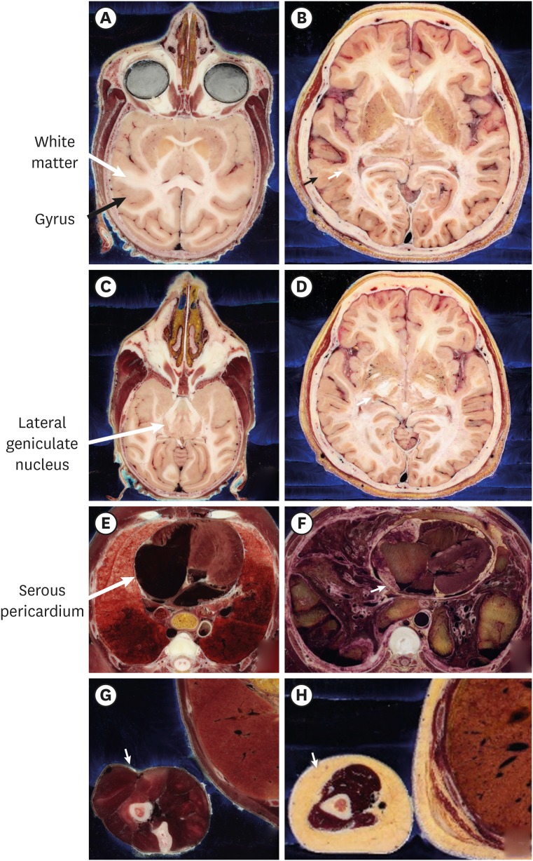 | Fig. 5Comparison of structures between a monkey and a human. In the head, (A) cerebral gyri of the monkey are simpler than (B) those of the human (black arrow). (A) The gray and white matter of the monkey are also simpler than (B) those of the human (white arrow). (A) The eyeball, optic nerve, (C) lateral geniculate nucleus (arrow), and hippocampus of the monkey are larger than (D) that of the human. (E) Because there is minimal fat in the monkey body, the rim of the heart and visceral layer of serous pericardium (arrow) of monkey are very clear, compared with (F) those of the human. (G) In the monkey, there is little fat (arrow) between skin and muscle, as well as between organs; (H) the human exhibits greater volumes of fat in both areas.
|
The total spatial volume of the segmented structures was 9,181,930 mm3. The volumes of gray matter (density, 1,039 kg/m3) and white matter (density, 1,043 kg/m3) in the cerebrum were 71,333.1 mm3 and 19,799.5 mm3, respectively; the weights of gray and white matter in the cerebrum were 74.11 g and 20.65 g, respectively (Fig. 4). The volume of the gray matter in the monkey was approximately 3.5-fold greater than volume of white matter, whereas the volume of gray matter in humans is two-fold greater than that of white matter. This is because monkeys require fewer and less varied connections (white matter) between gray matter, compared with humans.2324 The spatial volumes of all 167 structures could also be calculated.
Go to :
DISCUSSION
In the current study, segmented images were prepared from the sectioned images; 2D and 3D software packages were produced using both the sectioned and segmented images (Figs. 1-3). We established the utility of the segmented images of Visible Monkey as follows.
First, the segmented images were used to interpret the sectioned images. Structures on the sectioned images could not be interpreted without labels or the segmented images. In this study, we assigned structure names (Table 1) to the sectioned images based on the segmented images in the browsing software.14 A student could easily recognize the structure names using the browsing software (Fig. 1).
Second, the segmented images were used to reconstruct the surface models.14 The surface models could not be reconstructed without the boundaries of each structure, which comprised the framework of the surface models. In this study, we created segmented images (Fig. 1B), then produced the surface models after stacking the boundaries (Fig. 3).
Furthermore, the surface models can be used for analysis of the effects of electromagnetic radiation on the body in biophysics research. Surface models contain spatial information of each structure in STL format. If tissue properties (e.g., density or weight) are transfused with spatial information such as this study (Fig. 4), the energy absorption of the tissues (i.e., specific absorption rate) can be virtually calculated from electromagnetic radiation.24252627 In case of a monkey, in vivo experiment of the energy absorption can be performed. Finally, the result of virtual models of this study can be compared with the result of in vivo models to verify accuracy about effect of electromagnetic radiation by virtual models. If the accuracy of virtual models of monkey are proven, many virtual models of human in the world will be more positively used.
Third, the segmented images were used to reconstruct the volume models. Volume models of a single structure or small region are needed for medical training such as virtual ultrasonography of heart28 and virtual arthroscopy of knee.29 In case of the volume model of the entire body (Fig. 3A and B), the original resolution cannot be realized because of the limited capacity of a personal computer. In this study, we reconstructed volume models of a structure or small region using its segmented images (Fig. 3C).
Compared with CTs and MRIs, the sectioned images enable segmentation of more structures because of the improved color and resolution, whereas automatic and semiautomatic segmentations on the sectioned images are difficult because of this additional graphic information.1330 Nevertheless, in this study, some structures in sectioned images could be easily segmented for the following reasons.
First, in the sectioned images, the monkey brain was able to be segmented more easily than the human brain because of the simple gyri of the cerebrum. The number and curvature of monkey gyri are smaller and simpler, respectively (Fig. 5A and C), than those of the human gyri (Fig. 5B and D). Similarly, the curves of white matter in the monkey gyri are also simpler (Fig. 5A and C) than those of the human white matter (Fig. 5B and D). Consequently, we could semi-automatically segment the cerebral gyri of the monkey and automatically segment cerebral gray and white matter using Photoshop (Fig. 4B and C).
Second, segmentation of the monkey skin and muscle is easier than segmentation of the human skin and muscles because monkeys have minimal fat. Wild animals tend to be lean because they move frequently to survive. Although the monkey in the present study was reared in a laboratory, it had minimal fat in the skin and muscle. Also, the monkey had minimal fat around the visceral layer of serous pericardium (Fig. 5E and G), compared with the human (Fig. 5F and H). Therefore, we could easily segment the skin, muscle, and heart.
In 2019, the Visible Monkey project was launched based on sectioned images prepared from a monkey. The sectioned images first appeared on a computer; hence, the first study was named the “Rise of the Visible Monkey,”13 referring to the movie “Rise of the Planet of the Apes.” In the present study, segmentation of the Visible Monkey was performed to produce 2D and 3D software packages. This highlighted the utility of the Visible Monkey; hence, the study was named “Dawn of the Visible Monkey,” referring to the movie “Dawn of the Planet of the Apes.”
In the near future, virtual reality and augmented reality systems31 will be created based on 3D models with more detailed structures of the nervous system (e.g., nuclei and tracts of thalamus and hypothalamus, cranial nerves, spinal nerves with spinal ganglion, and blood vessels). To segment more detailed structures for virtual reality and augmented reality, anatomy experts must manually segment most structures, with some exceptions. This manual segmentation is difficult; therefore, the name of the future study may be “War for the Visible Monkey,” referring to the movie “War for the Planet of the Apes.”
In this study, to establish the utility of the sectioned images in Visible Monkey, 2D (browsing software) (Fig. 1) and 3D (surface models and volume models) (Figs. 2 and 3) software packages were produced. These software packages are expected to be useful for students of veterinary anatomy and radiology. Moreover, spatial information of the segmented structures can be analyzed based on the surface models, which may be useful for biophysical experiments.2425 Users will be able to download all data from this study after user certification by the Electronics and Telecommunications Research Institute.
Go to :
 XML Download