

 PDF
PDF Citation
 Print
Print


INTRODUCTION
Mucosa-associated lymphoid tissue (MALT) lymphoma is a distinct subtype of non-Hodgkin lymphomas that occurs in approximately 5% of non-Hodgkin lymphomas. Most MALT lymphomas are found in the gastrointestinal tract, with the stomach being the most common site. On the other hand, colonic MALT lymphoma is a rare disease that comprises only 2.5% of MALT lymphomas.1-3 Therefore, the etiology, clinical characteristics, treatment, and outcomes of colonic MALT lymphoma are not completely understood. The disease was previously treated by single or combination of surgery, endoscopic resection, conventional chemotherapy, rituximab, or radiation therapy.1-3 This paper reports a case of a 50-year-old woman with MALT lymphoma arising in the transverse colon that was treated successfully with radiation therapy alone with a review of the relevant literature.
Go to : 
CASE REPORT
A 50-year-old woman visited Chonnam National University Hwasun Hospital with a 2-year history of constipation. She denied any prior medical history or other symptoms, such as fever, weight loss, general weakness, anorexia, abdominal pain, hematochezia, and melena. She was afebrile, and her blood pressure and pulse were normal. Her abdomen was soft, not distended, and there was no tenderness. The liver and spleen were not palpable, and there were no palpable superficial lymph nodes. The laboratory examinations revealed the following: white blood cell count 5,000/mm3 (normal: 6,000-10,000), hemoglobin 13.0 g/dL (normal: 12-16), platelet count 287,000/mm3 (normal: 130,000-450,000), BUN 10.6 mg/dL (normal: 8-23), creatinine 0.6 mg/dL (normal: 0.5-1.3), serum albumin 4.2 g/dL (normal: 3.0-5.0), AST 22 U/L (normal: 5-37), ALT 13 U/L (normal: 5-40), ALP 72 U/L (normal: 39-117), and LDH 376 IU/L (normal: 218-472). The total bilirubin was 0.74 mg/dL with 0.35 mg/dL direct fraction (normal: 0.2-1.2/0.05-0.3). HBsAg was negative. The anti-HBs was positive, and anti-HCV was negative. The beta-2-microglobulin level was 1,924 μg/L (normal: 970-2,640).
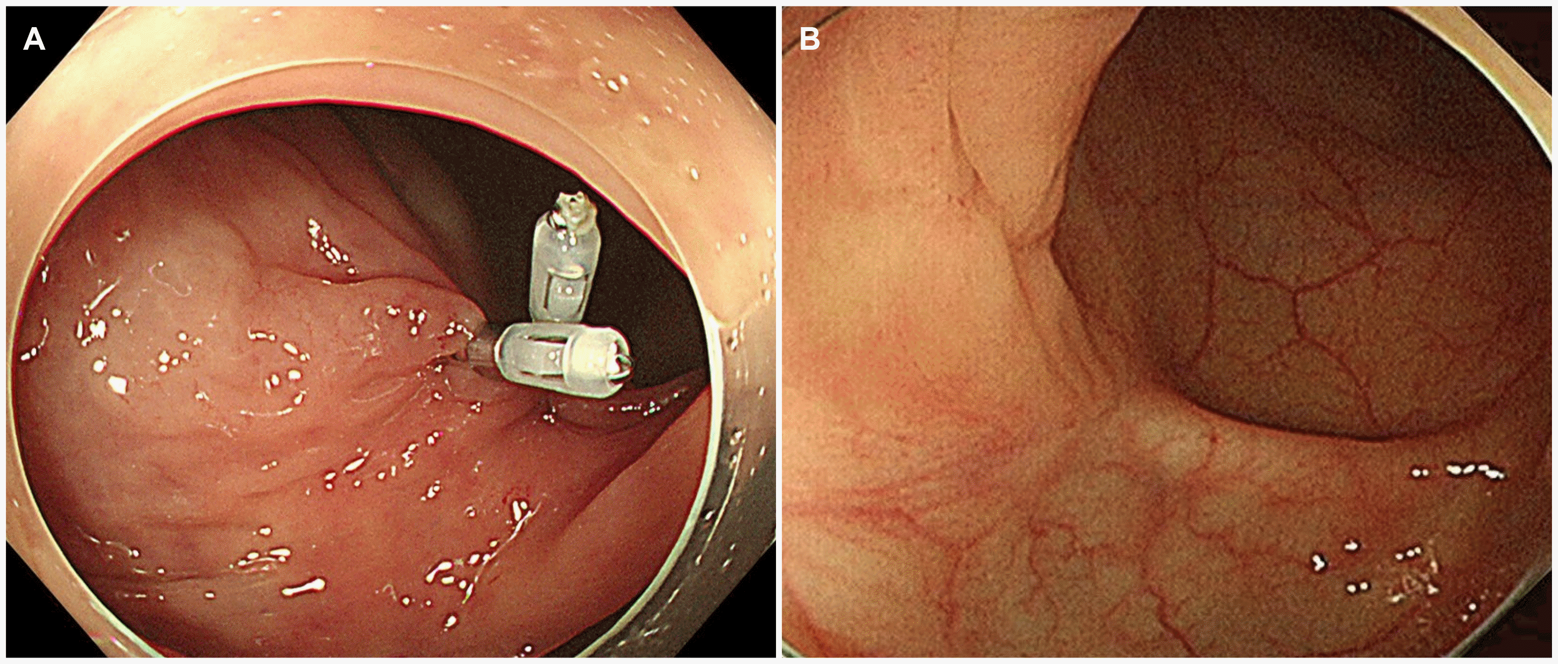
Contrast-enhanced neck, chest, abdomen, and pelvis CT was performed, and bone marrow aspiration was negative for lymphoma involvement. Esophagogastroduodenoscopy revealed chronic gastritis with intestinal metaplasia. The histology study and CLO test were negative for a Helicobacter pylori (H. pylori) infection. Colonoscopy showed a 20 mm sized subepithelial tumor with hyperemic mucosa arising in the transverse colon (Fig. 1), and a colonoscopy biopsy was performed. Routine histology and staining using hematoxylin-eosin revealed a diffuse and dense infiltrate of small-sized lymphocytes and a lymphoepithelial lesion in the colonic mucosa (Fig. 2A). An immunohistochemical examination was performed to clarify the nature of the cells. The tumor cells were positive for CD20 (Fig. 2B) and negative for CD3 and Bcl-6. In addition, the Ki-67 labeling index was 30%. The biopsy specimens were interpreted as MALT lymphoma. The tumor was diagnosed as a MALT lymphoma of the transverse colon and classified as stage IE according to the Ann Arbor staging classification. Radiation therapy was performed after a discussion with the authors’ multidisciplinary medical team, including a medical oncologist, radiation oncologist, gastroenterologist, and surgeon. Before treatment, the target of radiation therapy was indicated by hemoclips (Fig. 3A) because the lesion was not detected in the abdomen CT scan. The patient received 3,060 cGy in 17 fractions with external beam radiation therapy for 3 weeks. Two months after the radiation therapy was complete, a colonoscopy examination revealed complete resolution with the endoscopic and histologic disappearance of the lesion and a scar change of the lesion (Fig. 3B). The patient has been on a regular followed-up schedule at the out-patient clinic and has shown no evidence of recurrence at 24 months after radiation therapy.
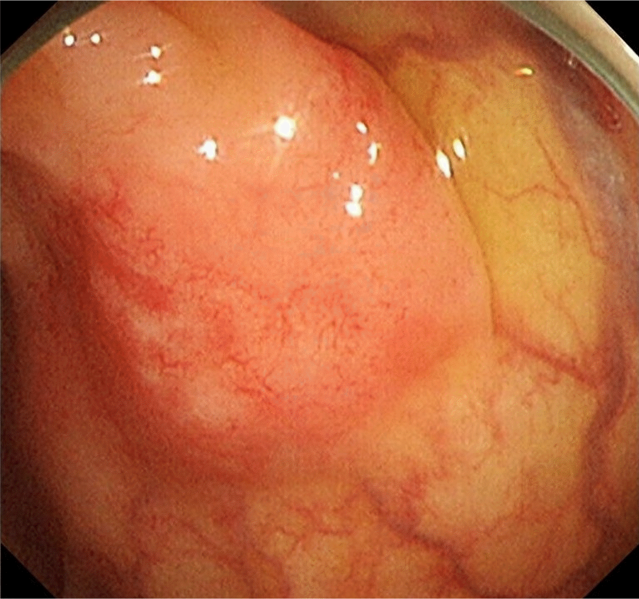 | Fig. 1Colonoscopy shows a 20 mm sized subepithelial tumor with hyperemic mucosa arising in the transverse colon.
|
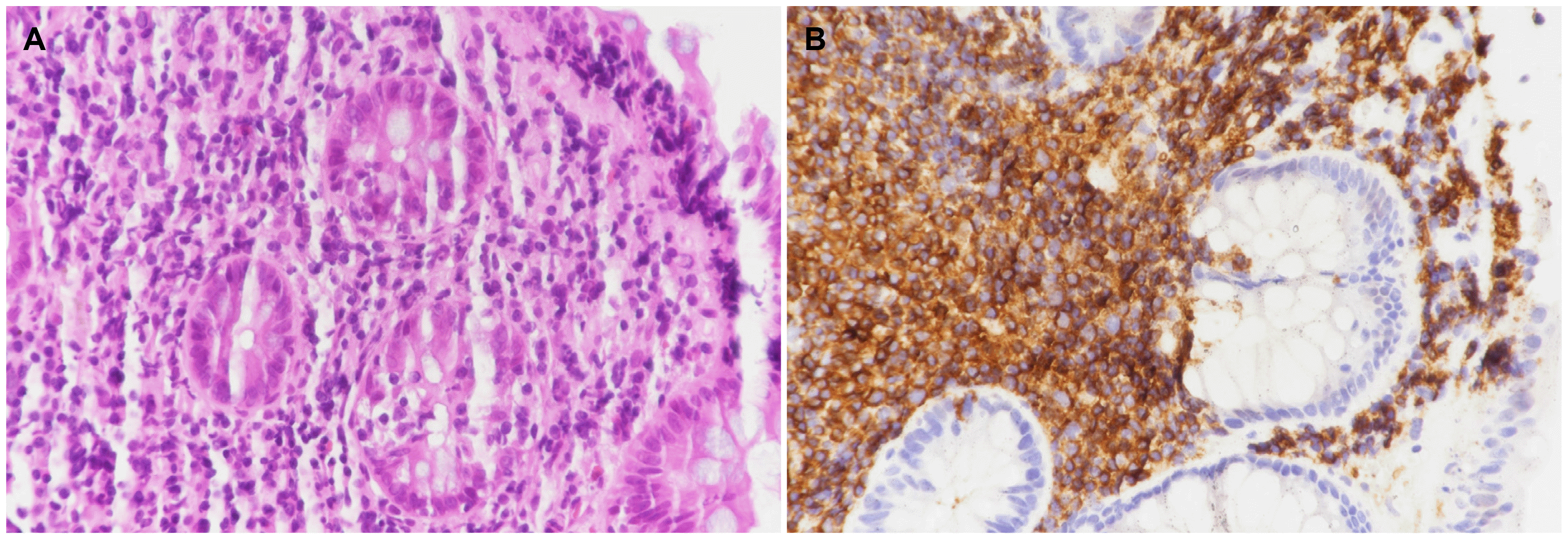 | Fig. 2Microscopic findings. (A) Endoscopic biopsy specimens show a diffuse and dense infiltration of small-sized lymphocytes and a lymphoepithelial lesion in the colonic mucosa (H&E staining, ×400). (B) Immunohistochemical staining shows infiltrative small-sized lymphocytes with positive for B-cell marker CD20 (×400).
|
Go to :
DISCUSSION
MALT lymphoma is a subtype of non-Hodgkin lymphoma that is classified as an extranodal marginal zone B cell lymphoma of the MALT type.4 The stomach is the most common site of MALT lymphoma involvement, but colon involvement is very rare.1-4
Colonic MALT lymphoma typically occurs in patients between 50 and 75 years. The gender predisposition ranges from equal to a female predominance. The clinical presentation is often asymptomatic, followed by abdominal discomfort and pain, hematochezia, tenesmus, mucoid stool, constipation, or palpable mass. The most common site is the rectum, followed by the right-side colon, sigmoid colon, and transverse colon. The main endoscopic appearance was a single lesion or multinodular polypoid lesions, followed by a subepithelial tumor, flat elevation, mucosal edema, erythema, and loss of vascularity.1-3 The patient in this study was a 50-year-old woman with an asymptomatic MALT lymphoma of the transverse colon, presenting as a subepithelial tumor found incidentally on screening colonoscopy.
Colonic MALT lymphomas are treated with various modalities, including single surgery or a combination of surgery with endoscopic resection, chemotherapy, rituximab alone, or radiation therapy. Colonic MALT lymphoma has an indolent nature and favorable clinical behavior. On the other hand, its rarity and indolent nature mean that the treatment and outcome of colonic MALT lymphoma are not well established.
Gastric MALT lymphoma constitutes approximately 70% of MALT lymphomas of the gastrointestinal tract. The disease is associated strongly with a H. pylori infection, and H. pylori eradication is a standardized therapy for localized gastric MALT lymphoma.4-6 Although several reports have shown that colonic MALT lymphomas can be successfully treated with H. pylori eradication,7-9 the relationship between colonic MALT lymphoma and H. pylori infection is unclear.
Radiation therapy is an effective modality in dealing with MALT lymphomas arising in other organs, and its recurrence is not always in the original area of disease.4 Therefore, early colonic MALT lymphoma may be treated safely by non-surgical management, avoiding the morbidity and mortality that is associated with surgical resections.10 In the present case, the lesion was stage IE and limited to the transverse colon. As MALT lymphoma has an indolent nature and was a localized lesion, definitive treatment with radiation therapy was planned after discussion with the multidisciplinary medical team. Because the tumor was not detected in the abdomen CT scan, the tumor was marked by hemoclips before radiation therapy. At 2 months after radiation therapy, the colonoscopy examination revealed complete resolution and scar change of the lesion. Twenty-four months after radiation therapy, the patient showed no tumor recurrence. Previously, limited cases of localized colonic MALT lymphomas were treated successfully with radiation therapy alone ranging from 30 to 45 Gy, but it did not appear to be used widely over the world.2,3,11-13
The present case and previous reports suggest that radiation therapy alone may be a feasible and safe treatment, allowing for organ preservation, and is considered to treat such a localized colonic MALT lymphoma.10 On the other hand, further follow-up will be needed to determine the long-term efficacy of this treatment approach and its side effects, such as radiation colitis.
Go to :
 XML Download