

 PDF
PDF Citation
 Print
Print


I. Introduction
Trauma to the oral region occurs frequently and accounts for 5% of all injuries for which people seek dental treatment1. The highest incidence of dentoalveolar trauma occurs in the 20- to 40-year age group2. Among all facial traumas analysed in one report2, the incidence of dental injuries with respect to the total number of facial injuries was as follows: 57.8% in play and household accidents, 50.1% in sports accidents, 38.6% in accidents at work, 35.8% in acts of violence, 34.2% in traffic accidents, and 31% in unspecified accidents. The overall incidence revealed was 48.25%. Dentoalveolar injury either in isolation or associated with other facial fracture such as zygomatic bone fracture or LeFort I fracture is prone to avulsion and displacement of teeth in adjacent structures, such as the soft tissue, maxillary antrum, nasal cavity, and respiratory tract. Such displaced teeth can be accidentally swallowed or spit out by the patient3. The following cases about traumatic dislocation of teeth have been reported. One case involved displacement of the mandibular lateral incisor into the nasal floor after panfacial fractures. Diagnostic computed tomography (CT) of the brain and nasal endoscopy were carried out to locate and retrieve the tooth, respectively4. Another case reported ingestion of two teeth, one reaching the bronchus and the other the liver, confirmed using fibre-optic bronchoscopy and CT of the abdomen5. A final case was of a patient who accidental swallowed a hypodermic needle during root canal treatment and that was removed safely by gastrointestinal endoscopy6.
Go to : 
II. Case Report
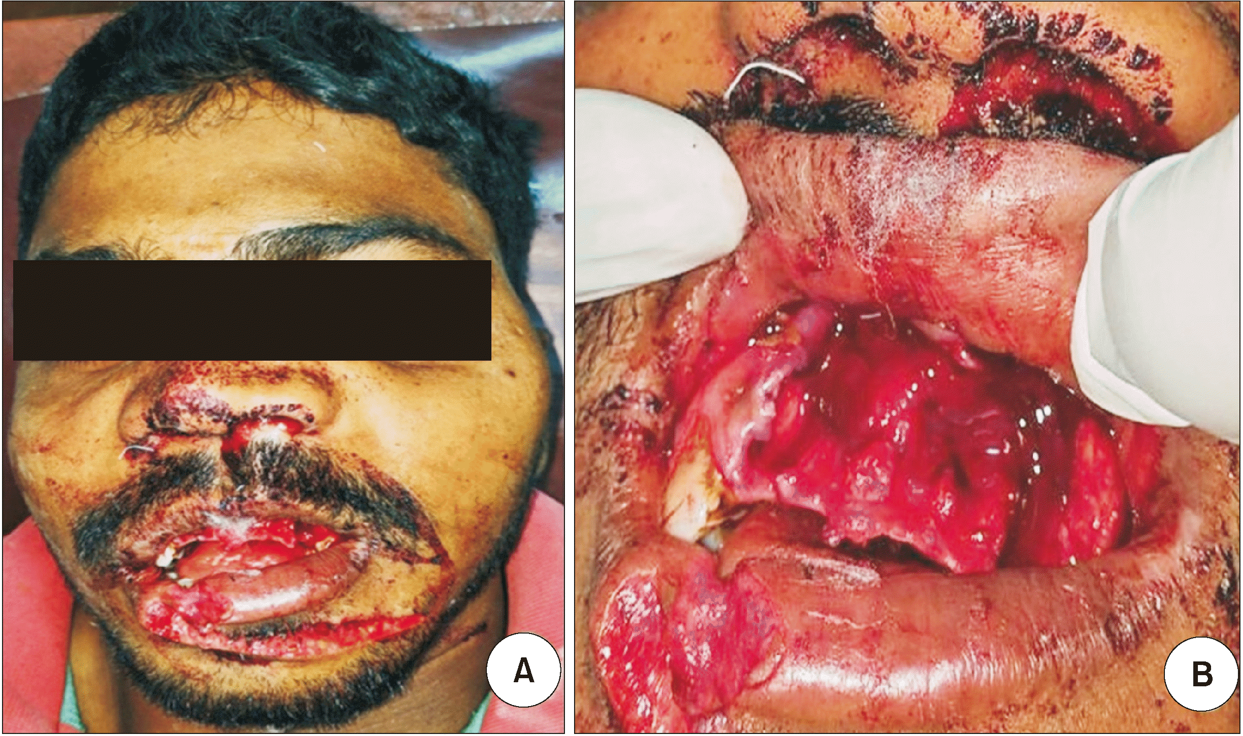
A 26-year-old male reported to the emergency department with chief complaints of laceration and pain in his face. The patient reported a fall from height off a lorry, during which he hit his face on the corner of the lorry and sustained injuries in the orofacial region. Clinical examination revealed bilateral epistaxis, a dentoalveolar fracture with a left zygomaticomaxillary complex fracture, and missing #11, #12, #21, #22, and #23 teeth.(Fig. 1) The patient reported that he spat out two teeth at the site of the incident.
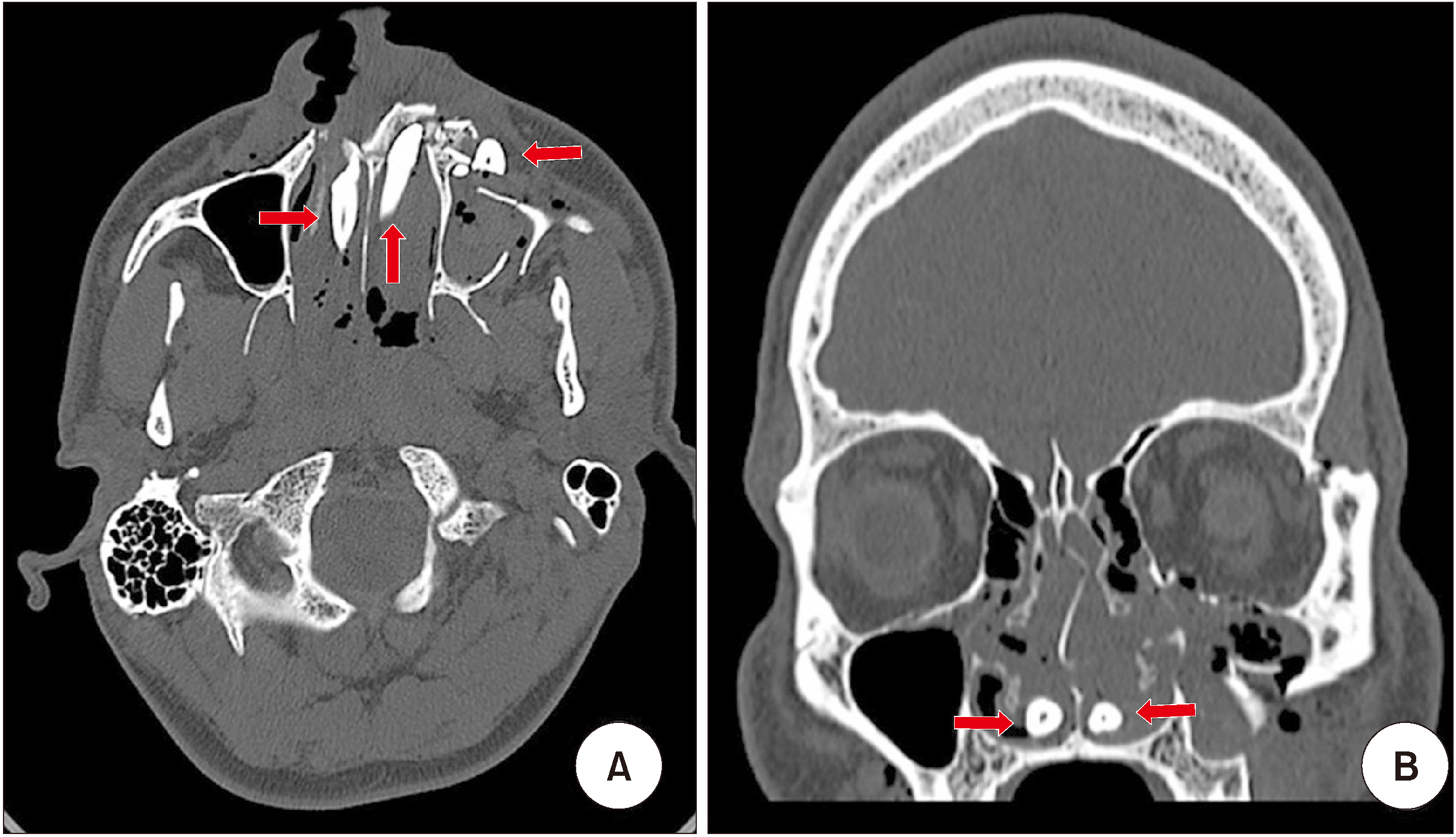
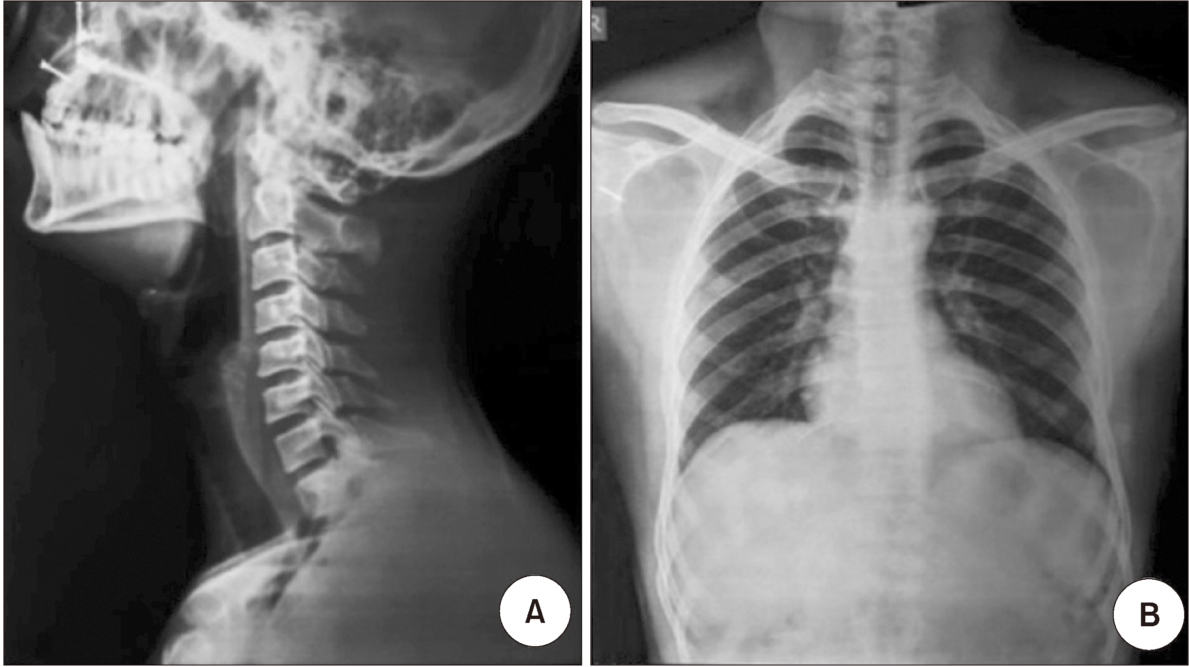
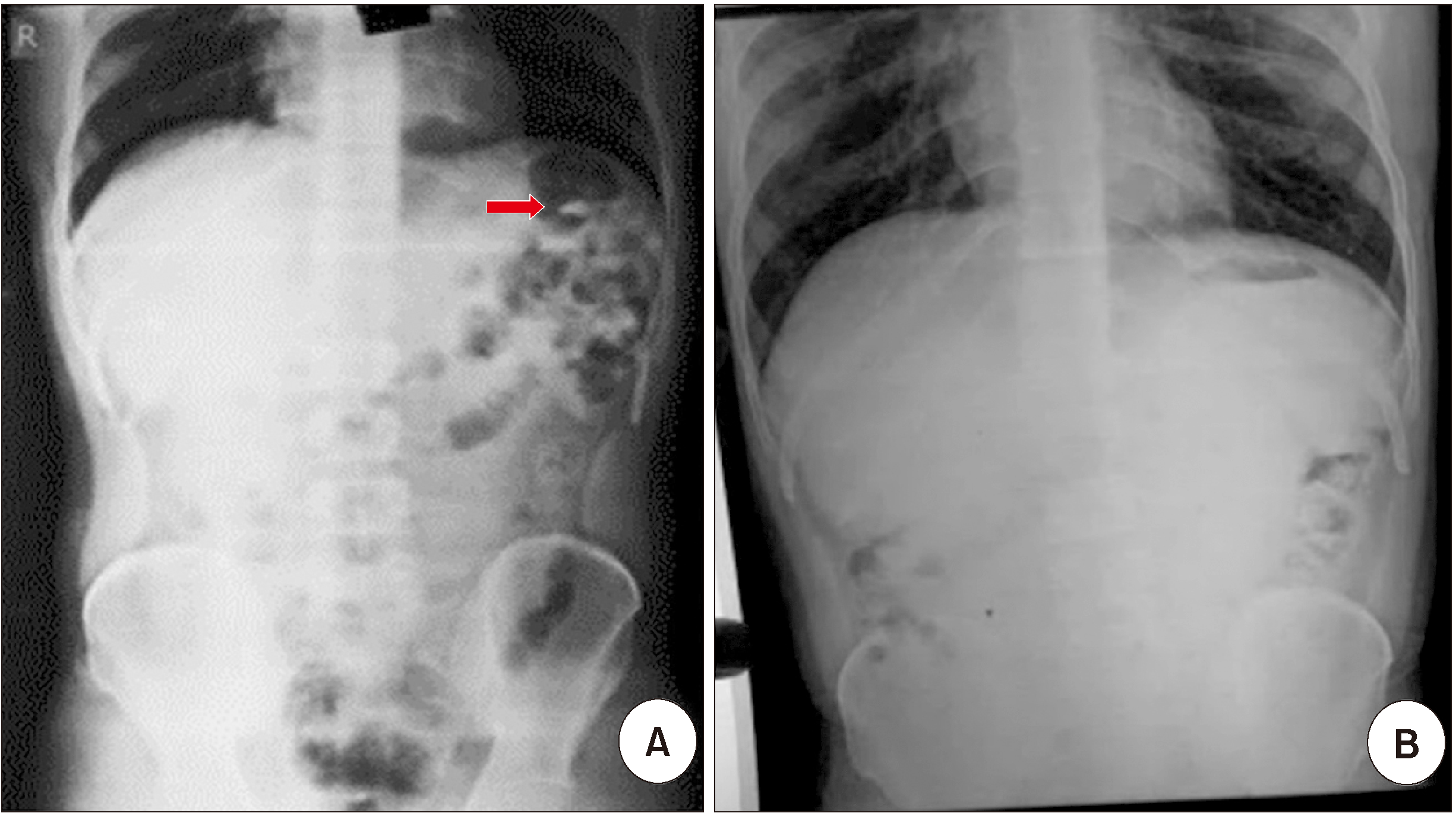
Plain CT scan of the brain revealed multiple dislocations of teeth #11 and #21 (Fig. 2) to the respective nasal cavity, and tooth #23 (Fig. 2. A) intruded completely above the socket and was displaced into the soft tissue near the left maxillary antrum. A lateral neck radiograph, anteroposterior (AP) chest radiograph (Fig. 3), and erect abdominal radiograph were collected to investigate the location of the other missing teeth, but did not reveal any abnormal findings or avulsed teeth.
The patient received primary management with anterior nasal packing for the bilateral epistaxis, and the left maxillary canine, which was displaced in the soft tissue near the left maxillary sinus, was removed using artery forceps.
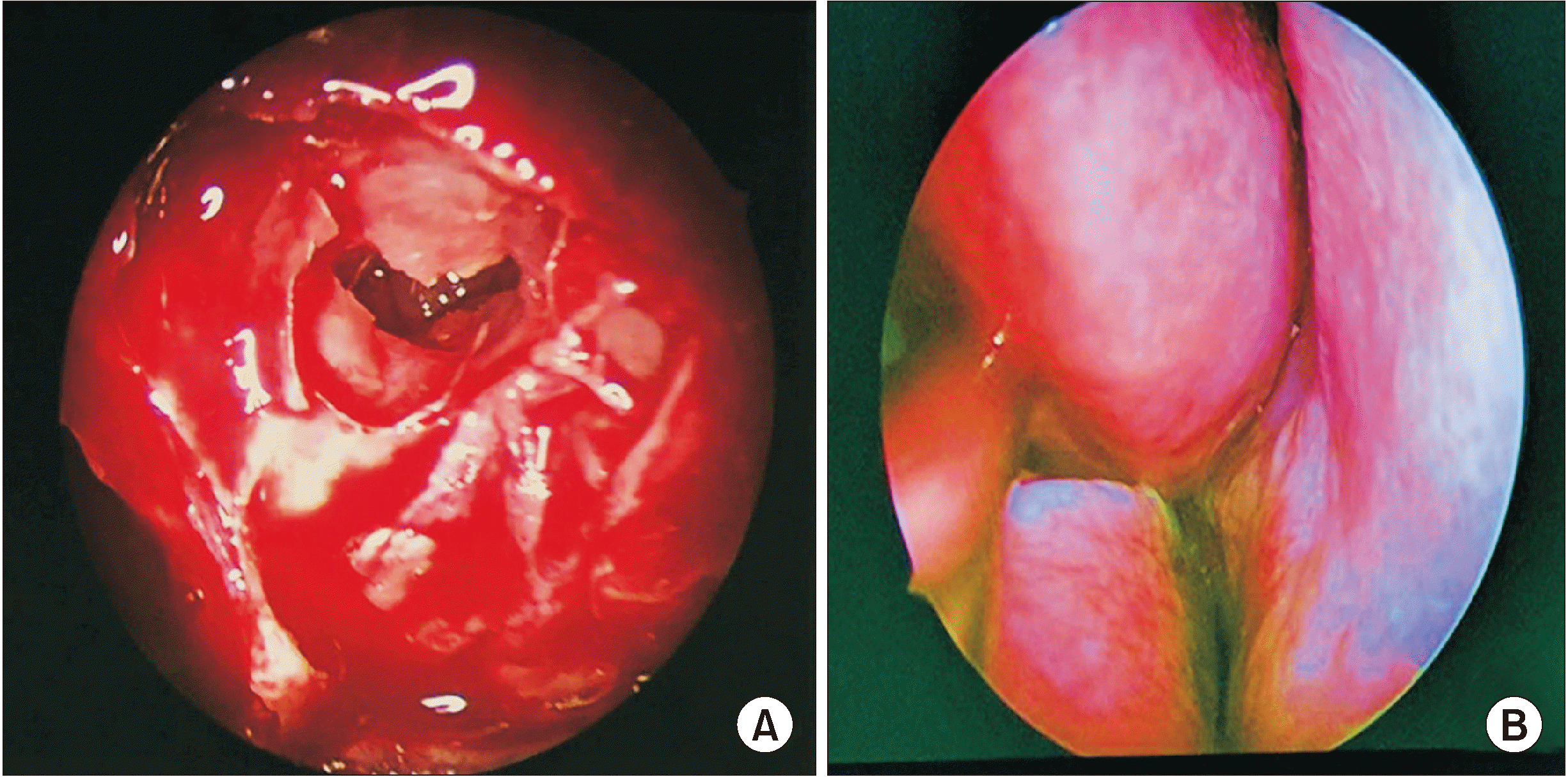
Open reduction and internal fixation of the zygomaticomaxillary complex fracture and endoscopic removal of teeth from the nasal cavity were planned. On the day of surgery, the patient reported loss of another tooth that day. Intraoperative nasal endoscopy (Fig. 4) did not reveal the missing teeth, which had been seen in the nasal cavity on the CT scan obtained in the emergency department. Postoperative repeat lateral neck, AP chest, and erect abdominal radiographs were collected. Although the neck and chest examinations did not reveal any teeth in the upper airway tract or chest, the radiograph of the abdomen revealed hyperdense structures, i.e., teeth, in the bowel.(Fig. 5. A) We hypothesized that, during primary management using anterior nasal packing, the teeth in the nasal cavities were displaced to the upper aero-digestive tract. One tooth was spat out by the patient on the day of the procedure and the other likely was ingested. Therefore, brain CT was not repeated. Conservative management for the tooth displaced to the bowel was planned using laxatives, and the patient was instructed to follow up. After laxative use for seven days, the patient returned for follow up. An erect abdominal radiograph (Fig. 5. B) did not reveal any teeth in the digestive tract. It was concluded that the patient naturally excreted the missing tooth.
Go to :
III. Discussion
Trauma to the oral region occurs frequently and accounts for 5% of all injuries for which people seek dental treatment1. Among all facial traumas, the overall incidence of dental injuries is 48.25%2. The present case involves midfacial trauma associated with dentoalveolar fracture and displaced teeth in the nasal cavity and digestive tract. An attempt is made to describe a protocol for primary management of midface trauma that includes a dentoalveolar component. Various investigations from basic techniques like X-ray to endoscopic evaluation should be performed to evaluate and manage possible displacement of intruded teeth. Conventional plain radiographs are the first line of investigation in maxillofacial trauma, but they are limited. Therefore, CT is preferred due to its accuracy of diagnosis.(Fig. 6)
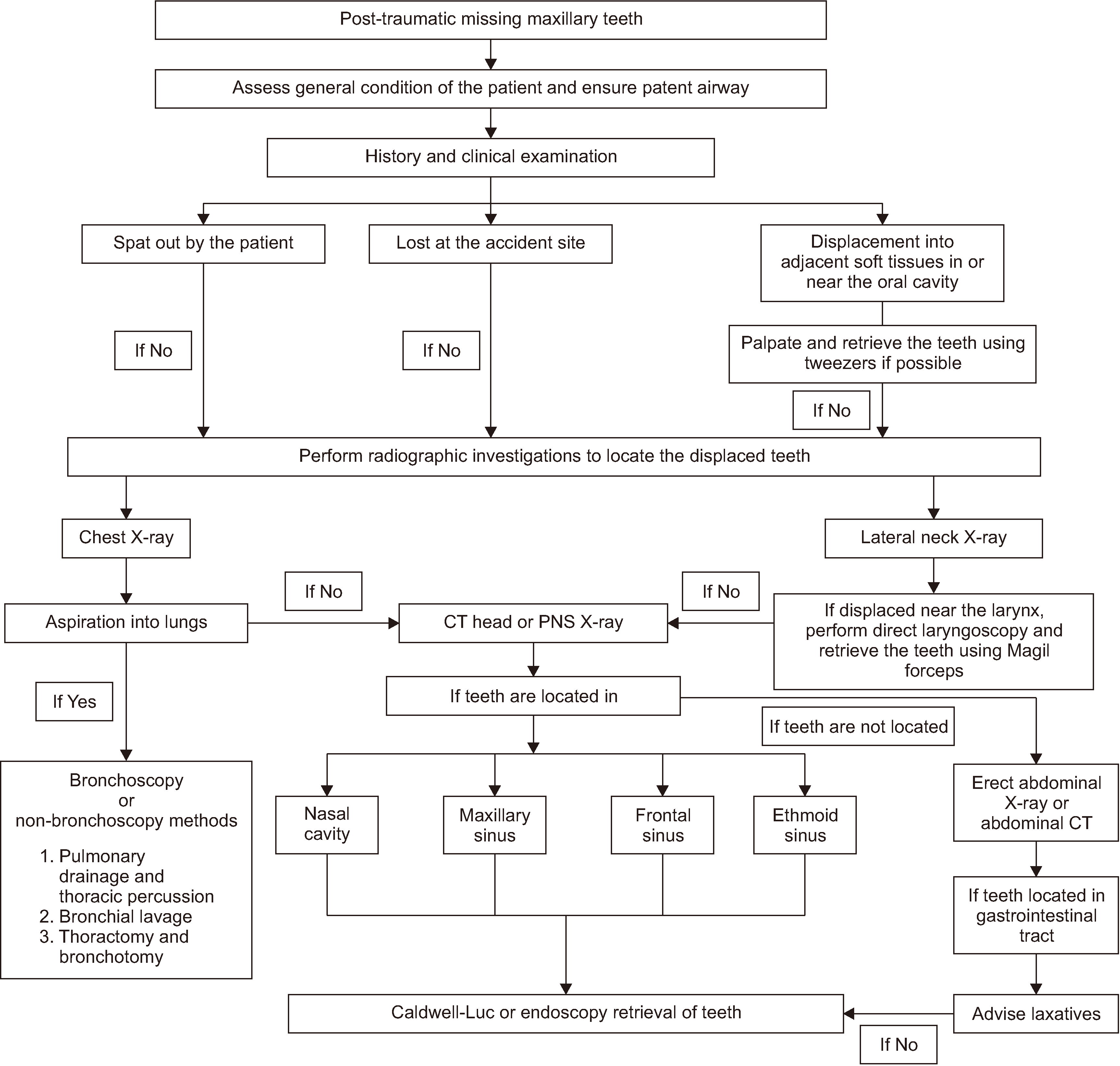
After trauma, intruded teeth missing from the oral cavity or a foreign body can be displaced along three paths: (a) expulsion, (b) aspiration, and (c) ingestion6.
In such cases, patient history is very important. In paediatric cases, history of tooth avulsion or expulsion plays a critical role. If the history is not clear, then a hierarchy of clinical examinations and a series of radiographic investigation are vital. Clinical examination involves palpation for missing teeth in the surrounding soft tissue and examination of the oropharynx. If teeth are palpable in the soft tissue near the cavity, every attempt should be made to remove them using artery forceps.
If teeth are displaced in the upper aspect of the trachea or oesophagus, they may be retrieved with simple instrumentation using haemostats, DeBakey forceps, Magill forceps, high-vacuum suction, and/or a laryngoscope7.
If the missing teeth are not located, a series of paranasal sinus view, lateral neck, AP chest8, and erect abdominal radiographs should be collected. In addition, abdominal CT can be performed. Finally, CT of the head and neck may reveal teeth displaced into the nasal cavity or adjacent paranasal sinuses9.
More serious than ingestion, aspiration of teeth must always be treated as an emergency situation. Early complications of aspiration include acute dyspnoea, asphyxia, cardiac arrest, and laryngeal oedema. Thin pointed teeth have higher risk of perforation and pneumothorax than those with a smoother shape.
Delayed removal of a foreign body beyond 24 hours may be associated with increased morbidity and longer hospital stay. Chronic retention of a foreign body can lead to formation of granulation tissue, inflammatory polyps around the foreign body, and obstruction of the bronchus10. Aspirated teeth can be removed using either a flexible or rigid bronchoscope; rarely is open thoracotomy needed11.
Ilyas et al.5 reported a case of ingestion of two teeth, one to the bronchus and the other to the liver. Fibre-optic bronchoscopy revealed a tooth in the posterior segment of the right lower lobe bronchus that was removed by a rigid bronchoscope. Removal of the tooth in the liver was not attempted, as the patient’s symptoms were relieved with conservative management.
Foreign bodies that reach the digestive tract usually can be managed with conservative treatment. After 7-10 days, teeth are naturally removed from the digestive tract by the peristaltic movement of the gastro-intestinal tract12. In some cases, however, endoscopic removal of teeth is warranted13. Jain et al.6 reported a case of accidental swallowing of a hypodermic needle by a patient during a root canal treatment, and the needle was safely removed by gastrointestinal endoscopy after 24 hours.
In conclusion, dentoalveolar fracture is common in cases of midface fracture. Teeth may intruded or can avulse and displace into surrounding soft tissues and associated structures, such as the nasal cavity, maxillary sinus, and oropharynx. Avulsed and displaced teeth also can be lost or ingested or aspirated by the patient. Proper history, adequate clinical examination, and a series of radiographs help in locating missing teeth. This paper reported a case of displaced teeth in the gastro-intestinal tract that was managed conservatively, and it provided a brief protocol for diagnosis and management of displaced teeth after maxillofacial trauma.
Go to :
 XML Download