This article has been
cited by other articles in ScienceCentral.
Abstract
Objectives
The purpose of this paper is to introduce an effective technique to easily obtain adequate amounts of keratinized gingiva and vestibular depth.
Materials and Methods
Free gingiva (vertical height 10 mm) was harvested on the palatal mucosa and a partial thickness flap was elevated on the recipient site with same width as the free gingiva graft. After a conventional suture, a titanium mesh covered the graft and was fixed with miniscrews. Titanium mesh was removed 4.1±2.5 weeks after surgery. The amount of keratinized gingiva and vestibular depth was measured at the final follow-up.
Results
Nine patients (males 4, females 5; 53.9±14.1 years) who underwent bone graft surgery before vestibuloplasty were included. No free gingival graft failure or complications were encountered in any of the patients. The relapse rate for vestibular depth (23.3%) was lower than that for keratinized gingiva (48.3%) after 34.4±14.4 months (P=0.010).
Conclusion
Vestibuloplasty with a free gingival graft using titanium mesh could be achieved with an acceptable amount of keratinized gingiva and an appropriate vestibular depth around dental implant.
Go to :

Keywords: Dental implant, Tissue graft, Titanium, Vestibuloplasty
I. Introduction
In the widespread study of dental implant success, an appropriate amount of keratinized gingiva (KG) around the implant has become a well-established success factor. A lack of KG could result in a shallow vestibular depth (VD) and positioning in the movable oral mucosa
1. VD is defined as the distance from the uppermost boundary of the attached gingiva to the lowest boundary of the mucobuccal fold, and as the functional space available for mastication and pronunciation. An appropriate VD is an important factor for maintaining good oral hygiene
2. Resulting VD may be shallow when extensive surgical procedures are performed, such as a bone graft
3. Although vestibuloplasty with free gingival graft (FGG) is a useful method for restoring deficient KG and VD, many studies have reported inadequate results because of scar formation and shrinkage after vestibuloplasty
4. Therefore, to maintain a successful implant, it is essential to consider an effective method that not only increases KG but also provides appropriate VD.
Titanium is a useful biocompatible material in dentistry because it can resist bacterial infections and withstand high temperatures and corrosion
5. In addition, when used with grafts, the sufficient rigidity of titanium mesh is expected to reduce dead space by exerting constant pressure over the graft. An approach to vestibuloplasty using titanium mesh and FGG was reported for the edentulous anterior mandible
6. The titanium mesh used for this technique was flexible enough to provide even pressure across the graft to prevent dead space, minimize the need for stitches, and reduce operation time
6. The aim of the current study was to analyze the KG and VD obtained around implants placed after vestibuloplasty using titanium mesh and FGG.
Go to :

II. Materials and Methods
The study was approved by the Institutional Review Board of Jeonbuk National University Hospital (IRB No. 2017-11-002-001), and the informed consent was waived by the IRB.
Data were collected through chart review of patients who showed a lack of both KG and VD (<1 mm;
Fig. 1) because of bone graft surgery with a muscle releasing incision for dental implantation and who underwent vestibuloplasty with a 10-mm-wide free palatal gingiva covered with titanium mesh at Jeonbuk National University Hospital between May 2013 and March 2017.
 | Fig. 1Lack of keratinized gingiva and vestibular depth due to bone graft and implant surgery. A. Patient #1. B. Patient #2. C. Patient #4. 
|
Baseline of VD and KG was set to 10 mm, which was the width of the graft. On follow-up, VD was measured from the highest margin of the gingiva to the lowest point of the vestibule, and the amount of KG was determined by measuring the vertical depth from the gingival crest to the movable alveolar mucosa. The amount of VD and KG was calculated as the average of the mesial, middle, and distal implant prosthetics.
Statistical analysis was performed using PASW Statistics for Windows (ver. 18.0; IBM, Armonk, NY, USA). Postoperative changes in the KG and VD were analyzed from the amount of relapse using a Wilcoxon signed rank test. Correlations between changes in KG and VD were analyzed using Mann–Whitney U test according to the jaw and sex. The measurement values are expressed as median [interquartile range], and P-values less than 0.05 were statistically significant.
1. Surgical procedure
The surgical procedure was performed as the described in a previous report
6. A horizontal incision was made at the mucogingival junction of the recipient site in patient #1 (
Fig. 1. A) and a vertical incision was made approximately 10 mm toward the vestibule from the mesiodistal end of the horizontal incision. A sharp dissection was made to prevent damage to the periosteum. The detached muscle was sutured with 4-0 Vicryl (Ethicon, Somerville, NJ, USA) at the vestibular fornix to create the recipient bed. A rectangular flap was designed on a hard palate and the thickness of the graft was approximately 1.5 mm.
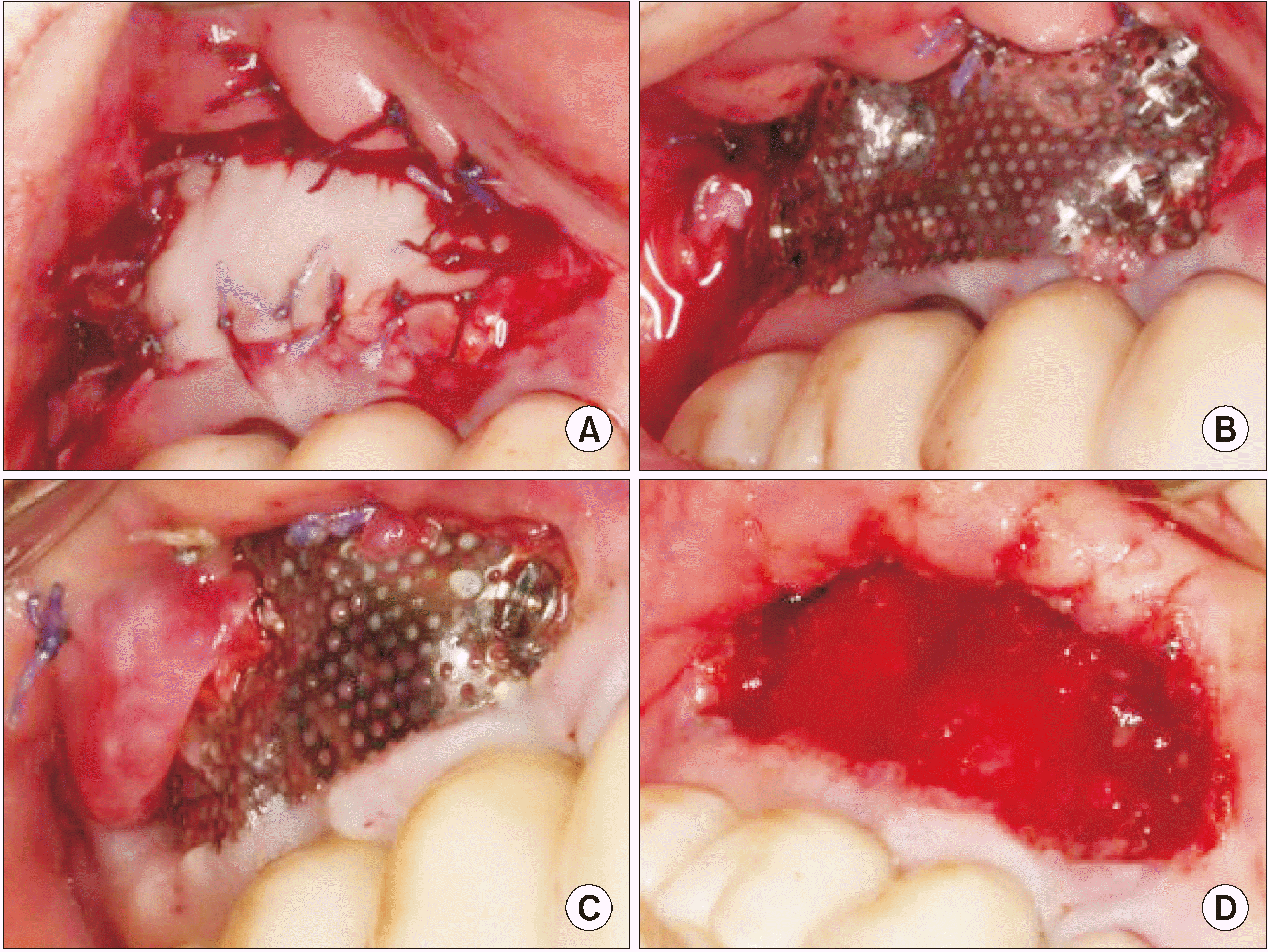
The graft was placed in the margin of the recipient bed and fixed by interrupted suture with a 5-0 Vicryl. Titanium mesh (depth 0.085 mm and hole size diameter 0.4 mm; Neo Biotech, Seoul, Korea) was applied after trimming it to the same size of the graft and was fixed with bone screws (Bone Screw System, diameter 1.5 mm and length 6 mm; Osung MND, Gimpo, Korea).(
Fig. 2. A, 2. B) The mesh was removed after four weeks to prevent relapse
7.(
Fig. 2. C) The soft tissue upturned above the mesh was trimmed before removing the mesh.(
Fig. 2. D) Clinical follow-ups took place after removal of the mesh.(
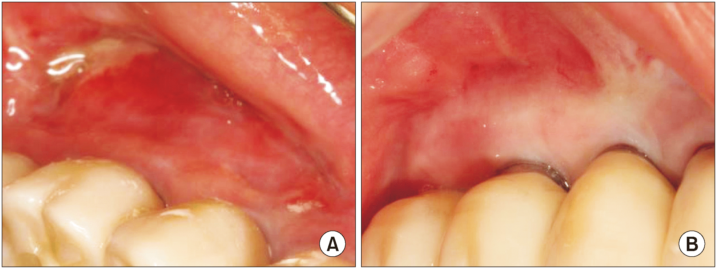
Fig. 3)
 | Fig. 2Vestibuloplasty with free gingival graft and titanium mesh. A. Suturing of the graft onto the recipient periosteum. B. Adaptation of titanium mesh with bone screws. C. Overgrowth of muscular fiber covering the titanium mesh 4.5 weeks postoperative. D. Removal of the titanium mesh and screws, and surplus soft tissue 4.5 weeks postoperative. 
|
 | Fig. 3Follow-up after removal of the titanium mesh. A. Incomplete gingival remodeling 5.5 weeks postoperative. B. Sufficient amount of keratinized gingiva and vestibular depth 10 weeks postoperative. 
|
Go to :

III. Results
Ten operations among nine patients (53.9±14.1 years, four males and five females) were included in this study. The patients underwent vestibuloplasty with the method described above 14.2±7.3 months after bone graft. Titanium mesh was removed 4.1±2.5 weeks after surgery.(
Table 1) There were no specific complications in any patients.
Table 1
Demographic and clinical characteristics of vestibuloplasty and free gingival graft covered with titanium mesh
|
Patient No. |
Sex |
Age (yr) |
General condition |
Location |
Titanium mesh maintenance period
(wk) |
Follow-up period (mo) |
Remaining KG at final follow-up (mm) |
Remaining VD at final follow-up (mm) |
|
1 |
F |
44 |
- |
Posterior maxilla |
4 |
7 |
6.3 |
6.3 |
|
2 |
M |
54 |
Hypertension (controlled) |
Posterior maxilla |
7 |
28 |
3.3 |
5.3 |
|
3 |
F |
73 |
Burning mouth syndrome |
Posterior maxilla |
4 |
34 |
8.0 |
8.0 |
|
4 |
F |
61 |
Hypertension (controlled) |
Posterior mandible |
1 |
40 |
6.0 |
9.0 |
|
5 |
M |
68 |
Diabetes (controlled) |
Posterior maxilla |
4 |
41 |
4.0 |
7.7 |
|
6 |
M |
66 |
Hypertension (controlled) |
Posterior maxilla |
1 |
49 |
5.0 |
10.0 |
|
7 |
M |
35 |
Smoking, depression |
Anterior maxilla |
2 |
41 |
5.3 |
7.7 |
|
|
Anterior mandible |
7 |
41 |
3.0 |
6.0 |
|
8 |
F |
51 |
- |
Posterior mandible |
7 |
50 |
8.0 |
10.0 |
|
9 |
F |
49 |
- |
Posterior maxilla |
4 |
13 |
6.0 |
0 |

The median amount of KG and VD remaining were 5.7 mm [3.0 mm] and 7.8 mm [3.8 mm], respectively, after 34.4±14.4 months. In terms of the relapse rate, there was a significant difference between KG and VD (48.3% [29.2%] and 23.3% [40.0%],
P=0.010).(
Table 2) Of the 10 operations, seven and three were performed in the maxilla and mandible, respectively. The relapse rates of KG and VD were similar in the jaw, but there was a significant difference between the relapse rates of KG and VD in the maxilla (50.0% [30.0%] and 23.3% [40.0%], respectively,
P=0.043).(
Table 2) The relapse rate of VD was similar between males and females, but the rate of KG varied significantly (55.0% [17.5%] among males and 36.6% [30.0%] among females, respectively,
P=0.036).(
Table 3)
Table 2
Comparison of the relapse of keratinized gingiva (KG) and vestibular depth (VD) at the final follow-up
|
Parameter |
Total |
P-value |
Maxilla (n=7) |
P-value |
Mandible (n=3) |
P-value |
|
Relapse of KG (%) |
48.3 [29.2] |
0.0101
|
50.0 [30.0] |
0.0432
|
46.7 [NA] |
- |
|
Relapse of VD (%) |
23.3 [40.0] |
23.3 [40.0] |
23.3 [NA] |

Table 3
Comparison of the relapse of keratinized gingiva (KG) and vestibular depth (VD) among the sex
|
Parameter |
Male (n=4) |
Female (n=5) |
P-value |
|
Relapse of KG (%) |
55.0 [17.5] |
36.6 [30.0] |
0.0361
|
|
Relapse of VD (%) |
23.3 [35.0] |
20.0 [38.3] |
0.395 |

Go to :

IV. Discussion
Recently, Halperin-Sternfeld et al.
2 reported that shallow VD is highly associated with a peri-implant tissue degeneration. Thus, effective vestibuloplasty could contribute to implant success. Because vestibuloplasty with FGG showed a lower relapse rate than allogeneic or collagen matrix graft
8, we devised a more effective vestibuloplasty with FGG method for reducing relapse. Vestibuloplasty with FGG and titanium mesh showed a lower relapse rate of 23.3% in terms of VD without complications compared to a previous study
9; therefore, this technique could be effective for obtaining adequate VD.
There was, however, a statistically significant difference in relapse rate between KG and VD. Many studies reported the largest shrinkage one to four weeks after FGG
7. Therefore, titanium mesh was maintained until 4 weeks to minimize relapse. The median relapse rate of KG in this study was still 48.3%. This relapse rate was similar to that of previous studies (38%-45%)
10. Therefore, the technique presented in this study could achieve an acceptable amount of KG despite the use of mesh over the graft.
In this study, it was possible to obtain rigid fixation with titanium mesh to minimize stitches, which could prevent one pathway of infection, reduce operation time, and allow close contact between the graft and periosteum without dead space. Therefore, this method could effective for lowering the technique sensitivity of FGG, and obtaining adequate KG and VD. In three patients (#4, #6, and #7 [anterior maxilla]), the mesh was removed early (1 week, 1 week, and 2 weeks, respectively) because of screw loosening; despite this, the results for both KG and VD were acceptable.(
Table 1) This suggests that titanium mesh may be most effective in the early period after vestibuloplasty.
Titanium is biocompatible and can withstand intraoral conditions of high temperatures and corrosion
5. It also has sufficient rigidity and flexibility to prevent extensive pressure on the mucosa. Because the mesh does not react physiologically, there were no serious complications associated with the mesh or the newly-grown soft tissue. In addition, the mesh could allow to separate the newly grown fibrous tissues from that of the gingival graft. Therefore, VD could be obtained more effectively than KG by trimming unnecessary tissue when removing the mesh.
One limitation of this study was that a single surgeon performed the protocol. In addition, the patients had an unequal follow-up duration, and the results were not compared to a control group without the mesh. Therefore, a well-designed prospective study with a larger number of patients will be needed to accurately assess the effectiveness of this method. In addition, further research is needed on implant prognosis and oral hygiene management in patients treated using this technique.
Go to :

V. Conclusion
There were no complications, such as graft failure, after vestibuloplasty with titanium mesh-covered FGG. The mesh did not adversely affect the prognosis of the grafted gingiva or the formation of KG. In addition, the mesh effectively prevented relapse in terms of VD. Vestibuloplasty with FGG using titanium mesh can be used to achieve the appropriate amount of KG and VD without complications.
Go to :


 PDF
PDF Citation
 Print
Print


 XML Download