

 PDF
PDF Citation
 Print
Print


I. Introduction
The term “lag screw” describes both a particular form of screw design and a surgical technique. “Lagging” refers to “covering up”1, as the bone under the head of the screw covers or lags the underlying bone2. While the lag screw technique has only a limited role in maxillofacial osteosynthesis, it is commonly used in orthopedic surgery3,4. It was originally introduced to oral and maxillofacial surgery by Brons and Boering5, who concluded that the lag screw compresses fracture segments, and hence is classified as a rigid internal type of fixation.
The original true lag screw fixation technique involved a unique fabrication design. There are no screw threads in the section of the screw shaft nearest to the head, with the shaft size in this area equal to the total screw diameter in the threaded zone6. The compressive force of the head of the screw against bone is thereby transmitted to the bone that is secured to the threaded screw portion. This technique is costly and not widely available in all countries, as many are not served by maxillofacial suppliers and manufacturing companies. Meanwhile, the modern lag screw technique uses a screw with threads along the entire length and is applied using a special technique that is different from the true lag technique and is therefore cheaper and more widely available7.
In the past, the main indication for compression using lag screws was in the management of oblique fractures, whereby one section of mandibular cortex is split longitudinally from another for some distance through the cancellous bone. In this context, the technique is referred to as the gliding sliding principal8. No additional instrumentation is required for this technique except for special true lag screws, with threads that engage only the inner boneplate9.
The cortical lag screw fixation technique (CLSFT) is performed using ordinary titanium cortical screws that are threaded for the entire length, rather than true lag screws10. The procedure depends on the drilling technique itself and requires two different drill diameters11.
The true lag screw technique has been used and studied for the treatment of anterior mandibular fractures and other locations of mandibular fractures, such as those at the mandibular angle12 and body13, and has also been used to reduce condylar fracture, as first described by Petzel and Bülles14 and utilized by others4,14-17. It is used in various surgeries such as genioplasties, sagittal split ramus osteotomies, bone graft fixation, and alveolar ridge augmentation18. Despite the many applications of the true lag technique, the CLSFT using normal threaded screws has not been thoroughly investigated as an alternative.
The goal of this study was to present cases of mandibular fracture treated with CLSFT in our institute in order to critically evaluate its indications, contraindications, techniques, limitations, and potential complications at different mandibular fracture sites.
Go to : 
II. Materials and Methods
1. Study design and setting
This study included 33 patients treated at Al-Azhar University Hospital, Faculty of Dental Medicine for Girls, Cairo, Egypt from October 2007 to October 2017. They suffered from mandibular fractures at different sites that were treated with CLSFT through open reduction and internal fixation. The present study was reviewed and ethically approved by the Al-Azhar University Ethical Committee Board (No. PD-P-020-004) and conducted in compliance with the human research ethics principles in the Declaration of Helsinki (2008). All patients provided written informed consent prior to surgery.
2. Selection criteria
The present study included all mandibular fracture cases fixated by double or single CLSFT during the study period. Selected patients were free from any systemic diseases that could interfere with surgery or bone healing. Any cases that were fixated by the true lag screw technique or any other means of fixation were excluded.
3. Study variables
The sites of the fractures were treated as predictors. The outcome variables were fracture characteristics (type, side, cause, and degree of displacement). Other categories of analysis included number of CLSFT screws, pattern of application, and duration of application.
4. Data collection
1) Preoperative evaluation
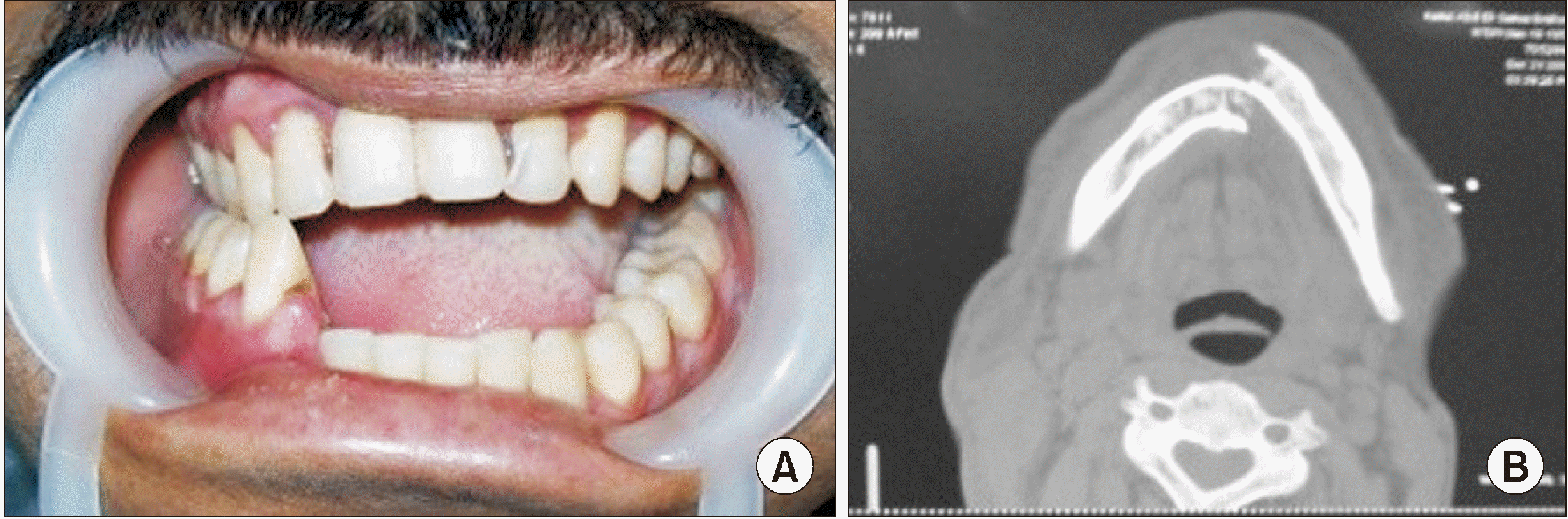
A complete history was acquired and registered for each patient according to a standard protocol. Systematically, the clinical review was conducted in two phases, extraoral and intraoral. Examinations were performed to detect potential lacerations, swelling, ecchymosis, facial asymmetry or malocclusion, deformity of the mandibular bony contour, and any restrictions of mandibular movement (opening, lateral movements, protrusive movements, and maximum interincisal openings).(Fig. 1. A)
General facial palpation was conducted with particular attention provided to the area around the mandible that was palpated bimanually, beginning with the condylar region and progressing over the mandible to identify any areas of tenderness, phase deformities, or bony crepitus. The lower lip and chin area were examined to detect any signs of nerve dysfunction, paresthesia, or anesthesia. The teeth were also palpated for tenderness or mobility on both sides of fracture lines.
2) Radiographic examination
Preoperative digital orthopantomogram was the standard radiography for initial assessment of the type of fracture line. Other views were requested as necessary, such as posteroanterior or computed tomography.(Fig. 1. B) These views were used to determine fracture direction and number of fracture lines. Diagnosis was based upon patient history and clinical and radiographic examinations, and the treatment plan was selected accordingly.
3) Preoperative preparations
At initial presentation, special preparations were taken before proceeding with the surgical phase of the treatment, including care of any soft tissue lacerations and wound debridement, and support and reduction of the fractured bony segments using both maxillary and segmented mandibular arch-bars at the fracture line or Ivy loops. If the fractures were severely displaced, gradual reduction was performed using elastics 24 hours before maxillofacial mandibular fixation (MMF).
Patients fasted for eight hours before surgery. All patients received 1.5 g of sulbactam/ampicillin (Unasyn 1.5 g vial; Pfizer, Cairo, Egypt). We administered 1,500 IM units of tetanus antitoxin (tetanus antitoxin; Pasteur Merieus, Lyon, France) in cases of soft tissue injuries contaminated by dirt. The patients were anesthetized using a cuffed tube and a mixture of fluthane/oxygen gas. The intraoral and extraoral regions were scrubbed using betadine (povidone-iodine; The Nile Company, Mundipharma, Switzerland).
5. Surgical technique of CLSFT
CLSFT was performed using ordinary titanium cortical screws threaded to the whole length. The procedure used depended on the drilling technique. We used two different drill diameters: a smaller drill diameter in the distal fragment hole, or the traction hole10, and a larger drill equal to the diameter of the outer screw threads in the outer fractured segment so that when the screw was tightened the threads would not engage the outer cortex. We also took care to maintain sufficient distance between the countersink and the fracture site to allow for satisfactory compression11.(Fig. 2. A)
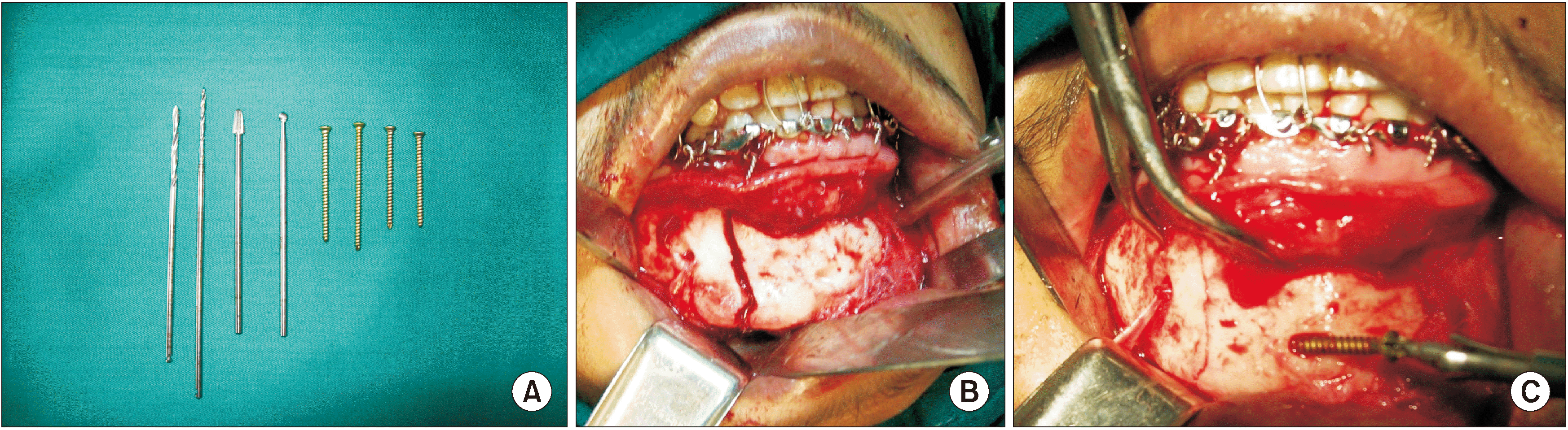 | Fig. 2A. Photograph showing drills and screws used in cortical lag screw fixation technique (2.4 mm and 1.8 mm drill bits, pear and round shaped burs for countersinking and 2.4 mm screws of different lengths). B. Intraoperative exposure of a linear parasymphyseal fracture. C. Fracture reduction and pre-compression using bone clamp forceps and screwing of the first screw.
|
In the region of the symphysis and parasymphysis, two cortical titanium (2.4 mm diameter and 28-40 mm length; Synthes Maxillofacial, Oberdorf, Switzerland) were used. In our technique, the traction hole was first drilled using a smaller drill bit (1.8 mm) that crossed the fracture line from the outer buccal cortex to reach the opposite cortex, and then the gliding hole in the near cortex was drilled using a larger drill bit (2.4 mm). Preload was applied with bone clamps.(Fig. 2. B, 2. C)
Visualizing the exit site of the drill bit on the buccal side of the far segment is important to maintain an appropriate distance from the fracture line. The screw length was measured by means of a depth gauge. A second lag screw was similarly placed from the same side or in the opposite direction to the first screw. Screws were tightened by hand using a cross-head screwdriver until complete reduction and compression of the fracture was obtained. To avoid palpability the head of the screw was counter-sunk.(Fig. 3. A, 3. B) Just one CLSFT screw could be applied at the body area. The fracture line was maintained strictly as a broad oblique surface fracture line and drilling was performed for the parasymphysis area.(Fig. 3. C)
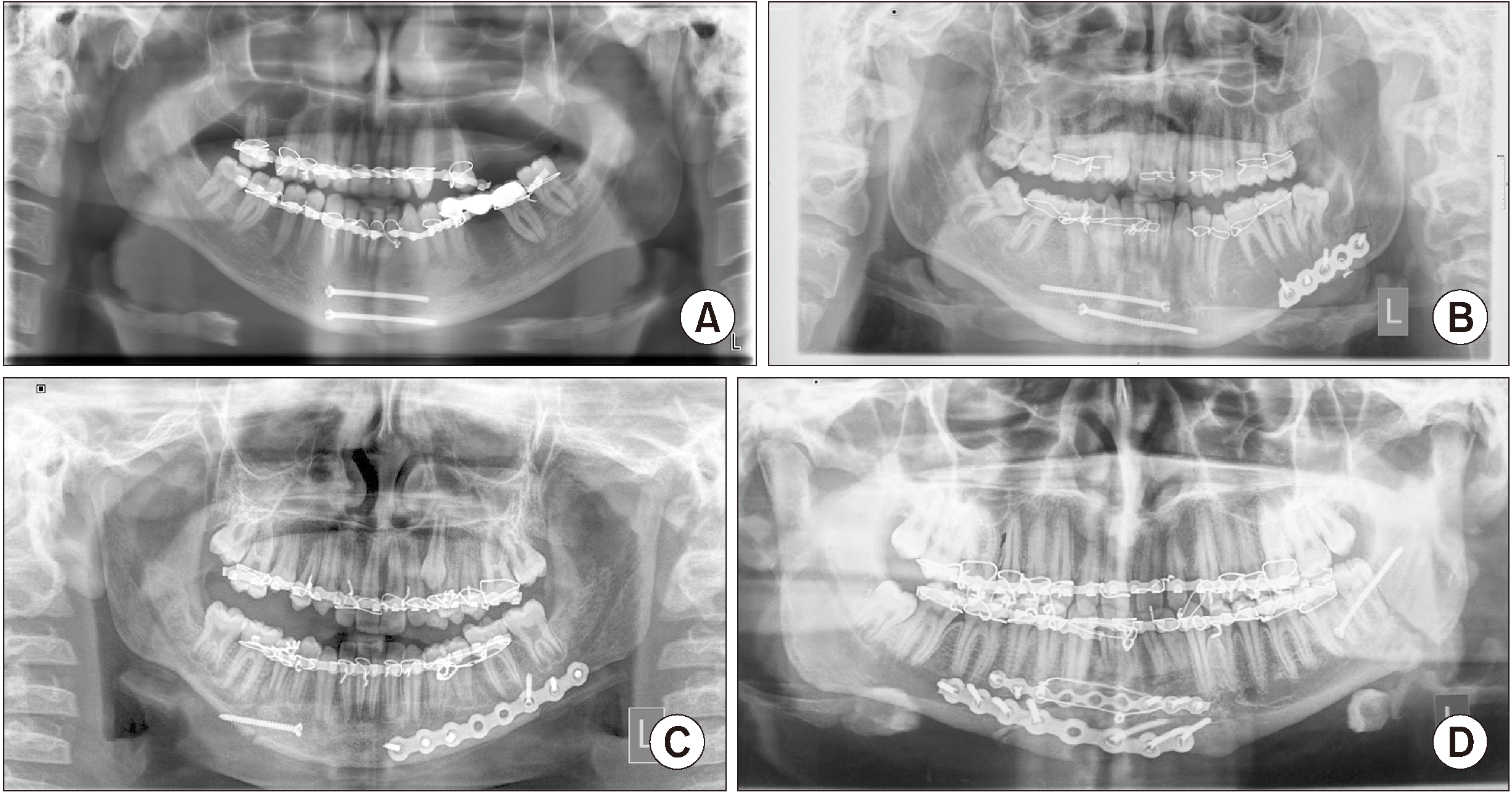 | Fig. 3A. Postoperative digital orthopantomogram radiograph showing perfect reduction and fixation of the symphyseal fracture using double lag screws in the same direction. B. Reduction and fixation of parasymphyseal fracture using double lag screws in the opposite direction. C. Perfect reduction and fixation of body fracture using a single lag screw. D. Reduction and fixation of angle fracture using a single lag screw.
|
At the angle region, a single lag screw was placed via intraoral posterior vestibular incision. The screw was tightened until adequate compression was achieved. The technique used for fixation of CLSFT in the angle region was previously described in detail by Elsayed et al.12 and applied in the present study in the same manner.(Fig. 3. D)
Deep resorbable sutures (2 horizontal mattress sutures) were inserted in the mentalis muscle to prevent lip and chin ptosis, then the mucosa was continuously repaired with resorbable sutures, and a pressure dressing was applied to the area to prevent the development of hematoma and maintain the mentalis muscle position. The MMF was removed at the end of the operation, and occlusion was rechecked.
6. Treatment of concomitant mandibular fractures
Concomitant mandibular fractures were found in 15 cases (45.5%). One patient suffered from an associated clavicle fracture. Two patients suffered from an associated left humerus fracture and one patient suffered from rib fracture and pneumothorax preoperatively at the time of admission. The intraoral vestibular approach was used for all cases associated with concomitant mandibular fractures and fractures were fixed with double miniplates or a single heavy plate.
Unfavorable displaced subcondylar fractures were found in 2 patients in addition to anterior mandibular fractures. Subcondylar fractures were approached intraorally and fixed with double miniplates. One patient had a sagittal subcondylar fracture that was treated conservatively.
CLSFT was applied in two anterior concomitant maxillary fractures, where the fracture occurred in the area of the anterior maxillary fissure exactly in the midline, and the compression achieved successfully reduced the two maxillary bones and made the fracture line unnoticeable after reduction. The drilling technique was performed exactly as described for fractures of the anterior mandibular symphysis.
7. Data analysis
Descriptive characteristics for the study variables were tabulated and analyzed using chi-square tests for comparisons of fracture sites and associations with investigated categorical variables of postoperative complications and surgical duration. P<0.05 were considered significant. Statistical analysis was performed using SPSS (ver. 16.0; SPSS, Chicago, IL, USA).
Go to :
III. Results
In this study, the surgical procedures included 33 internal fixations of mandibular fractures in 33 patients of both sexes (27 males and 6 females) who met our criteria for inclusion in the study and were fixated via CLSFT. The numbers of CLSFT screws used in the present investigation were two for the symphysis and parasymphysis and one for body, angle, and anterior maxillary regions.
Patient age ranged from 13 to 58 years (mean, 30.9±11.5 years). The fracture etiologies were road traffic accidents in 19 patients, assault in 3 patients, falls in 9 patients, and animal kicks in 2 patients. Table 1 provides all details about age, sex, etiology, fracture site, and concomitant fractures. The intraoral approach was used in all cases. The mean time from incision to fracture fixation did not exceed one hour, and operative time in the body and angle regions was shorter than in anterior sites where two screws were inserted (P=0.03).
Table 1
Descriptive characteristics of the fracture site variables and demographic characteristics of the study sample
![]()
Electrocauterization was required in only one patient due to excessive bleeding in the molar region during the dissection of the vestibular incision. All patients had teeth involved in the fracture line and these were retained in all cases except in one case at the time of reduction, as the lower canine involved was grossly broken and mobile, necessitating its extraction.
Table 1 indicates that the CLSFT principal mainly applied in the areas of the symphysis and parasymphysis (51.5%) followed by the angle and body regions (30.3% and 18.2%, respectively). The sample was 81.8% male, but the sex ratio did not differ significantly between sites (P=0.07).
1. Preoperative clinical and radiographic findings
Fractures were treated within 1 to 10 days of injury incidence. At the time of admission all patients were conscious, alert without any systemic problems except four patients who were first admitted to the ICU (intensive care unit) in coma after trauma, and then to the neurosurgical department for monitoring and controlling neurological conditions. All patients had intraoral lacerations related to the fracture site, and 4 also had extraoral skin lacerations. Ten cases had severe swelling in the fracture area with remarkable facial asymmetry and severe clinically and radiographically obvious displacement of fracture segments, whereas moderate swelling was noticed in 9 patients and mild swelling was observed in 14 patients.
Post-traumatic malocclusion was identified preoperatively in all cases as patients suffered from varying degrees of occlusal disturbances (malocclusion and step deformity, mobility, or open bite). All of the patients had limited mandibular movement. The maximum preoperative interincisal mouth opening was 10-20 mm. Two patients presented with lower lip numbness related to the fracture side.
2. Postoperative clinical findings
Minimal edema was immediately observed postoperatively in 60.6% of cases that started to resolve on the fourth day and disappeared on the seventh day. In general, all incisions healed uneventfully. Incision lines that were hidden became fainter by the end of the follow-up period. Postoperatively, proper occlusion was achieved in all patients and maintained during the follow-up period except for one patient with an angle fracture who was stabilized via single CLSFT and required MMF for 2 weeks in conjunction with lag fixation.(Table 2)
Table 2
Correlations between lag screw osteosynthesis site and outcome variables
| Variable | Category | Total | Body | Parasymphyseal | Symphyseal | Angle | P-value |
|---|---|---|---|---|---|---|---|
| Mandibular fracture site | Site | 33 (100) | 6 (18.2) | 9 (27.3) | 8 (24.2) | 10 (30.3) | - |
| Surgical time (min) | Range, 30-60 min | 45.36±9.54 | 32.50±9.55 | 53.67±5.11 | 48.80±9.94 | 40±0 | 0.035* |
| Double lag screw fixation technique | 17 (51.5) | 0 | 9 (27.3) | 8 (24.2) | 0 | ||
| Postoperative edema | Mild | 20 (60.6) | 3 (9.1) | 8 (24.2) | 6 (18.2) | 3 (9.1) | 0.10 |
| Moderate | 11 (33.3) | 3 (9.1) | 1 (3.0) | 2 (6.1) | 5 (15.2) | ||
| Severe | 2 (6.1) | 0 | 0 | 0 | 2 (6.1) | ||
| Wound dehiscence | Yes | 1 (3.0) | 0 | 0 | 0 | 1 (3.0) | 0.49 |
| No | 32 (97.0) | 6 (18.2) | 9 (27.3) | 8 (24.2) | 9 (27.3) | ||
| Postoperative infection | Yes | 1 (3.0) | 0 | 0 | 0 | 1 (3.0) | 0.4 |
| No | 32 (97.0) | 6 (18.2) | 9 (27.3) | 8 (24.2) | 9 (27.3) | ||
| Postoperative paresthesia | Yes | 4 (12.1) | 0 | 1 (3.0) | 1 (3.0) | 2 (6.1) | 0.7 |
| No | 29 (87.9) | 6 (18.2) | 8 (24.2) | 7 (21.2) | 8 (24.2) | ||
| Postoperative malocclusion | 1 (3.0) | 0 | 0 | 0 | 1 (3.0) | 0.49 |
![]()
Stability of osteosynthesis devices was tested immediately intraoperatively, using bimanual mobility tests of the fractured segments. It was very difficult to place a second screw on more posterior angle fracture lines. Palpation of the inferior border revealed properly aligned segments in all patients with no step deformities except in two cases of parasymphyseal fractures, as the step was only present at the inferior border and did not affect occlusion.
Four cases (12.1%) had lower lip numbness related to the side of the fracture but this number was not significant (P=0.7). By the end of the second week, considerable improvement was reported by these patients. At end of the study, after a period of six months, all affected patients had regained normal mental nerve function without permanent effects.
Patient compliance was good and none reported any discomfort from the screws, so they were maintained in place until the end of the study period. Loose screws were not observed in any patient. Regaining of the interincisal mouth opening was rapid and the maximum mouth opening ranged from 25-28 mm immediately after the operation to 39-45 mm after one month. All concomitant fractures healed uneventfully without complications.
3. Postoperative radiographic findings
The immediate postoperative radiographs, taken in the first week, showed considerable narrowing of the fracture gap. One month postoperatively, the fracture lines were easily identified. At three months postoperatively, the fracture lines were unidentifiable in all cases. Six month postoperative radiographs revealed satisfactory bone healing and fracture lines could not be identified in any cases. None of the patients showed nonunion or malunion.
Go to :
IV. Discussion
Here, we present our experience in managing maxillofacial fractures using the CLSFT instead of true lag or other heavy plates and screws. We present cases managed with CLSFT in order to critically evaluate technique indications and limitations of its implementation at various sites of mandibular fracture that demonstrate the best outcomes of clinical practice.
Although true lag screws are more common in orthopedic surgery19, 2.4 mm cortical screws threaded along the entire length were used in the current study. This diameter differed from that used for fixing parasymphyseal fractures by Kallela et al.20,21, who used 2.7 mm and 2.0 mm screws. In the lag screwing technique, the application of a single lag screw in the symphysis area may cause fracture segments to rotate and for this reason 2 lag screws were used in the symphysis region in the present study. However, due to the presence of neurovascular structures and teeth apices, only single lag screws were applied in the body and angle areas. The lag screws must be of sufficient length, longer than 20 mm. In the current study, the length of the screws used ranged from 28-40 mm.
The most commonly indicated conditions for the application of the CLSFT in the present study were symphyseal and parasymphyseal fractures (51.5%) followed by fractures of the body and angle (48.5%), and our findings agreed with those of Tiwana et al.22 and Terheyden et al.10. No condylar fractures were fixed with lag screws in this study sample.
Advantages of CLSFT include that it provides very strong, rigid fixation when properly applied and guarantees perfectly stabilized fractures, achieving primary bone healing with minimal use of osteosynthesis devices and lower costs in terms of operative time and material price23. However, the CLSFT is sensitive, as it would be difficult to counter-sink screws when the outer cortex is comminuted or fragmented16 and therefore it is not suggested in every case15.
The technique used for CLSFT placement in this study was different from that described by Ellis and Ghali6, Niederdellmann et al.24 and Kallela et al.20, as we first drilled the traction hole using a smaller drill bit (1.8 mm) that crossed the fracture line from the outer buccal cortex to the opposite cortex, followed by drilling of the gliding hole in the near cortex using a larger drill bit (2.4 mm).
Using this technique, we found that the eccentricity at the traction hole was minimal, therefore reducing the occurrence of malalignment that may occur during screw tightening. This technique was previously used in another study25. However, in more commonly-used techniques, drilling of the gliding hole is performed first in the near cortex followed by drilling of the traction hole through the gliding hole, which requires mandatory use of drill guides.
Placement of the incision in the oral cavity allows for superb exposure of a large part of the facial skeleton and the advantage of a fully hidden scar, as well as a rapid and safe mandibular vestibular approach. The intraoral approach provides good surgical exposure in all directions, allowing internal fixation of all concomitant mandibular fractures such as associated subcondylar and other side fractures. This eliminates the time needed for dissection in extraoral approaches. The fracture site was visualized simultaneously with dental occlusion, reducing the risk of postoperative malocclusion. This technique follows Ellis and Graham26 and several other studies27,28 that recommend the use of intraoral incisions. On the other hand, Forrest29 inserted lag screws via small incisions transmucosally or percutaneously after fracture reduction. Meanwhile, it has been reported that the intraoral approach to angle fractures is the most difficult, requiring more time for the use of transbuccal trocar or right-angled screwdrivers for screw fixation, but the intraoral approach used with the lag screw technique does not result in difficulty in fixation of the lag screw except for cases requiring the insertion of a second screw, as the mandibular canal was approximate and the space available was very limited12.
In the current study, we noted that the more recent the fracture, the easier the reduction and the faster the fixation because granulation tissue was easily removed and the fracture was easily mobilized and reduced. All patients were treated within 1-10 days of the date of injury. This finding is in agreement with those of previous studies12,30,31, which reported that earlier intervention is better than delayed management. Males made up most of the sample in the current study (81.8%), which follows a number of previous studies12,32 showing that males are more susceptible to mandibular fractures than females.
In the present study, dehiscence of the wound did not occur in the anterior and body regions. This may be due to the very low profile (only the screw head) placed under the wound, which allows a relaxed suturing technique. Clinically, complications were minimal in except for 12.1% of patients who reported mental nerve effects. However, mental nerve recovery did not require prolonged time. This effect may be attributed to the mental nerve and soft tissues stretching during surgery. Moreover, the lag screw was applied more rapidly than other techniques are typically applied23. In the present study, the mean time required to place two lag screws was 53.67±5.099 minutes. The duration and severity of neurosensory disability were directly related to the amount of trauma inflicted on the mental nerve33.
Occlusion was obtained in all cases after CLSFT in the present study. However, in angle fractures it appears that a single lag screw is not sufficient, and these patients required 2 weeks of MMF in 3% of cases. However, 100% bone healing was obtained in all cases, and this agrees with the main goal of fixation as outlined by Miles et al.34.
In this study, no infections were identified postoperatively in the symphyseal and parasymphyseal regions. No complications such as fracture segment malpositioning or severance of neurovascular structures or teeth were found in any case. This finding is consistent with those of several studies27,28,35,36 reporting that rigid fixation produces better, uncomplicated healing outcomes than traditional methods. However, others concluded that postoperative fracture line infection rate did not differ according to the technique used. Such results are contrary to those of Kallela et al.20 and Ellis and Ghali6, who reported 11.7% and 4.9% infection rates in parasymphyseal fractures, respectively.
Correct repositioning of fracture segments during surgery was obtained in all groups, and radiographic observation at postoperative one month revealed no interfragmentary gaps between bone ends, which means that no formation of callus was observed. This is consistent with the principle of direct bone healing, which allows absolute immobilization as in rigid fixation so that the full strength of the bone is restored more rapidly.
Forrest29 treated five patients suffering from anterior mandibular fractures using lag screws and concluded that lag screws can be applied with minimum exposure, thus reducing morbidities such as swelling, scarring, and nerve injury. Moreover, Nyárády et al.37 treated 468 patients suffering from alveolar fractures using transgingival lag screws and recommended this technique when the blood supply is jeopardized and MMF cannot be used.
Huang et al.38 evaluated clinical results in 12 patients who suffered from anterior mandibular fractures that were fixed using lag screws. They concluded that lag screw fixation can achieve good stability, appropriate compression with reduced surgical time, and promote osseous healing without the need for plate adaptation.
The need for affordable equipment in developing countries requires the use of the cheapest osteosynthesis fixation techniques possible12. To avoid waiting for the purchase of appropriate plates and screws, the use of CLSFT is the cheapest option available in these situations. The single long CLSFT used in the current investigation costs 600 Egyptian pounds, which is equivalent to $38 USD and therefore costs much less than bone plates. Such cost savings are beneficial in low socioeconomic status countries.
The success rate reported in this study maybe be related to early presentation to operation. The limitation that we found in this study was that the removal of these devices could be very difficult after healing, as is usually indicated with other fixation devices (plates and screws are usually removed after 6 months). Although this may be a problem, in the present study none of the patients returned to have these screws removed.
Go to :
V. Conclusion
Our clinical experience indicates that CLSFT is the most cost-effective and rapid technique for fixation of mandibular fractures, producing good treatment results with very limited complications. However, this technique is sensitive and requires surgical expertise. It may be applied to most mandibular fractures with specialized characteristics, as follows:
• Fresh sagittal split (true symphyseal) fractures.
• Single straight line, parasymphysis fractures directed anteriorly at the inferior border were more accessible. Fracture segments that were posteriorly directed and comminuted or cracked were not favorable.
• Long chins with curved or curled anterior regions are ideal for the application of two lag screws.
• Standardized indications of oblique transverse surface fractures in the anterior, body, and angle regions.
• CLSFT is not indicated in comminuted fractures, old, or delayed fractures, or in missing segment or gap fractures (in cases where compression is contraindicated).
Go to :
 XML Download