

 PDF
PDF Citation
 Print
Print


Numerous surgical methods have been practiced in the management of odontogenic keratocysts (OKC); however, much confusion still exists as to which methods provide the lowest recurrence rates (RR) without causing significant morbidity. These methods are divided into conservative and aggressive/radical approaches or a combination of the two. Conservative methods include simple enucleation and marsupialization/decompression1. Adjuvant methods such as peripheral ostectomy, cryotherapy (liquid nitrogen) and Carnoy’s solution are considered aggressive forms of treatment which have shown more promising outcomes1. Radical methods involve mainly resection which yields the lowest RR however causes significant morbidity1.
It is currently considered that enucleation alone is an inadequate form of treatment and needs to be used in combination with adjuvant methods to lower RR2,3. Epithelial remnants and satellite/daughter cysts can easily be left behind after enucleation which leads to high RR (20.8%-26.1%)4.
Decompression is a modified marsupialization technique which causes the cyst to decrease significantly in size and the cystic lining becomes thicker resembling oral mucosa that allows for easier enucleation2. This method decreases the levels of IL-1α which regulates epithelial cell proliferation in OKC; hence, there is immune-histochemical evidence that decompression is superior to enucleation alone2. de Castro et al.4 reported RR of OKC treated by decompression followed by enucleation (11.9%) to be significantly lower than enucleation alone (20.8%).
Peripheral ostectomy is an aggressive form of adjuvant therapy where methylene blue is utilized to stain any cystic remnants and a rosehead bur is used to remove these2. Cryotherapy with liquid nitrogen causes cell necrosis of the cystic lining as with chemical curettage with Carnoy’s solution2. All these methods reduce RR when compared to enucleation or marsupialization/decompression alone; however, these methods can cause damage to adjacent structures2. Resection provides the least recurrences (RR, 0%-8.4%)5,6.
Data from five large systematic reviews reporting on RR for different treatment modalities were combined (Table 1) and these formed the basis for developing a management protocol for OKCs1,4
-7.(Fig. 1) The protocol highlights the role of advanced imaging in OKC management. Magnetic resonance imaging (MRI) is the only non-invasive modality that characterizes OKC accurately, preoperatively. T2-weighted images show hyper-intensity with signal drop out which is highly suggestive of OKC (85% sensitivity)8. Computed tomography (CT) can detect cortical perforations8.
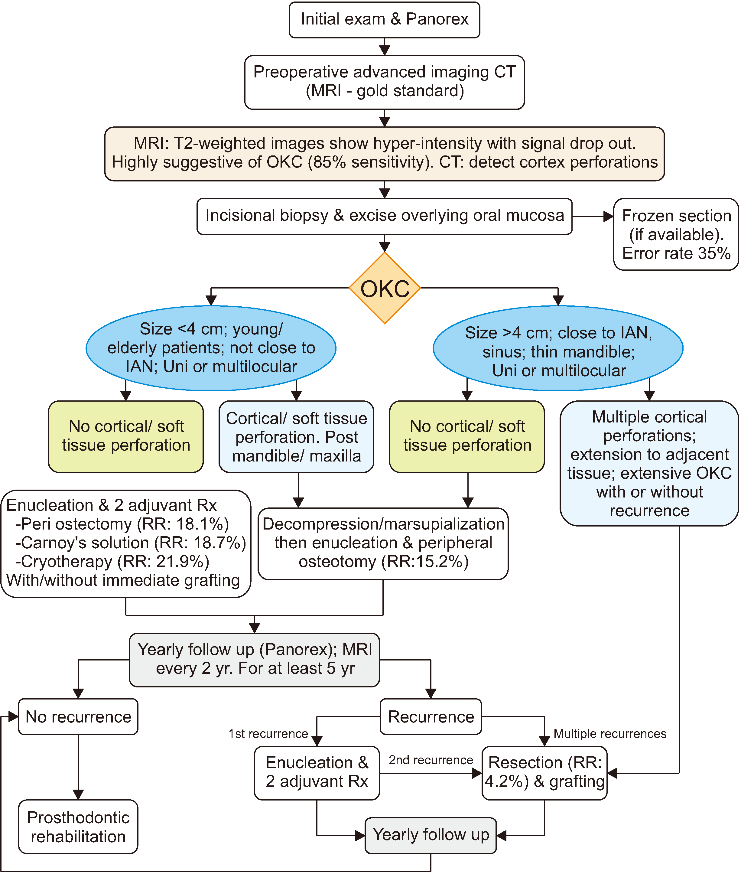 | Fig. 1Management protocol for odontogenic keratocysts (OKC). (CT: computed tomography, MRI: magnetic resonance imaging, IAN: inferior alveolar nerve, Rx: treatment, RR: recurrence rates)
|
Table 1
Summary of recurrence rates for different surgical methods in the management of odontogenic keratocysts
| Study | Enucleation alone | Enucleation & peri ostectomy | Enucleation & Carnoy’s solution | Enucleation & cryotherapy | Marsupialization/decompression alone | Decompression & residual cystectomy | Resection |
|---|---|---|---|---|---|---|---|
| Al-Moraissi et al.1 (2017) | 23.10 | 17.40 | 11.50 | 14.50 | 32.30 | 14.60 | 8.40 |
| de Castro et al.4 (2018) | 20.80 | NA | NA | NA | 18.50 | 11.90 | NA |
| Chrcanovic and Gomez7 (2017) | 22.50 | 18.60 | 5.30 | 20.90 | 28.70 | 18.60 | 2.20 |
| Johnson et al.5 (2013) | 25.60 | NA | 7.90 | 30.30 | NA | 15.80 | 6.30 |
| Kaczmarzyk et al.6 (2012) | 26.09 | 18.18 | 50 | NA | 40 | NA | 0 |
| Total | 23.60 | 18.10 | 18.70 | 21.90 | 29.90 | 15.20 | 4.20 |
![]()
Other important factors in lowering the RR of OKC include excision of the overlying oral mucosa, especially at the time of biopsy, as studies have shown that approximately all recurrent OKCs contain epithelial islands and micro-cysts in the mucosa overlying areas of cortical perforation9.
The surgical approach is determined by size, patient age, proximity to vital structures, accessibility, soft tissue/cortical perforation, and if the lesion is recurrent. Age is an important factor in the recurrence of OKC, with younger patients (especially those in their second to third decade of life) experiencing higher RR that should be managed more aggressively10. Decompression followed by enucleation has been shown to reduce RR (15.2%). If this technique is followed by an adjuvant method such as application of Carnoy’s solution and peripheral ostectomy, it can result in very low recurrence and is an acceptable first line of treatment6. If the lesion is accessible and not large in size, then enucleation of the cyst should be performed along with 2 adjuvant methods. Resection should be reserved for multiple recurrent lesions, multiple cortical perforations, and soft tissue extension.
OKC should be monitored closely with yearly panoramic radiographs2. MRI should be performed every 2 years to detect early recurrences8. Follow-up should be for at least 10 years2. Bone grafting and rehabilitation can be performed early in cases of resection, a thin mandibular cortex, and large lesions that were treated with cryotherapy2. Bone grafting should be delayed in recurrent lesions2.
In conclusion, an evidence-based protocol for management of OKCs has been proposed. In order to reduce recurrence and morbidity, a systematic approach should be implemented based on presenting factors and sound evidence-based treatment options. This protocol can also be used as a valuable educational tool for patients.
 XML Download