Journal List > J Korean Assoc Oral Maxillofac Surg > v.46(5) > 1145906

Nguyen, Sodnom-Ish, Choi, Jung, Cho, Hwang, and Kim: Salivary biomarkers in oral squamous cell carcinoma

Abstract

In disease diagnostics and health surveillance, the use of saliva has potential because its collection is convenient and noninvasive. Over the past two decades, the development of salivary utilization for the early detection of cancer, especially oral cavity and oropharynx cancer has gained the interest of the researcher and clinician. Until recently, the oral cavity and oropharynx cancers are still having a five-year survival rate of 62%, one of the lowest in all major human cancers. More than 90% of oral cancers are oral squamous cell carcinoma (OSCC). Despite the ease of accessing the oral cavity in clinical examination, most OSCC lesions are not diagnosed in the early stage, which is suggested to be the main cause of the low survival rate. Many studies have been performed and reported more than 100 potential saliva biomarkers for OSCC. However, there are still obstacles in figuring out the reliable OSCC salivary biomarkers and the clinical application of the early diagnosis protocol. The current review article discusses the emerging issues and is hoped to raise awareness of this topic in both researchers and clinicians. We also suggested the potential salivary biomarkers that are reliable, specific, and sensitive for the early detection of OSCC.

I. Introduction

A biomarker is defined as ‘a biological molecule found in blood, other body fluids, or tissues that is a sign of a normal or abnormal process, or of a condition or disease’ by the National Cancer Institute 1 . The biomarker, also called a molecular marker, has a wide range of applications in diagnosis, monitoring of treatment, and the prognosis of a disease or condition. Despite attempts to classify biomarkers of cancer, a consensus has not been established. Mishra and Verma 1 have suggested that the classification of biomarkers can be based on the disease state, biomolecules, or other criteria.(Fig. 1)
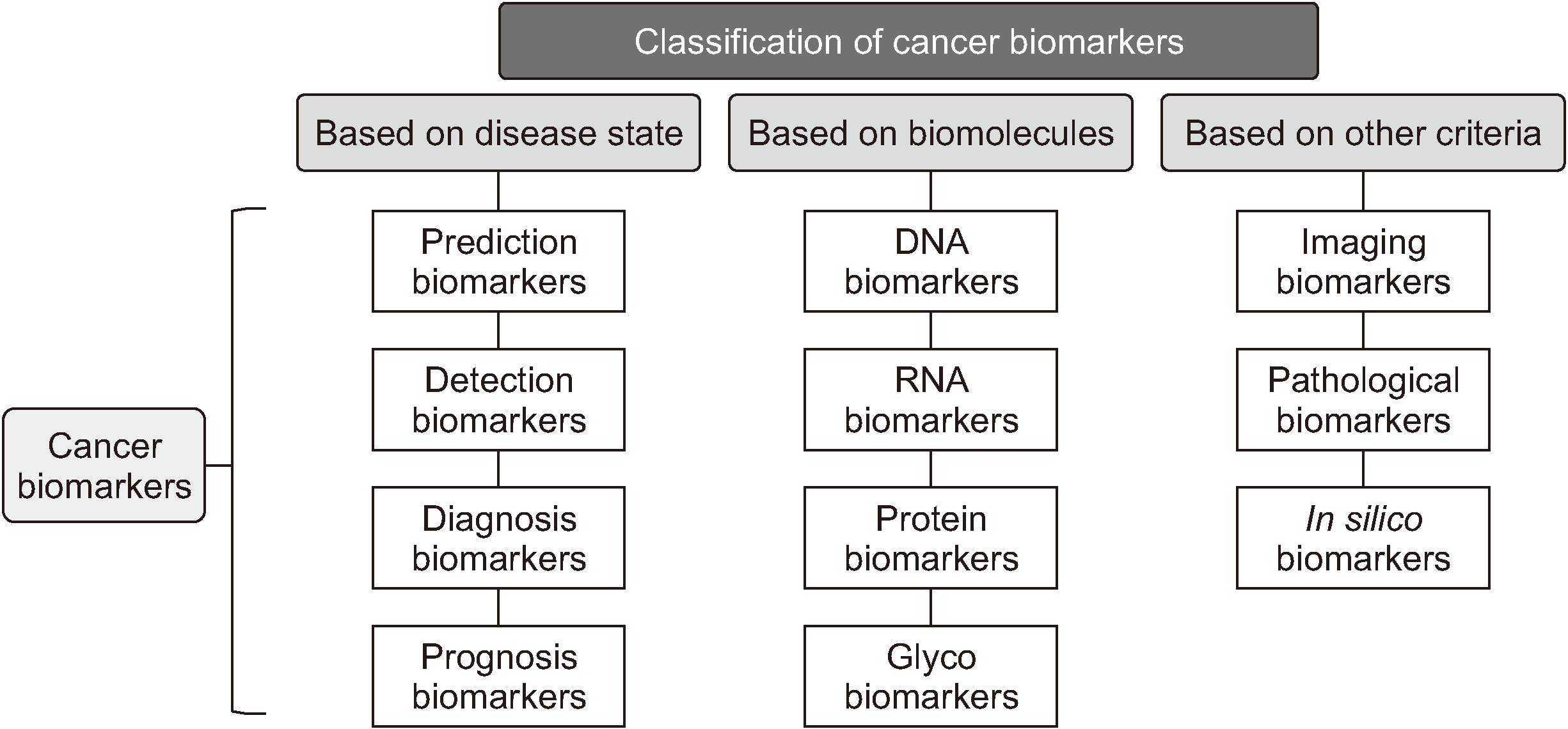
A peer-review analysis by the World Health Organization International Agency for Research on Cancer (WHO IARC) reported that the global estimated rate for oral cavity cancer was 2.7 per 100,000 in 2012 2,3 . Oral cancers are also the eighth most common causes of cancer-related deaths worldwide 4 . Oral squamous cell carcinoma (OSCC) accounts for over 90% of oral cancers and is considered to be a rising global public health issue because of its high incidence and low survival rate 5,6 .
In the efforts to reduce OSCC-related mortality, enhancing and innovating screening and early detection technologies has been suggested as the most effective and fastest developing strategy 7 . In this area, the liquid biopsy came up as a noninvasive diagnostic technique that is based on the detection of tumor markers in body fluids. In addition to blood, saliva has a role as an auxiliary tool in oral cancer diagnosis 8 . Furthermore, recent studies have revealed that saliva sampling can be a more effective method of detecting specific OSCC biomarkers 9 .
Recently, diagnostic markers are the focus of our clinical and experimental studies. A diagnostic cancer marker can be specific to stage, tissue, relapse, follow-up, or age 9 , and present at any stage during cancer development. This review article introduces an updated list of reported OSCC salivary biomarkers up until 2019 and discusses the current clinical application of salivary biomarkers.

II. Salivary Biomarkers

Whole saliva also contains a variety of non-organic and organic substances from the serum, gingival crevicular fluid, as well as oral microorganisms and their products10,11. In addition to a diversity of biomarkers for many diseases, saliva’s collection is noninvasive and convenient, and the transportation and storage are easy, therefore saliva sampling is cost-effective and efficient 12 . These advantages demonstrate that saliva is a potential body fluid for laboratory tests compared to serum and tissue samples.
Biomarkers are detected and determined by various molecular techniques. For the genomic biomarkers (including DNA, mitochondrial DNA [mt.DNA], RNA, messenger RNA [mRNA], microRNA [miRNA]), the utilized techniques can be DNA microarrays, polymerase chain reaction (PCR), Southern blot analysis, restriction fragment length polymorphism (RFLP), and cross-linking immunoprecipitation (CLIP). The proteomic biomarkers class includes proteins, peptides, antibodies, and can be analyzed by liquid chromatography, Western blot analysis, protein sequencing, protein arrays, and immunofluorescence. The metabolomics biomarkers (including carbohydrates, enzymes, metabolites, liquids) are determined by liquid chromatography, nuclear magnetic resonance, enzyme assays, and mass spectrometry 11 . One of the earliest developed saliva biomarkers, human papillomavirus (HPV) markers, have been used as the diagnostic biomarker in the cervical cancer screening program and vaccine development 1 .
The identification of reliable salivary biomarkers for the OSCC screening has been enhanced thanks to the easy and noninvasive collection of saliva compared to the drawing of blood 9 . The underlying tissue changes in the disease process can be classified as genomic, proteomic, or metabolomic expression.(Fig. 2) With the development of salivaomics, a lot of researches have been performed and more than 100 potential saliva biomarkers for OSCC have been reported in the literature 13 . Salivary diagnostic has been an attractive potential modality screening, early detection and prognosis evaluation for researchers and clinicians 14 .
The classification of biomarkers can be based on the disease state, biomolecules, or other criteria 1 . Currently, we are paying attention to the screening and early detection of OSCC, and diagnostic markers are in the focus of our clinical and experimental studies. Diagnostic markers may be present at any stage of cancer development.(Table 1) The expression of Eph and/or ephrin are common in various primary tumors, including OSCCs. Among the various biological functions of ephrin and Eph receptors in cancer, their involvement in EFNB2/EphB4 signaling is thought to be associated with angiogenesis, differentiation, and development. Therefore, EFNB2 gene expression is suggested to be a useful biological marker in prognostic evaluation in patients with OSCC 15 . Shpitzer et al. 16 reported that the levels of 8-oxoguanine DNA glycosylase, phosphorylated-Src, and mammary serine protease inhibitor (Maspin) were found to decrease in the saliva of OSCC patient. Several studies reported that interleukin (IL)-6 and IL-8, which are well-known as post-inflammatory cytokines, significantly increase in patients with OSCC and therefore suggesting their value as a diagnostic marker of oral malignant and premalignant lesions 17,18 . Arellano-Garcia et al. 19 reported that both IL-8 and IL-1β were found to significantly increase in OSCC patients.
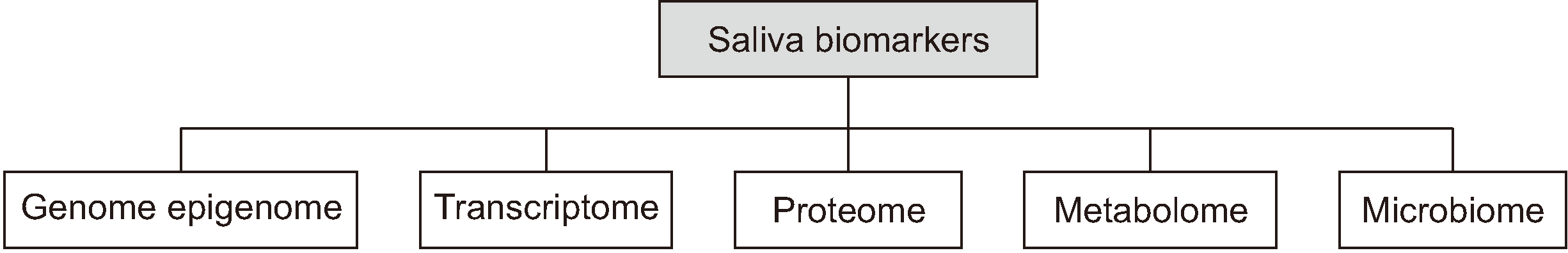
A diagnostic cancer marker can be specific to tissue, stage, follow-up, relapse, and age. Despite the attempts made to classify cancer biomarkers, a consensus has not been established. However, for the diagnosis and comprehension of the OSCC genomic architecture, more recent efforts have focused on new and noninvasive methods using human saliva sampling, which include proteomic (Table 2)20-25, proteins (Table 3)20,25-36, transcriptomic (Table 4)20,37-41, and metabolomic (Table 5)20,42-44 biomarkers.

III. Liquid Biopsy of OSCC

Laboratory examination is an essential and high accurate method for disease diagnosis and prognosis. Among the available laboratory test, liquid biopsy is a less invasive method that limits the need for acquiring tissue 45 . In the past 20 years, liquid biopsy has become an essential examination in multiple areas of oncology, based on the detection of circulating tumor cells (CTCs), circulating tumor DNA (ctDNA), and circulating tumor RNA (ctRNA), proteins, and exosomes 8 . Liquid biopsy samples include blood, urine, saliva, and other bodily fluids such as seminal plasma, pleural effusions, cerebrospinal fluid, sputum, and stool samples 46 .
Saliva has complex components that originate from the major and minor salivary glands, as well as from the oropharynx, gastrointestinal reflux, gingival crevicular fluid, and blood. The analysis of saliva components has been considered an effective method for monitoring the status of health 47 because changes in the compounds that constitute saliva can reflect the physiological and pathological status of the body.

1. Blood biomarkers for OSCC

Currently, the most common liquid biopsy is blood. Approximately, 5 to 10 mL of blood is all that is needed for a liquid biopsy. Blood biomarkers are classified as CTCs, ctDNA, ctRNA, proteins, and exosomes, which can be used for differential diagnosis, prognosis, cancer recurrence detection, tumor evolution monitoring, and treatment efficacy in various types of tumors8,48. Circulating cell-free DNA (cfDNA) is released into the bloodstream from apoptotic or necrotic cells. cfDNAs can originate from nonmalignant host cells and tumor cells, thus including ctDNA. There are also authors suggesting that tumor exosomes can play an important role in immune suppression and enhancing tumor development, and plasma exosomes can be the next generation of biomarkers in head and neck cancer progression evaluation8.

2. Standard saliva collections

There are a lot of methods are for the whole saliva collecting, such as draining, spitting, suction, and swabs49. A variety of commercial devices and methods for collecting saliva from individual glands have also been developed20,49.(Table 6) The clinician should be properly trained on performing saliva sample collection to achieve the best performance and samples. Despite the variety of choices, only one type of collection device should be used in one study50,51. The eligible participants need to be educated and given the appropriate instructions before the collecting procedure. The sample volume needs to be sufficient, and the type of container needs to be prepared beforehand accordingly. Besides, sample labeling and handling protocols must be well-designed and carried out consistently52.
Saliva components can vary or remain stable at age53. It is reported that children and adults also can have differences in the salivary level of a specific protein, peptide, and proteome54. The unstimulated saliva secretion was higher in healthy men compared with women55, indicating gender-dependent secretion. Due to these variables, patients should be categorized into different age groups and gender to minimize statistical errors.
The position of the mouth during saliva collection is important from the basis of salivary glands location and their secretion patterns49. While saliva secreted from the major salivary glands contains common components, the concentrations of each component and some specific contents can vary from one gland to another. On the other hand, components of the saliva from the minor glands mainly include mucins and lipase51.
Besides, saliva components and origin depend on the resting state or stimulated state of the individual. For example, cortisol, alpha-amylase, and secretory IgA levels in the saliva are affected by stimulation49. The complex in the composition of saliva and its dependence on various conditions should be considered and evaluated thoroughly by the investigator. This consistency in sampling procedure is essential to achieve valuable data.(Table 7)

3. Saliva preservation with detailed information

The saliva samples can be kept at room temperature for a maximum of 30 to 90 minutes for the immediate analysis56. Thomadaki et al.10 recommended that the samples are frozen at or below –20ºC immediately after collection, due to the low temperature can slow down the degradation of the salivary proteome. Salivary specimens can also be stored at –80ºC for several years with little or no degradation56. In RNA analysis sampling, an RNase inhibitor should be added in the supernatant fractions before storing it at –80ºC57. Shirtcliff et al.58 suggested that the collection method was an important cause of the unsystematic error. Samples obtained by spitting contain more bacteria than drooling samples, which can affect the analysis results of saliva compounds56.

IV. Relations with Other Serum Biomarkers

As with saliva, blood is a complex fluid that contains a wide range of molecular components, including various antibodies, growth factors, enzymes, and hormones. Therefore, blood serum and plasma are the traditional and conventional sources of liquid biopsy and biomarker examination. Although used widely, blood sampling and analysis can often be expensive, problematic, and invasive.
Comparatively, sampling saliva has many advantages over blood, including the following 52 : 1) The saliva sampling procedure is easier and can be self-collected, 2) the procedure is noninvasive, 3) samples are safer to handle and 4) are easier to transport and store because saliva requires simpler manipulation than blood, and 5) the procedure is cost-efficient both for patients and the investigators.
Despite these advantages, the use of saliva sampling as a diagnostic tool is still under controversy. The greatest obstacle in the development of a salivary diagnostic protocol is that while most biomarkers detected in the blood serum are also can be found in saliva, but their levels are so low that they barely can contribute to the diagnosis. For example, the normal level of IgG (5 to 30 mg/mL vs 5 to 30 μg/mL) and IgM (0.5 to 1 mg/mL vs 5 to 10 μg/mL) also have a huge difference in serum and saliva analysis 59 . However, the link between blood and saliva at a molecular level may also suggest that saliva can be a potential alternative to blood- and tissue-based diagnostics.
While there are reports about many saliva biomarkers, the correlation of their levels in the blood and saliva are various, and there are a scarce number of publications on the blood levels of specific biomarkers found in saliva, or how the saliva collection technique affects the quality and diagnosis value of specific biomarkers. Williamson et al. 59 analyzed the correlation of biomarkers among passive drool saliva, filter paper saliva, and plasma samples in healthy adults. The author found that between passive drool and filter paper saliva samples, statistically significant correlations were found among 16 cytokines, including IL1β, IL-1ra, IL-4, IL-7, IL-8, IL-9, IL-10, IL-12, IL-13, IL-15, granulocyte colony-stimulating factor (G-CSF), interferon gamma (IFN-γ), IFN-γ-inducible protein 10 (IP-10), monocyte chemoattractant protein 1 (MCP-1), macrophage inflammatory protein-1β (MIP-1β), and vascular endothelial growth factor (VEGF). When plasma and passive drool saliva samples were compared, only 3 cytokines, IL-6, IFN-γ, and MIP-1β, were statistically significantly correlated 49 . Glutathione (reduced glutathione [GSH] and oxidized glutathione [GSSG]) is an anti-oxidant that responds to both xenobiotic and endogenous compounds. Ngamchuea et al. 60 found a weak correlation between salivary and whole blood glutathione content (GSH+2GSSG) in healthy participants. Almadori et al. 26 suggested that salivary glutathione levels may be an index of oxidative stress at the level of the upper airways and in particular of the oral cavity and pharynx. A study by Sharma et al. 61 showed a significant and gradual increase in serum and salivary L-fucose between control and oral potentially malignant disorders (OPMDs) or oral cancer (OC) samples. The authors suggested that L-fucose can be used as a reliable biomarker and saliva can be used as a diagnostic fluid for the screening and early detection of OC. A significant positive correlation was found between serum and salivary Cyfra 21-1, serum Cyfra 21-1, and CK19 mRNA expression and between salivary Cyfra 21-1 and CK19 mRNA expression 62 . The magnesium concentration was low in both the blood plasma and saliva of OSCC when individuals with potentially malignant disorders were compared to healthy subjects. Thus, the magnesium ion concentration in blood plasma and saliva could be considered as a tumor marker, playing an important role in carcinogenesis 63 . Oral pre-malignancy and malignancy patients were reported to have lower serum albumin levels compared to healthy individuals. Otherwise, salivary albumin levels were found to increase in oral pre-malignancy and oral malignancy cases compared to healthy individuals. A study by Metgud and Patel 64 suggested that albumin may play a role in early diagnosis and prognosis of oral premalignant and malignant tissues.

V. Suggested Salivary Biomarkers, Present and Future

The saliva of patients with OSCC has been studied for biomarkers and many potential biomarkers in genomic, proteomic, and metabolomics have been found in the last decade 27,65,66 . However, most of them have been confined to the laboratory and not expanded into clinics due to sensitivity and specificity, as well as technical requirements and costs. Analysis of the salivary proteome is a feasible strategy for salivary biomarker discovery, and several representative proteins could be suggested as potential salivary markers for OSCC diagnosis.
The protease components of saliva were found to correlate with diverse oral diseases67-69. Due to its high sensitivity and specificity, the combination of cathepsin V/kallikrein5/ADAM9 was a promising biomarker for the early diagnosis of OSCC. The levels of matrix metalloproteinase (MMP)-1, MMP-2, MMP-10, MMP-12, a disintegrin and metalloprotease 9 (ADAM 9), a disintegrin and metalloprotease with thrombospondin type 13 motifs (ADAMST13), cathepsin V and kallikrein 5 in the saliva of OSCC patients were significantly increased in comparison with those of other groups 45 .
The high salivary level of complement factor H (CFH), fibrinogen alpha chain (FGA), and alpha-1-antitrypsin (SERPINA1) was reported to correlate with advanced stages of OSCC. These proteins (CFH, FGA, and SERPINA1) were determined to be a potential biomarker for the early detection and prognosis of OSCC 70 .
It is also important to review biomarkers based on carcinogenesis-related factors. A change in the level of each factor has its clinical significance, including early detection with oxidases and stress-related markers, differential diagnosis, monitoring of the tumor with inflammation-related or angiogenesis-related markers, or predicting distant metastasis with metastasis-related markers. Therefore, we also classified our candidate saliva biomarkers using carcinogenesis-related factors to evaluate the potential clinical application of each marker.(Table 1)
Despite attempts to classify cancer biomarkers, a consensus has not been established. The classification of biomarkers can be based on the disease state, biomolecules, and other criteria. Herein, we have classified candidates for salivary biomarkers in oral cancer based on biomolecules and carcinogenesis-related factors. Due to the distinct characteristics of each type of biomolecule, it is essential to review the biomarkers using biomolecular characteristics. Each type of biomolecule requires specific saliva collection devices and analysis techniques. We classified our candidate biomarkers based on biomolecular evidence.(Table 7)

VI. Conclusion with Future Trends

Salivary biomarkers are a very promising noninvasive approach to oral cancer detection. In monitoring the disease progression and patient’s response to therapeutics, salivary biomarkers show many advantages because of the noninvasive and cost-effective sampling methods. However, the current challenges in salivary biomarker research are standardizing sample collection, improving sample processing and storage, and reduce the wide variability in cancerous and non-cancerous individuals. Useful biomarkers in the screening and early diagnosis of OSCC are still under research but will be defined in the near future.

References

1. Mishra A, Verma M. 2010; Cancer biomarkers: are we ready for the prime time? Cancers (Basel). 2:190–208. https://doi.org/10.3390/cancers2010190 . DOI: 10.3390/cancers2010190. PMID: 24281040. PMCID: PMC3827599.
crossref
2. Shield KD, Ferlay J, Jemal A, Sankaranarayanan R, Chaturvedi AK, Bray F, et al. 2017; The global incidence of lip, oral cavity, and pharyngeal cancers by subsite in 2012. CA Cancer J Clin. 67:51–64. https://doi.org/10.3322/caac.21384 . DOI: 10.3322/caac.21384. PMID: 28076666.
crossref
3. Conway DI, Purkayastha M, Chestnutt IG. 2018; The changing epidemiology of oral cancer: definitions, trends, and risk factors. Br Dent J. 225:867–73. https://doi.org/10.1038/sj.bdj.2018.922 . DOI: 10.1038/sj.bdj.2018.922. PMID: 30412558.
crossref
4. Patel RS, Clark JR, Dirven R, Wyten R, Gao K, O'Brien CJ. 2009; Prognostic factors in the surgical treatment of patients with oral carcinoma. ANZ J Surg. 79:19–22. https://doi.org/10.1111/j.1445-2197.2008.04791.x . DOI: 10.1111/j.1445-2197.2008.04791.x. PMID: 19183373.
crossref
5. Gupta N, Gupta R, Acharya AK, Patthi B, Goud V, Reddy S, et al. 2016; Changing Trends in oral cancer - a global scenario. Nepal J Epidemiol. 6:613–9. https://doi.org/10.3126/nje.v6i4.17255 . DOI: 10.3126/nje.v6i4.17255. PMID: 28804673. PMCID: PMC5506386.
crossref
6. Johnson NW, Jayasekara P, Amarasinghe AA. 2011; Squamous cell carcinoma and precursor lesions of the oral cavity: epidemiology and aetiology. Periodontol 2000. 57:19–37. https://doi.org/10.1111/j.1600-0757.2011.00401.x . DOI: 10.1111/j.1600-0757.2011.00401.x. PMID: 21781177.
crossref
7. Ilbawi AM, Anderson BO. 2015; Cancer in global health: how do prevention and early detection strategies relate? Sci Transl Med. 7:278. https://doi.org/10.1126/scitranslmed.3008853 . DOI: 10.1126/scitranslmed.3008853. PMID: 25761886.
crossref
8. Lousada-Fernandez F, Rapado-Gonzalez O, Lopez-Cedrun JL, Lopez-Lopez R, Muinelo-Romay L, Suarez-Cunqueiro MM. 2018; Liquid biopsy in oral cancer. Int J Mol Sci. 19:1704. https://doi.org/10.3390/ijms19061704 . DOI: 10.3390/ijms19061704. PMID: 29890622. PMCID: PMC6032225.
crossref
9. Cheng YS, Rees T, Wright J. 2014; A review of research on salivary biomarkers for oral cancer detection. Clin Transl Med. 3:3. https://doi.org/10.1186/2001-1326-3-3 . DOI: 10.1186/2001-1326-3-3. PMID: 24564868. PMCID: PMC3945802.
crossref
10. Thomadaki K, Helmerhorst EJ, Tian N, Sun X, Siqueira WL, Walt DR, et al. 2011; Whole-saliva proteolysis and its impact on salivary diagnostics. J Dent Res. 90:1325–30. https://doi.org/10.1177/0022034511420721 . DOI: 10.1177/0022034511420721. PMID: 21917601. PMCID: PMC3188460.
crossref
11. Arglebe C. 1981; Biochemistry of human saliva. Adv Otorhinolaryngol. 26:97–234. https://doi.org/10.1159/000395291 . DOI: 10.1159/000395291. PMID: 7020374.
crossref
12. Kaczor-Urbanowicz KE, Martin Carreras-Presas C, Aro K, Tu M, Garcia-Godoy F, Wong DT. 2017; Saliva diagnostics - current views and directions. Exp Biol Med (Maywood). 245:459–72. https://doi.org/10.1177/1535370216681550 . DOI: 10.1177/1535370216681550. PMID: 27903834. PMCID: PMC5367650.
crossref
13. Hu S, Loo JA, Wong DT. 2007; Human saliva proteome analysis and disease biomarker discovery. Expert Rev Proteomics. 4:531–8. https://doi.org/10.1586/14789450.4.4.531 . DOI: 10.1586/14789450.4.4.531. PMID: 17705710.
crossref
14. Wong DT. 2012; Salivaomics. J Am Dent Assoc. 143(10 Suppl):19S–24S. https://doi.org/10.14219/jada.archive.2012.0339 . DOI: 10.14219/jada.archive.2012.0339. PMID: 23034834.
crossref
15. Tachibana M, Tonomoto Y, Hyakudomi R, Hyakudomi M, Hattori S, Ueda S, et al. 2007; Expression and prognostic significance of EFNB2 and EphB4 genes in patients with oesophageal squamous cell carcinoma. Dig Liver Dis. 39:725–32. https://doi.org/10.1016/j.dld.2007.05.013 . DOI: 10.1016/j.dld.2007.05.013. PMID: 17611172.
crossref
16. Shpitzer T, Hamzany Y, Bahar G, Feinmesser R, Savulescu D, Borovoi I, et al. 2009; Salivary analysis of oral cancer biomarkers. Br J Cancer. 101:1194–8. https://doi.org/10.1038/sj.bjc.6605290 . DOI: 10.1038/sj.bjc.6605290. PMID: 19789535. PMCID: PMC2768098.
crossref
17. Rajkumar K, Ramesh Kumar A, Ramyamalini V, Nandhini G, Dinesh Kumar T, Ashwini BK, et al. 2010; Estimation of serological and salivary biomarkers in patients vith oral squamous cell carcinoma, premalignant lesions & conditions. SRM J Res Dent Sci. 1:14–9.
18. St John MA, Li Y, Zhou X, Denny P, Ho CM, Montemagno C, et al. 2004; Interleukin 6 and interleukin 8 as potential biomarkers for oral cavity and oropharyngeal squamous cell carcinoma. Arch Otolaryngol Head Neck Surg. 130:929–35. https://doi.org/10.1001/archotol.130.8.929 . DOI: 10.1001/archotol.130.8.929. PMID: 15313862.
crossref
19. Arellano-Garcia ME, Hu S, Wang J, Henson B, Zhou H, Chia D, et al. 2008; Multiplexed immunobead-based assay for detection of oral cancer protein biomarkers in saliva. Oral Dis. 14:705–12. https://doi.org/10.1111/j.1601-0825.2008.01488.x . DOI: 10.1111/j.1601-0825.2008.01488.x. PMID: 19193200. PMCID: PMC2675698.
crossref
20. Khurshid Z, Zafar MS, Khan RS, Najeeb S, Slowey PD, Rehman IU. 2018; Role of salivary biomarkers in oral cancer detection. Adv Clin Chem. 86:23–70. https://doi.org/10.1016/bs.acc.2018.05.002 . DOI: 10.1016/bs.acc.2018.05.002. PMID: 30144841.
crossref
21. de Jong EP, Xie H, Onsongo G, Stone MD, Chen XB, Kooren JA, et al. 2010; Quantitative proteomics reveals myosin and actin as promising saliva biomarkers for distinguishing pre-malignant and malignant oral lesions. PLoS One. 5:e11148. https://doi.org/10.1371/journal.pone.0011148 . DOI: 10.1371/journal.pone.0011148. PMID: 20567502. PMCID: PMC2887353.
crossref
22. Wang Q, Gao P, Wang X, Duan Y. 2014; Investigation and identification of potential biomarkers in human saliva for the early diagnosis of oral squamous cell carcinoma. Clin Chim Acta. 427:79–85. https://doi.org/10.1016/j.cca.2013.10.004 . DOI: 10.1016/j.cca.2013.10.004. PMID: 24144867.
crossref
23. Camisasca DR, da Rós Gonçalves L, Soares MR, Sandim V, Nogueira FC, Garcia CH, et al. 2017; A proteomic approach to compare saliva from individuals with and without oral leukoplakia. J Proteomics. 151:43–52. https://doi.org/10.1016/j.jprot.2016.07.029 . DOI: 10.1016/j.jprot.2016.07.029. PMID: 27478070.
crossref
24. Flores IL, Santos-Silva AR, Coletta RD, Leme AF, Lopes MA. 2016; Low expression of angiotensinogen and dipeptidyl peptidase 1 in saliva of patients with proliferative verrucous leukoplakia. World J Clin Cases. 4:356–63. https://doi.org/10.12998/wjcc.v4.i11.356 . DOI: 10.12998/wjcc.v4.i11.356. PMID: 27900324. PMCID: PMC5112355.
crossref
25. Hu S, Arellano M, Boontheung P, Wang J, Zhou H, Jiang J, et al. 2008; Salivary proteomics for oral cancer biomarker discovery. Clin Cancer Res. 14:6246–52. https://doi.org/10.1158/1078-0432.CCR-07-5037 . DOI: 10.1158/1078-0432.CCR-07-5037. PMID: 18829504. PMCID: PMC2877125.
crossref
26. Almadori G, Bussu F, Galli J, Limongelli A, Persichilli S, Zappacosta B, et al. 2007; Salivary glutathione and uric acid levels in patients with head and neck squamous cell carcinoma. Head Neck. 29:648–54. https://doi.org/10.1002/hed.20579 . DOI: 10.1002/hed.20579. PMID: 17274058.
crossref
27. Jou YJ, Lin CD, Lai CH, Chen CH, Kao JY, Chen SY, et al. 2010; Proteomic identification of salivary transferrin as a biomarker for early detection of oral cancer. Anal Chim Acta. 681:41–8. https://doi.org/10.1016/j.aca.2010.09.030 . DOI: 10.1016/j.aca.2010.09.030. PMID: 21035601.
crossref
28. Katakura A, Kamiyama I, Takano N, Shibahara T, Muramatsu T, Ishihara K, et al. 2007; Comparison of salivary cytokine levels in oral cancer patients and healthy subjects. Bull Tokyo Dent Coll. 48:199–203. https://doi.org/10.2209/tdcpublication.48.199 . DOI: 10.2209/tdcpublication.48.199. PMID: 18360107.
crossref
29. Duffy SA, Taylor JM, Terrell JE, Islam M, Li Y, Fowler KE, et al. 2008; Interleukin-6 predicts recurrence and survival among head and neck cancer patients. Cancer. 113:750–7. https://doi.org/10.1002/cncr.23615 . DOI: 10.1002/cncr.23615. PMID: 18536030.
crossref
30. Zhong LP, Chen GF, Xu ZF, Zhang X, Ping FY, Zhao SF. 2005; Detection of telomerase activity in saliva from oral squamous cell carcinoma patients. Int J Oral Maxillofac Surg. 34:566–70. https://doi.org/10.1016/j.ijom.2004.10.007 . DOI: 10.1016/j.ijom.2004.10.007. PMID: 16053879.
crossref
31. Krishna Prasad RB, Sharma A, Babu HM. 2013; An insight into salivary markers in oral cancer. Dent Res J (Isfahan). 10:287–95.
32. Franzmann EJ, Reategui EP, Pedroso F, Pernas FG, Karakullukcu BM, Carraway KL, et al. 2007; Soluble CD44 is a potential marker for the early detection of head and neck cancer. Cancer Epidemiol Biomarkers Prev. 16:1348–55. https://doi.org/10.1158/1055-9965.EPI-06-0011 . DOI: 10.1158/1055-9965.EPI-06-0011. PMID: 17627000.
crossref
33. Kumar M, Srivastava G, Kaur J, Assi J, Alyass A, Leong I, et al. 2015; Prognostic significance of cytoplasmic S100A2 overexpression in oral cancer patients. J Transl Med. 13:8. https://doi.org/10.1186/s12967-014-0369-9 . DOI: 10.1186/s12967-014-0369-9. PMID: 25591983. PMCID: PMC4324434.
crossref
34. Righini CA, de Fraipont F, Timsit JF, Faure C, Brambilla E, Reyt E, et al. 2007; Tumor-specific methylation in saliva: a promising biomarker for early detection of head and neck cancer recurrence. Clin Cancer Res. 13:1179–85. https://doi.org/10.1158/1078-0432.CCR-06-2027 . DOI: 10.1158/1078-0432.CCR-06-2027. PMID: 17317827.
crossref
35. Reddy I, Sherlin HJ, Ramani P, Premkumar P, Natesan A, Chandrasekar T. 2012; Amino acid profile of saliva from patients with oral squamous cell carcinoma using high performance liquid chromatography. J Oral Sci. 54:279–83. https://doi.org/10.2334/josnusd.54.279 . DOI: 10.2334/josnusd.54.279. PMID: 23047040.
crossref
36. Al Kawas S, Rahim ZH, Ferguson DB. 2012; Potential uses of human salivary protein and peptide analysis in the diagnosis of disease. Arch Oral Biol. 57:1–9. https://doi.org/10.1016/j.archoralbio.2011.06.013 . DOI: 10.1016/j.archoralbio.2011.06.013. PMID: 21774913.
crossref
37. Li Y, St John MA, Zhou X, Kim Y, Sinha U, Jordan RC, et al. 2004; Salivary transcriptome diagnostics for oral cancer detection. Clin Cancer Res. 10:8442–50. https://doi.org/10.1158/1078-0432.CCR-04-1167 . DOI: 10.1158/1078-0432.CCR-04-1167. PMID: 15623624.
crossref
38. Elashoff D, Zhou H, Reiss J, Wang J, Xiao H, Henson B, et al. 2012; Prevalidation of salivary biomarkers for oral cancer detection. Cancer Epidemiol Biomarkers Prev. 2:664–72. https://doi.org/10.1158/1055-9965.EPI-11-1093 . DOI: 10.1158/1055-9965.EPI-11-1093. PMID: 22301830. PMCID: PMC3319329.
crossref
39. Tang H, Wu Z, Zhang J, Su B. 2013; Salivary lncRNA as a potential marker for oral squamous cell carcinoma diagnosis. Mol Med Rep. 7:761–6. https://doi.org/10.3892/mmr.2012.1254 . DOI: 10.3892/mmr.2012.1254. PMID: 23292713.
crossref
40. Liu CJ, Lin SC, Yang CC, Cheng HW, Chang KW. 2012; Exploiting salivary miR-31 as a clinical biomarker of oral squamous cell carcinoma. Head Neck. 34:219–24. https://doi.org/10.1002/hed.21713 . DOI: 10.1002/hed.21713. PMID: 22083872.
crossref
41. Park NJ, Zhou H, Elashoff D, Henson BS, Kastratovic DA, Abemayor E, et al. 2009; Salivary microRNA: discovery, characterization, and clinical utility for oral cancer detection. Clin Cancer Res. 15:5473–7. https://doi.org/10.1158/1078-0432.CCR-09-0736 . DOI: 10.1158/1078-0432.CCR-09-0736. PMID: 19706812. PMCID: PMC2752355.
crossref
42. Wei J, Xie G, Zhou Z, Shi P, Qiu Y, Zheng X, et al. 2011; Salivary metabolite signatures of oral cancer and leukoplakia. Int J Cancer. 129:2207–17. https://doi.org/10.1002/ijc.25881 . DOI: 10.1002/ijc.25881. PMID: 21190195.
crossref
43. Sugimoto M, Wong DT, Hirayama A, Soga T, Tomita M. 2010; Capillary electrophoresis mass spectrometry-based saliva metabolomics identified oral, breast and pancreatic cancer-specific profiles. Metabolomics. 6:78–95. https://doi.org/10.1007/s11306-009-0178-y . DOI: 10.1007/s11306-009-0178-y. PMID: 20300169. PMCID: PMC2818837.
crossref
44. Ishikawa S, Sugimoto M, Kitabatake K, Sugano A, Nakamura M, Kaneko M, et al. 2016; Identification of salivary metabolomic biomarkers for oral cancer screening. Sci Rep. 6:31520. https://doi.org/10.1038/srep31520 . DOI: 10.1038/srep31520. PMID: 27539254. PMCID: PMC4990923.
crossref
45. Feng Y, Li Q, Chen J, Yi P, Xu X, Fan Y, et al. 2019; Salivary protease spectrum biomarkers of oral cancer. Int J Oral Sci. 11:7. https://doi.org/10.1038/s41368-018-0032-z . DOI: 10.1038/s41368-018-0032-z. PMID: 30602733. PMCID: PMC6315043.
crossref
46. Peng M, Chen C, Hulbert A, Brock MV, Yu F. 2017; Non-blood circulating tumor DNA detection in cancer. Oncotarget. 8:69162–73. https://doi.org/10.18632/oncotarget.19942 . DOI: 10.18632/oncotarget.19942. PMID: 28978187. PMCID: PMC5620327.
crossref
47. Zhang CZ, Cheng XQ, Li JY, Zhang P, Yi P, Xu X, et al. 2016; Saliva in the diagnosis of diseases. Int J Oral Sci. 8:133–7. https://doi.org/10.1038/ijos.2016.38 . DOI: 10.1038/ijos.2016.38. PMID: 27585820. PMCID: PMC5113094.
crossref
48. Gröbe A, Blessmann M, Hanken H, Friedrich RE, Schön G, Wikner J, et al. 2014; Prognostic relevance of circulating tumor cells in blood and disseminated tumor cells in bone marrow of patients with squamous cell carcinoma of the oral cavity. Clin Cancer Res. 20:425–33. https://doi.org/10.1158/1078-0432.CCR-13-1101 . DOI: 10.1158/1078-0432.CCR-13-1101. PMID: 24218516.
crossref
49. Bhattarai KR, Kim HR, Chae HJ. 2018; Compliance with saliva collection protocol in healthy volunteers: strategies for managing risk and errors. Int J Med Sci. 15:823–31. https://doi.org/10.7150/ijms.25146 . DOI: 10.7150/ijms.25146. PMID: 30008593. PMCID: PMC6036086.
crossref
50. Golatowski C, Salazar MG, Dhople VM, Hammer E, Kocher T, Jehmlich N, et al. 2013; Comparative evaluation of saliva collection methods for proteome analysis. Clin Chim Acta. 419:42–6. https://doi.org/10.1016/j.cca.2013.01.013 . DOI: 10.1016/j.cca.2013.01.013. PMID: 23384500.
crossref
51. Navazesh M. 1993; Methods for collecting saliva. Ann N Y Acad Sci. 694:72–7. https://doi.org/10.1111/j.1749-6632.1993.tb18343.x . DOI: 10.1111/j.1749-6632.1993.tb18343.x. PMID: 8215087.
crossref
52. Yoshizawa JM, Schafer CA, Schafer JJ, Farrell JJ, Paster BJ, Wong DT. 2013; Salivary biomarkers: toward future clinical and diagnostic utilities. Clin Microbiol Rev. 26:781–91. https://doi.org/10.1128/CMR.00021-13 . DOI: 10.1128/CMR.00021-13. PMID: 24092855. PMCID: PMC3811231.
crossref
53. Nassar M, Hiraishi N, Islam S, Otsuki M, Tagami J. 2014; Age-related changes in salivary biomarkers. J Dent Sci. 9:85–90. https://doi.org/10.1016/j.jds.2013.11.002 . DOI: 10.1016/j.jds.2013.11.002.
crossref
54. Cabras T, Pisano E, Boi R, Olianas A, Manconi B, Inzitari R, et al. 2009; Age-dependent modifications of the human salivary secretory protein complex. J Proteome Res. 8:4126–34. https://doi.org/10.1021/pr900212u . DOI: 10.1021/pr900212u. PMID: 19591489.
crossref
55. Fenoll-Palomares C, Muñoz Montagud JV, Sanchiz V, Herreros B, Hernández V, Mínguez M, et al. 2004; Unstimulated salivary flow rate, pH and buffer capacity of saliva in healthy volunteers. Rev Esp Enferm Dig. 96:773–83. https://doi.org/10.4321/s1130-01082004001100005 . DOI: 10.4321/S1130-01082004001100005. PMID: 15584851.
crossref
56. Chiappin S, Antonelli G, Gatti R, De Palo EF. 2007; Saliva specimen: a new laboratory tool for diagnostic and basic investigation. Clin Chim Acta. 383:30–40. https://doi.org/10.1016/j.cca.2007.04.011 . DOI: 10.1016/j.cca.2007.04.011. PMID: 17512510.
crossref
57. Henson BS, Wong DT. 2010; Collection, storage, and processing of saliva samples for downstream molecular applications. Methods Mol Biol. 666:21–30. https://doi.org/10.1007/978-1-60761-820-1_2 . DOI: 10.1007/978-1-60761-820-1_2. PMID: 20717775.
crossref
58. Shirtcliff EA, Granger DA, Schwartz E, Curran MJ. 2001; Use of salivary biomarkers in biobehavioral research: cotton-based sample collection methods can interfere with salivary immunoassay results. Psychoneuroendocrinology. 26:165–73. https://doi.org/10.1016/s0306-4530(00)00042-1 . DOI: 10.1016/S0306-4530(00)00042-1.
crossref
59. Williamson S, Munro C, Pickler R, Grap MJ, Elswick RK Jr Jr. 2012; Comparison of biomarkers in blood and saliva in healthy adults. Nurs Res Pract. 2012:246178. https://doi.org/10.1155/2012/246178 . DOI: 10.1155/2012/246178. PMID: 22619709. PMCID: PMC3350846.
crossref
60. Ngamchuea K, Batchelor-McAuley C, Cowen PJ, Williams C, Gonçalves LM, Compton RG. 2016; Can saliva testing replace blood measurements for health monitoring? Insights from a correlation study of salivary and whole blood glutathione in humans. Analyst. 14:4707–12. https://doi.org/10.1039/c6an01139j . DOI: 10.1039/C6AN01139J. PMID: 27278110.
crossref
61. Sharma M, Sharma E, Prabhu V, Pai VR, D'souza JM, Harish S, et al. 2020; Salivary L-fucose as a biomarker for oral potentially malignant disorders and oral cancer. J Cancer Res Ther. 16:546–50. https://doi.org/10.4103/jcrt.JCRT_552_17 . DOI: 10.4103/jcrt.JCRT_552_17. PMID: 32719265.
crossref
62. Singh P, Barpande SR, Bhavthankar JD, Mandale MS, Bhagwat AU. 2017; Serum Cyfra 21-1 levels in oral squamous cell carcinoma patients and its clinicopathologic correlation. Indian J Dent Res. 28:162–8. https://doi.org/10.4103/0970-9290.207789 . DOI: 10.4103/0970-9290.207789. PMID: 28611326.
crossref
63. Aziz NZ, Arathi K, Prasad BG, Desai D, Shetty SJ, Shahid M. 2018; Evaluation of magnesium levels in blood and saliva of oral squamous cell carcinoma and potentially malignant disorders by xylidyl blue method. J Oral Maxillofac Pathol. 22:147. https://doi.org/10.4103/jomfp.JOMFP_34_17 .
crossref
64. Metgud R, Patel S. 2014; Serum and salivary levels of albumin as diagnostic tools for oral pre-malignancy and oral malignancy. Biotech Histochem. 89:8–13. https://doi.org/10.3109/10520295.2013.793394 . DOI: 10.3109/10520295.2013.793394. PMID: 23738795.
crossref
65. Ai J, Smith B, Wong DT. 2010; Saliva ontology: an ontology-based framework for a Salivaomics knowledge base. BMC Bioinformatics. 11:302. https://doi.org/10.1186/1471-2105-11-302 . DOI: 10.1186/1471-2105-11-302. PMID: 20525291. PMCID: PMC2898059.
crossref
66. Wu CC, Chu HW, Hsu CW, Chang KP, Liu HP. 2015; Saliva proteome profiling reveals potential salivary biomarkers for detection of oral cavity squamous cell carcinoma. Proteomics. 15:3394–404. https://doi.org/10.1002/pmic.201500157 . DOI: 10.1002/pmic.201500157. PMID: 26205615.
crossref
67. Flannery CR. 2006; MMPs and ADAMTSs: functional studies. Front Biosci. 11:544–69. https://doi.org/10.2741/1818 . DOI: 10.2741/1818. PMID: 16146752.
crossref
68. Kim JM, Kang SW, Shin SM, Su Kim D, Choi KK, Kim EC, et al. 2014; Inhibition of matrix metalloproteinases expression in human dental pulp cells by all-trans retinoic acid. Int J Oral Sci. 6:150–3. https://doi.org/10.1038/ijos.2013.63 . DOI: 10.1038/ijos.2013.63. PMID: 24008270. PMCID: PMC4170145.
crossref
69. Edwards DR, Handsley MM, Pennington CJ. 2008; The ADAM metalloproteinases. Mol Aspects Med. 29:258–89. https://doi.org/10.1016/j.mam.2008.08.001 . DOI: 10.1016/j.mam.2008.08.001. PMID: 18762209. PMCID: PMC7112278.
crossref
70. Chu HW, Chang KP, Hsu CW, Chang IY, Liu HP, Chen YT, et al. 2019; Identification of salivary biomarkers for oral cancer detection with untargeted and targeted quantitative proteomics approaches. Mol Cell Proteomics. 18:1796–806. https://doi.org/10.1074/mcp.RA119.001530 . DOI: 10.1074/mcp.RA119.001530. PMID: 31253657. PMCID: PMC6731081.
crossref

Fig. 1
Classification of biomarkers. Adapted from the article of Mishra and Verma 1 (Cancers [Basel] 2010;2:190-208) in accordance with the Creative Commons Attribution 3.0 Unported (CC BY 3.0) license.
jkaoms-46-301-f1.tif
Fig. 2
Variety of biomarkers found in saliva.
jkaoms-46-301-f2.tif
Table 1
Candidates for salivary biomarkers in oral squamous cell carcinoma based on carcinogenesis-related factors
Angiogenesis-related markers Inflammation-related markers Metastasis-related markers Oxidase stress-related markers Metabolism-related marker
-CD31
-EFNB2
-ANGPT1, ANGPT2
-VEGF
-miR125
-IL-6
-IL-8
-IL-1b
-CD44
-Maspin
-S100P
-8-OHdG DNA damage marker (8-OHdG)
-Glutathione
-Non-organic compound: Na, Ca, F, Mg
-Fucose
-Albumin
-Actin and myosin
-L-phenylalanine

(VEGF: vascular endothelial growth factor, IL: interleukin)

Table 2
Description of oral disease proteomic analysis using unstimulated whole saliva (USWS)20
Disease Type of saliva Proteomic approach Proteins identified References
Oral squamous cell carcinoma USWS Mass spectrometry (MS) and western blotting Increased abundance of myosin and actin. de Jong et al. 21
USWS Using shotgun proteomics approach (RP-HPLC, CP-LC with TOF and immunoassay) Detection of 52 protein that presented in diseased samples but absent in healthy samples Hu et al. 25
USWS Using ultraperformance liquid chromatography-mass spectrometry (UPLC-MS) with hydrophilic interaction chromatography mode ↑Level of choline, betaine and pipecolinic acid
↑Level of L-carnitine
Wang et al.22
Oral leukoplakia USWS Two-dimensional gel electrophoresis, mass spectrometry, immunohistochemistry 22 spots very abundant among them apolipoprotein A1, alpha-amylase, cystatins, keratin 10, lysozyme precursor, and CK10 were relevant to the study. Camisasca et al. 23
Proliferative verrucous leukoplakia USWS Mass spectrometry Angiotensinogen (AGT) and dipeptidyl peptidase 1 (DPP1) Flores et al. 24
Premalignant lesions USWS Western blotting, mass spectrometry Salivary actin and myosin de Jong et al. 21
Table 3
Protein biomarkers in USWS for the detection of OSSC20
Candidate biomarkers Techniques Clinical significance References
Interleukin (IL)-6, IL-8, IL-1α, IL-1β, TNF-α, tissue polypeptide antigen (TPA), Cyfra 21-1, cancer antigen 125 (CA 125), telomerase, Mac-2 binding protein (M2BP) ELISA Proinflammatory and proangiogenic cytokines found to be indicators of carcinogenic transformation from oral precancerous lesions to oral cancer.
Cyfra 21-1, CA 125, and TPA markers attend in telomerase activity in tumor cells and are responsible for the maintenance of telomere length.
M2BP helps in the detection of OSCC.
Katakura et al. 28
Duffy et al. 29
Zhong et al. 30
Krishna Prasad et al. 31
CD44, CD59, Profilin, MRP14 Immunoblot CD44 and CD59 are the very high sensitive cancer and benign diseases differentiate markers.
MRP14 is a calcium-binding protein with a sensitivity of 90% and a specificity of 83% in cancer detection.
Hu et al. 25
Franzmann et al. 32
Glutathione HPLC Epidemiological marker for chemoprevention identifies the risk of development of OSCC. Almadori et al. 26
Mac-2 binding protein (M2BP), Squamous cell carcinoma antigen 2, involucrin, calcyclin, cathepsin-G, azurocidin, transaldolase, carbonic anhydrase I, calgizzarin, myeloblastin, vitamin D-binding protein ELISA, shotgun proteomics M2BP is for detection of OSCC this biomarker gives a sensitivity of 90% and a specificity of 83%, and all of them serve as a clinical tool for the noninvasive diagnosis of OSCC. Hu et al. 25
Immunoglobulin heavy chain constant region gamma (IgG), S100 calciumbinding protein, cofilin-1, transferrin, fibrin LC/MS IgG known to be an inhibitor of apoptosis, S100A2, an 11.4 kDa protein that is a prognostic biomarker for OSCC, cofilin proteins are involved in cancer progression, metastasis, and angiogenesis. Transferrin levels in saliva are associated with the size and stage of cancer. Fibrin in OSCC is involved in several carcinogenic processes. Jou et al. 27
Kumar et al. 33
α-1-antitrypsin (AAT) 2DE AAT is useful for the prediction and determining the aggression of OSCC. Righini et al. 34
Secretory leukocyte peptidase inhibitor (SLPI), cystatin A, keratin 36, thioredoxin, haptoglobin (HAP), salivary zinc finger, protein 510 peptide, a-amylase, and albumin MS-based proteomics SLPI, cystatin A, keratin 36 are potentially involved in the preventive treatment of OSCC. Thioredoxin mRNA levels are elevated in oral cancers and in other cancers as well. Salivary zinc finger, protein 510 peptide, a-amylase, and albumin are useful in the early detection of OSCC. Reddy et al. 35
Al Kawas et al. 36
Table 4
Transcriptomic biomarkers identified in USWS for OSSC detection20
Candidate biomarkers Techniques Clinical significance References
Interleukin (IL)-1 , IL-8 ELISA Angiogenesis, cell adhesion, chemotaxis, immune response, replication, signal transduction, proliferation, inflammation, and apoptosis Li et al. 37
Elashoff et al. 38
Dual specificity phosphatase 1 (DUSP1) Quantitative PCR (qPCR) and microarrays followed by qPCR Oxidative stress, protein modification, signal transduction Li et al. 37
H3 histone family 3A (H3F3A) qPCR and microarrays followed by qPCR DNA binding activity Li et al. 37
Long noncoding HOTAIR qPCR and microarrays followed by qPCR Expression of HOTAIR is associated with p53 gene and causes DNA damage Tang et al. 39
miR-125a, miR-200a, miR-31 qPCR and microarrays followed by qPCR Posttranscriptional regulation by RNA silencing complex, cellular growth, and elevated levels in proliferation in OSCC Liu et al. 40
Park et al. 41

(USWS: unstimulated whole saliva, OSCC: oral squamous cell carcinoma, ELISA: enzyme-linked immunosorbent assay, PCR: polymerase chain reaction)

Table 5
Metabolomics biomarkers identified in USWS for OSCC detection20
Candidate biomarkers Techniques Clinical significance References
Cadaverine, alanine, serine, glutamine, piperidine, taurine piperidine, choline, pyrroline hydroxycarboxylic acid, beta-alanine, alpha-aminobutyric acid betaine, tyrosine, leucine+isoleucine, histidine, tryptophan, glutamic acid, threonine, carnitine, pipercolic acid, lactic acid, phenylalanine and valine Capillary electrophoresis time-of-flight mass spectrometry (CE-TOF-MS) and HPLC with quadrupole/TOF MS Facilitates the clinical detection of OSCC and improves its diagnosis and prognosis. They have a high level of predictive value and serves as a stratification tool. Wei et al. 42
Sugimoto et al. 43
Hypoxanthine, guanine, guanosine, trimethylamine N-oxide, spermidine, pipecolate, methionine CE-TOF-MS Discrimination of controls from OSCC patients and all of these metabolites have elevated levels in saliva, and hence can be used in noninvasive oral cancer screening. Ishikawa et al. 44

(USWS: unstimulated whole saliva, OSCC: oral squamous cell carcinoma)

Table 6
Commercially available saliva collection devices and their key advantages20
Device name Company (city, country) Dose/volume Patent No. Characteristics
OraSure OraSure Tech. (Bethlehem, PA, USA) The pad draws oral fluid, rich in antibodies US8062908B2 U.S. Food and Drug Administration (FDA) approved for HIV-1 testing. Easy and safe for public health screening, life insurance risk assessment, and good for outreach community programs.
Quantisal Immunalysis Co. (Pomona, LA, USA) 1 mL±10% US8641642B2 Contains cellulosic (paper-based) absorbent material for collection of saliva.
Rapid saliva absorption. Buffer allows high recovery of drugs, including marijuana (THC) at room temperature.
FDA cleared for forensics, criminal justice and other applications.
Salivette Sarstedt AG & Co. KG (Nümbrecht, Germany) 1.1±0.3 mL US9113850B2 Wide application range, including detection of HIV antibodies, oxidative stress steroid hormones for general wellness.
Available as either cotton or polyester rolls or sponges, and each configuration includes a sample transport tube.
UltraSal-2 Oasis Diagnostics Co. (Vancouver, WA, USA) Up to 24 mL of the whole saliva US9113850B2 Large amount of saliva (24 mL).
Spit into two vials.
The device includes two collection tubes connected to a single mouthpiece into which the user expectorates. The mouthpiece can be tilted/rotated during collection to direct saliva into one or the other of the two tubes.
Mainly for drug testing purposes.
Greiner Bio-One SCS Greiner Bio-one (Monroe, NC, USA) 4 mL of a tartrazine US 20090017442 Only device with internal dye (tartrazine) for 2 minutes used as a saliva quantification tool. Uses a colorimetric method.
RNAPro•SAL Oasis Diagnostics Co. (Vancouver, WA, USA) 1.0 mL of saliva in 1-3 min US, EU
7,618,591
7,927,548
8,273,305
Simultaneous collection of RNA and proteins, including cell-free DNA, cell-free RNA, and exosomes. Large DNA and interfering factors removed. Useful for exploration of the salivary transcriptome and the salivary proteome. Built-in Sample Volume Adequacy Indicator (SVAI).
Pure•SAL Oasis Diagnostics Co. (Vancouver, WA, USA) 4.0 mL US, EU
7,618,591
7,927,548
8,273,305
Collection of cell-free DNA, cell RNA total RNA, or proteins. Major impurities removed by a built-in filtration system. Built-in SVAI.
Super•SAL Oasis Diagnostics Co. (Vancouver, WA, USA) 1.0 mL US, EU
7,618,591
7,927,548
8,273,305
Whole saliva collection system. Absorbent pad material removes interfering mucinous material. Shorter collection time due to the higher surface area of pad material exposed to fluid in the oral cavity.
Versi•SAL Oasis Diagnostics Co. (Vancouver, WA, USA) A maximum sample volume of 1.4 mL Patents pending Whole saliva collection system. Absorbent pad material removes interfering mucinous material.
Oral fluid collection device is currently used for general purpose saliva collection for downstream testing in the laboratory.
Applications include hormone testing for general wellness, substance abuse testing, cotinine (nicotine), infectious diseases, and others
Pedia•SAL Oasis Diagnostics Co. (Vancouver, WA, USA) A passive collection process with soother design to not only collect but also relax the infant. Patents pending Device for passive saliva collection from neonates and infants. Based on pacifier design. Collect whole saliva.
DNA•SAL Oasis Diagnostics Co. (Vancouver, WA, USA) 3 mL US D627882 Saliva DNA Collection Kit – uses a buccal cell scrape followed by an oral rinse.
SimplOFy Oasis Diagnostics Co., Vancouver, WA, USA 2.0 mL of whole saliva US20180235206;
US20170071582
Saliva DNA Collection Kit for genomic DNA – consumer-oriented device, collects whole saliva by expectoration (spitting).
Micro•SAL Oasis Diagnostics Co. (Vancouver, WA, USA) 0.5 mL US and EU Patents including US
7,618,591
7,927,548
8,273,305
Device for collection of saliva from infants and neonates. Separate configuration available for collection from small animals.
OraGene Dx DNA Genotek (Ottawa, ON, Canada) 2 mL 7,482,116; 8,221,381; D631,554 S; D640,795 S; 9,079,181; 9,523,115 Saliva DNA collection kit for consumer-based genomic DNA collection.
Expectoration (spitting) technique
OraGene Discover DNA Genotek (Ottawa, ON, Canada) 1 mL OGR-500, OGR-500.005 kits: D640,795 S; 9,079,181; 9,523,115 Saliva DNA collection device for research applications
OraGene RNA DNA Genotek (Ottawa, ON, Canada) 2 mL 8,221,381; 9,207,164; 10,000,795 Saliva RNA collection device
ORAcollect DNA Genotek (Ottawa, ON, Canada) 1 mL 7,482,116; 8,728,414; D631,350 S and patent pending Swab-based device for oral DNA collection. Pediatric version available as a separate product
ORACOL Malvern Medical Developments (Worcester, UK) 1 mL whole saliva US8641642B2 Foam/sponge device on a stick, mainly used for infectious disease antibody testing particularly measles, HIV, hepatitis A and B, mumps, syphilis, and rubella, but has also been used for substance abuse testing.
SalivaBio SalivaBio LLC (State College, PA, USA) 2 mL EP2745112A1 Range of products for collecting whole saliva from adults, children, and neonates.
i-Swab Mawi DNA Tech. (Hayward, CA, USA) 1 mL US20140243706A1 Collection of salivary DNA using swab-based materials
Saliva DNA Collection Device Norgen Biotek Co. (Thorold, ON, Canada) 2 mL Patents pending Expectoration (spitting) technique.
Table 7
Candidates for salivary biomarkers in oral cancer: based on biomolecules
Genomic markers Salivary transcriptome markers (mRNA) Salivary protein markers Other markers
-Mutation p53 genes codon 63
-Promoter hypermethylation of:
+DAPK gene
+TIMP3 gene
+p16 gene
+MGMT gene
-Cyclin D1 gene amplification
-Decrease in mammary serine protease inhibitor (Maspin)
-IL-8
-IL-1
-S100P
-SAT (spermidine/spermine N1-acetyltransferase)
-miR 125, miR 31, miR 200a
-Elevated CD44
-IL-6
-Intermediate filament protein (Cyfra 21-1)
-8-OHdG DNA damage marker (8-OHdG)
-Albumin
-Glutathione
-Actin and myosin
-L-phenylalanine
-EFNB2, ANGPT1, ANGPT2, CD31, VEGF
-Presence of HPV and EBV
-Salivary non-organic compound: Na, Ca, F, Mg
-Fucose
TOOLS
Similar articles