This article has been
cited by other articles in ScienceCentral.
Abstract
Objectives
The aim of this study was to evaluate the relationship between masticatory muscle thickness and mandibular morphology in young Korean adults with normal occlusion and mandibular prognathism.
Patients and Methods
Multidetector computed tomography (MDCT) was used to measure the masticatory muscle thickness on the right side in 100 Korean young adults (50 normal occlusion group, 50 mandibular prognathism group). Cephalometric analysis was done to measure mandibular morphology. Pearson correlation analysis was done to investigate the relationship between the masticatory muscle thickness and mandibular morphometry.
Results
The four masticatory muscles showed positive correlation with intergonial width in all subjects. All muscles, except temporalis, positively correlated with height of the ramus and mandibular length. Positive correlation was also observed in all muscles, except medial pterygoid, with thickness of the ramus. In the normal occlusion group, all four masticatory muscles showed positive correlation with intergonial width and ramus thickness. Positive correlation was also observed in all muscles (except lateral pterygoid) with mandibular length. Masseter and lateral pterygoid positively correlated with height of the ramus. In the mandibular prognathism group, all masticatory muscles, except lateral pterygoid, showed positive correlation with intergonial width. The masseter muscle showed negative correlation with ANB.
Conclusion
The results suggest a positive correlation of the thickness of masticatory muscles with both horizontal and vertical dimensions of the mandible. However, thickness of the masseter was found to decrease in patients with increasing severity of mandibular prognathism.
Go to :

Keywords: Multidetector computed tomography, Masticatory muscle, Normal occlusion, Mandibular prognathism, Korean young adult
I. Introduction
The masticatory muscles are responsible for the chewing movements of the mandible. These muscles originate in the skull and insert into the angle of the mandible; they are innervated by the mandibular division of the trigeminal nerve. There are four masticatory muscles: the masseter, temporalis, medial pterygoid, and lateral pterygoid.
Wolff’s law states that the shape and internal structure of bone is closely related to muscle function. This explains the effect of muscle thickness on bone morphology. The interaction between the orofacial muscles and cranial bones acts as a regulator of craniofacial growth
1
. Although bone is a hard tissue, its shape continues to change during the process of remodeling, with concomitant changes in facial morphology. The morphology of bones is affected by adjacent soft tissues, including muscles, and physical stresses such as masticatory function play an important role in this
2,3
. Therefore, in the study of mandibular morphology, it is necessary to investigate the role of masticatory muscles to understand their interactions with the skull.
Various studies have investigated the relationship between the size of masticatory muscles and facial morphology using tools such as magnetic resonance imaging (MRI), computed tomography (CT), and ultrasonography. Raadsheer et al.
4
reported a positive correlation of masseter thickness with intergonial and bizygomatic facial width, but a negative correlation of masseter thickness with anterior facial height and mandibular length. Benington et al.
5
showed a negative correlation between masseter muscle volume and gonial angle and a positive correlation between masseter muscle volume and ramus height on ultrasonography. On the other hand, van Spronsen et al.
6
observed limited correlations between jaw muscles and craniofacial morphology on MRI.
Mandibular prognathism is the most common maxillofacial deformity in South Korea and is often treated with orthognathic surgery
7
. Ariji et al.
8
reported that patients with mandibular prognathism had significantly morphological variation of the masseter muscle compared to normal subjects. Moreover, significantly different functional activity has been demonstrated between patients with mandibular prognathism and normal subjects
9
.
The purpose of this study was (1) to investigate the relationship between masticatory muscle thickness and morphology of the mandible in young Korean adults with normal occlusion and mandibular prognathism using CT and (2) to assess differences in this relationship between the two groups.
Go to :

II. Patients and Methods
The study was approved by the Institutional Review Board of Dankook University Dental Hospital (IRB No. DKUDH IRB 2020-05-001), and the informed consent was waived by the IRB.
The subjects were young Korean adults who visited the Dankook University Dental Hospital (Cheonan, Korea) from May 2015 to October 2019. Multidetector computed tomography (MDCT) and cephalometric images of 50 patients with normal occlusion and 50 patients with mandibular prognathism were analyzed. Patients with any pre-existing syndrome, a history of surgery due to facial fracture, or a history of orthognathic surgery were excluded.
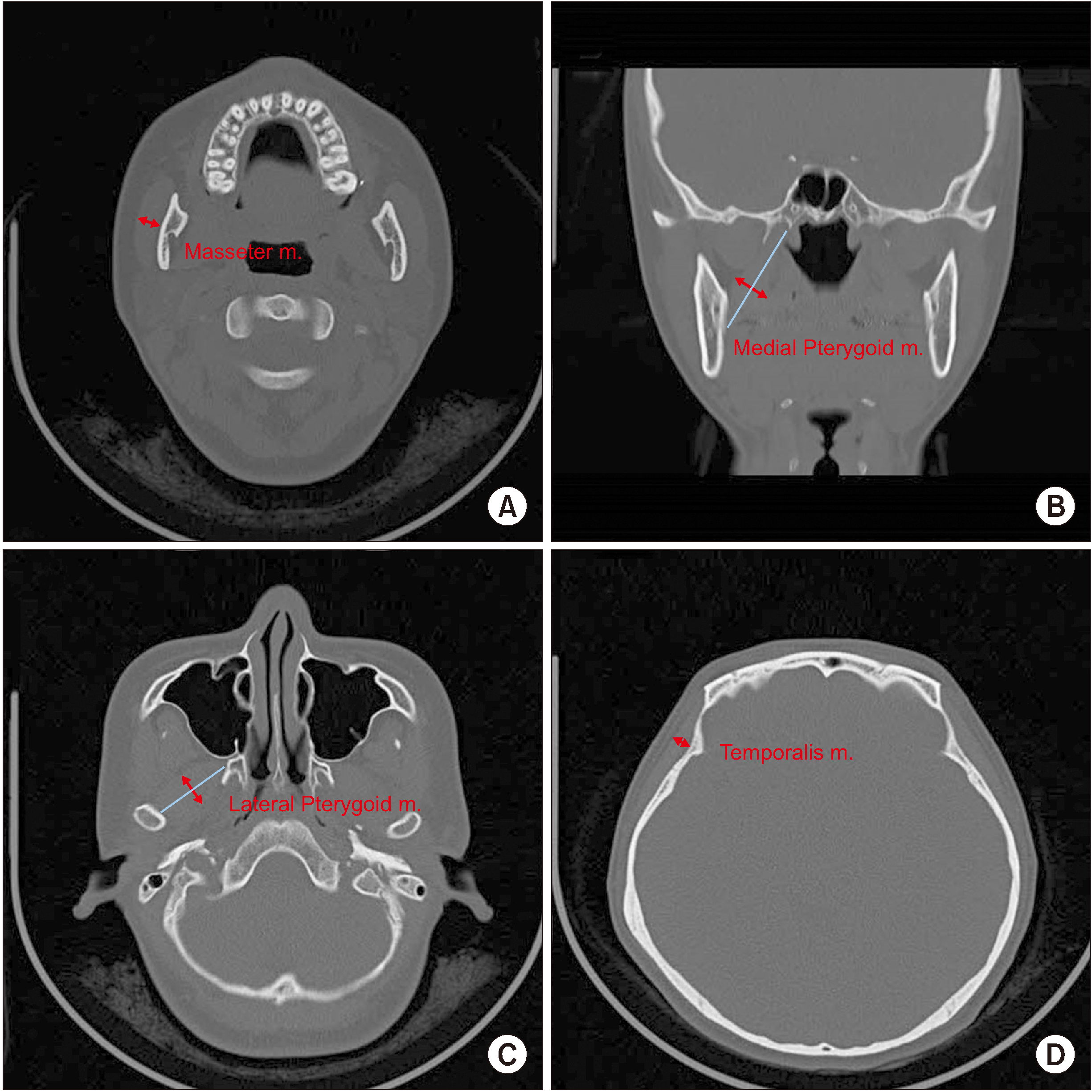
MDCT images were obtained with the Somatom Emotion 6 scanner (Siemens, Erlangen, Germany) and cephalometric images were taken using Orthophos 3 (Sirona, Bensheim, Germany). The sample was limited to young adults between 18 years and 30 years of age. They were classified into the normal occlusion group and mandibular prognathism group based on the occlusal relationship of the maxillary and mandibular first molars. The normal occlusion group included 21 males and 29 females, and the mandibular prognathism group included 29 males and 21 females. Each patient was placed in the supine position and asked to place their mandible in the resting position with no force on the masticatory muscles while obtaining MDCT images. MDCT cross-sectional slice thickness was 2 mm. For cephalometric images, the head was positioned and fixed so that the clinical Frankfort horizontal plane was parallel to the floor, and the mandible was positioned in centric relation, with the teeth lightly touching each other. For analysis of the acquired MDCT images, the axial plane was set parallel to the occlusal plane and the occlusal plane was set as the reference horizontal plane. The thickness of the masseter and lateral pterygoid muscles was measured as the maximum length perpendicular to the direction of the muscle on the axial view of the CT. Thickness of the temporalis was measured as the maximum length in the plane in contact with the supraorbital margin on the axial view of the CT. The thickness of the medial pterygoid was measured as the maximum length perpendicular to the direction of the muscle on the coronal view of the CT.(
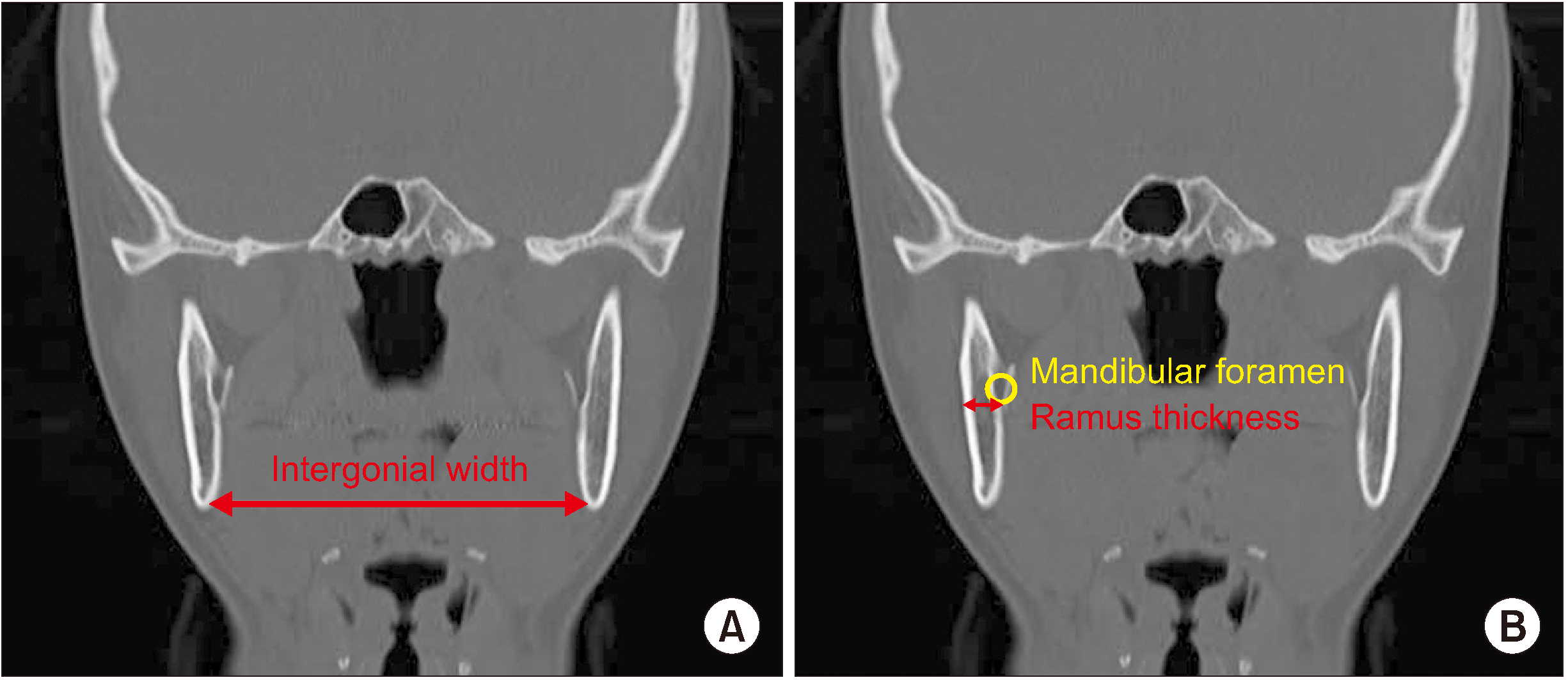
Fig. 1) Intergonial width was measured as the distance between bilateral gonions on the coronal view. Ramus thickness was measured at the tangent below the mandibular foramen, where it first appeared on the coronal view.(
Fig. 2) Muscle and ramus thickness was measured on the right side of each patient.
 | Fig. 1A. Thickness of the masseter muscle (m.) measured in axial view. B. Thickness of the medial pterygoid muscle (m.) measured in coronal view. C. Thickness of the lateral pterygoid muscle (m.) measured in axial view. D. Thickness of the temporalis muscle (m.) measured in axial view. 
|
 | Fig. 2Intergonial width (A) and ramus thickness (B) measured in coronal view. 
|
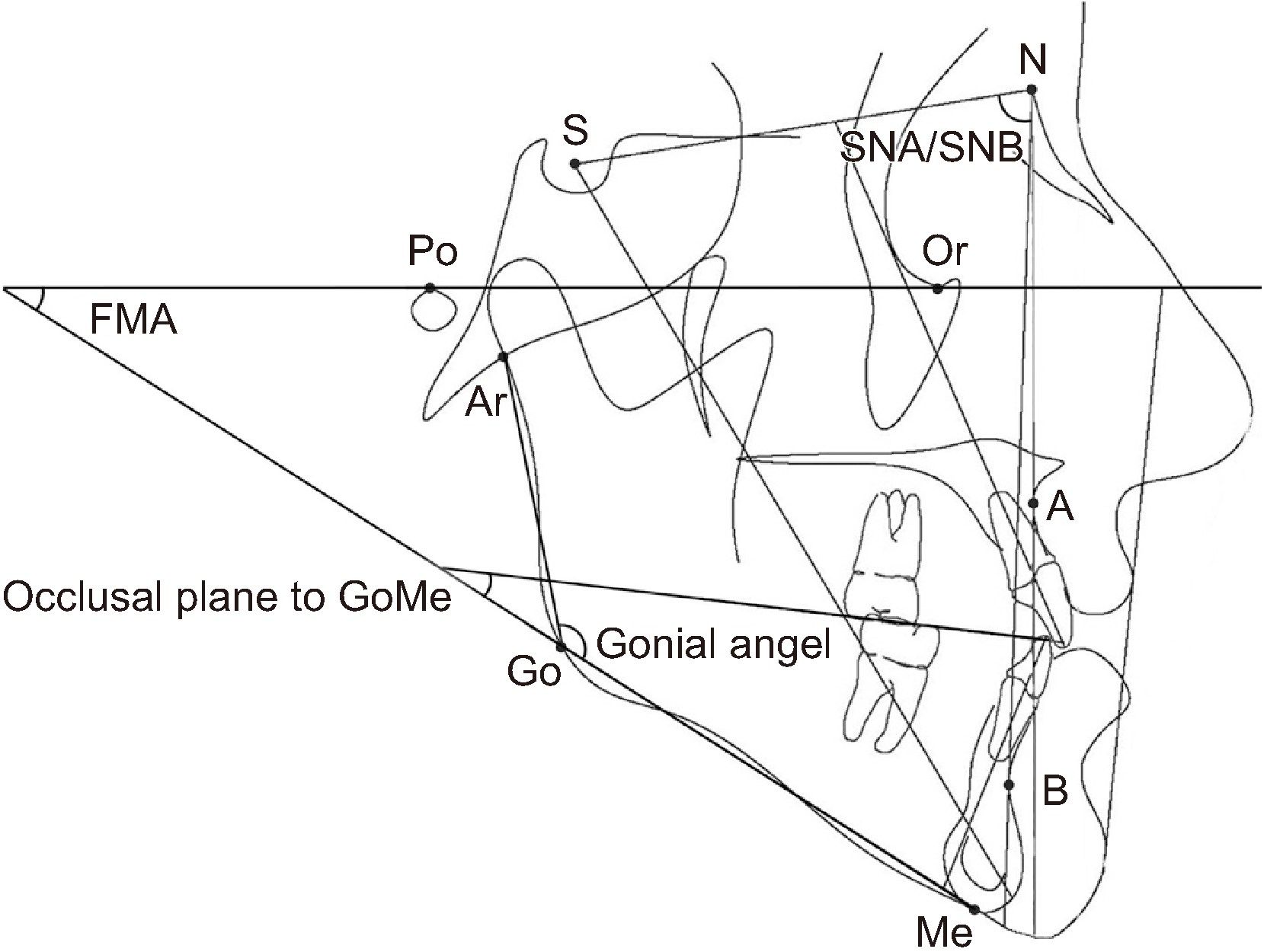
Sella (S), nasion (N), point A (A), point B (B), menton (Me), gonion (Go), articulare (Ar), orbitale (Or), and porion (Po) were marked for cephalometric analysis. SNA, SNB, ANB, gonial angle (Ar-Go-Me), occlusal plane to GoMe, FMA (FH plane-GoMe), ramus height (Go-Ar), and mandibular length (Go-Me) were measured.(
Fig. 3)
 | Fig. 3Landmarks used for measurement: S (sella), N (nasion), A (point A), B (point B), Me (menton), Go (gonion), Ar (articulare), Or (orbitale), Po (porion). Linear measurements: ramus height (Go-Ar), mandibular length (Go-Me). Angular measurements: SNA, SNB, gonial angle (Ar-Go-Me), occlusal plane to GoMe, FMA (FH plane-GoMe). 
|
Student’s t-test and Mann–Whitney U-test were performed after testing for normality, to compare the measured values of each group. Pearson correlation analysis was performed to confirm the relationship between masticatory muscle thickness and each measured value of the mandible. A P<0.05 was chosen as the level of significance for all tests. Analyses were performed using statistical software (IBM SPSS 26.0; IBM, Armonk, NY, USA).
Go to :

III. Results
First, masticatory muscle thickness was compared based on sex in all 100 patients.(
Table 1) The thickness of all four masticatory muscles was significantly different between males and females and was greater in males. Subsequently, masticatory muscle thickness was compared between the normal occlusion and mandibular prognathism groups. In males, there was a significant difference in masseter thickness between the two groups, while the same was observed in the lateral pterygoid in females.
Table 1
Mean and SD of masticatory muscles of 100 subjects
|
Masticatory muscle |
Sex |
Mean±SD |
Group |
Mean±SD |
|
MAS (mm) |
M |
14.98±1.94*
|
A |
15.73±1.98*
|
|
|
|
B |
14.43±1.75*
|
|
F |
12.75±1.82*
|
A |
12.85±2.15 |
|
|
|
B |
12.60±1.27 |
|
MPM (mm) |
M |
14.61±1.58*
|
A |
14.73±1.32 |
|
|
|
B |
14.52±1.76 |
|
F |
13.47±1.27*
|
A |
13.58±0.84 |
|
|
|
B |
13.31±1.71 |
|
LPM (mm) |
M |
15.34±1.56*
|
A |
15.59±1.40 |
|
|
|
B |
15.15±1.66 |
|
F |
13.85±1.35*
|
A |
13.51±1.22*
|
|
|
|
B |
14.31±1.42*
|
|
TEM (mm) |
M |
6.55±0.97*
|
A |
6.66±1.14 |
|
|
|
B |
6.48±0.85 |
|
F |
5.85±0.83*
|
A |
5.94±0.73 |
|
|
|
B |
5.73±0.96 |

Morphometric values of the mandible were compared according to sex in all 100 patients.(
Table 2) Significant differences between males and females were found in SNB, height and thickness of the ramus, mandibular length, occlusal plane to GoMe, and intergonial width. Following this, probable differences in the measurements were assessed based on sex between the normal occlusion and mandibular prognathism groups. Males showed significant differences in SNB, ANB, ramus height, and occlusal plane angle to GoMe between normal and affected groups, while females showed significant differences in SNB and ANB between groups.
Table 2
Mean and SD of measurement values of the mandible of 100 subjects
|
Measurement |
Sex |
Mean±SD |
Group |
Mean±SD |
|
SNA (°) |
M |
81.50±4.15 |
A |
82.81±3.45 |
|
|
|
B |
80.55±4.40 |
|
F |
80.39±4.26 |
A |
80.41±4.31 |
|
|
|
B |
80.36±4.31 |
|
SNB (°) |
M |
83.41±4.59*
|
A |
80.99±3.65*
|
|
|
|
B |
85.16±4.46*
|
|
F |
81.05±5.88*
|
A |
78.26±4.72*
|
|
|
|
B |
84.90±5.17*
|
|
ANB (°) |
M |
–1.91±4.50 |
A |
1.82±3.18*
|
|
|
|
B |
–4.61±3.19*
|
|
F |
–0.66±4.27 |
A |
2.15±2.82*
|
|
|
|
B |
–4.54±2.50*
|
|
Ramus height (Go-Ar) (mm) |
M |
54.75±6.06*
|
A |
57.69±4.92*
|
|
|
|
B |
52.63±5.98*
|
|
F |
49.91±5.57*
|
A |
50.04±5.58 |
|
|
|
B |
49.74±5.67 |
|
Mandibular length (Go-Me) (mm) |
M |
77.90±5.23*
|
A |
78.91±4.53 |
|
|
|
B |
77.17±5.65 |
|
F |
73.96±5.63*
|
A |
73.29±5.16 |
|
|
|
B |
74.87±6.25 |
|
Gonial angle (Ar-Go-Me) (°) |
M |
126.47±6.79 |
A |
124.38±7.92 |
|
|
|
B |
127.98±5.50 |
|
F |
125.98±7.01 |
A |
126.19±7.43 |
|
|
|
B |
125.68±6.56 |
|
Occlusal plane angle to GoMe (°) |
M |
19.42±5.91*
|
A |
17.08±5.36*
|
|
|
|
B |
21.12±5.78*
|
|
F |
15.99±4.56*
|
A |
15.79±4.43 |
|
|
|
B |
16.27±4.82 |
|
FMA (FH plane-GoMe) (°) |
M |
27.25±6.00 |
A |
26.38±5.96 |
|
|
|
B |
27.89±6.05 |
|
F |
27.55±6.74 |
A |
28.35±6.08 |
|
|
|
B |
26.43±7.57 |
|
Ramus thickness (mm) |
M |
8.60±1.25*
|
A |
8.90±1.12 |
|
|
|
B |
8.38±1.32 |
|
F |
8.10±1.24*
|
A |
8.16±1.17 |
|
|
|
B |
8.03±1.36 |
|
Intergonial width (mm) |
M |
95.12±5.33*
|
A |
96.35±4.13 |
|
|
|
B |
94.23±5.96 |
|
F |
88.90±4.74*
|
A |
89.20±4.60 |
|
|
|
B |
88.49±5.02 |

Pearson correlation analysis was used to determine the relationship between masticatory muscle thickness and mandibular measurement values in all patients.(
Tables 3,
4) There were significant positive correlations among the thicknesses of the four masticatory muscles. Masseter thickness was positively correlated with height and thickness of the ramus, mandibular length, and intergonial width. Thickness of the medial pterygoid had a positive correlation with ramus height, mandibular length, and intergonial width, while the lateral pterygoid was positively correlated with SNB, height and thickness of the ramus, mandibular length, and intergonial width. Temporalis thickness correlated positively with thickness of the ramus and intergonial width.
Table 3
Correlation between the four masticatory muscles
|
Group |
MAS |
MPM |
LPM |
TEM |
|
MAS |
Total |
|
0.416**
|
0.560**
|
0.422**
|
|
A |
|
0.508**
|
0.609**
|
0.489**
|
|
B |
|
0.389**
|
0.550**
|
0.336*
|
|
MPM |
Total |
0.416**
|
|
0.440**
|
0.424**
|
|
A |
0.508**
|
|
0.379**
|
0.441**
|
|
B |
0.389**
|
|
0.509**
|
0.428**
|
|
LPM |
Total |
0.560**
|
0.440**
|
|
0.406**
|
|
A |
0.609**
|
0.379**
|
|
0.508**
|
|
B |
0.550**
|
0.509**
|
|
0.316*
|
|
TEM |
Total |
0.422**
|
0.424**
|
0.406**
|
|
|
A |
0.489**
|
0.441**
|
0.508**
|
|
|
B |
0.336*
|
0.428**
|
0.316*
|
|

Table 4
Correlation between the four masticatory muscles and measurement values of the mandible
|
Group |
MAS |
MPM |
LPM |
TEM |
|
SNA |
Total |
0.165 |
0.144 |
0.091 |
0.039 |
|
A |
0.152 |
0.158 |
0.088 |
0.185 |
|
B |
0.170 |
0.138 |
0.125 |
–0.112 |
|
SNB |
Total |
0.150 |
0.161 |
0.204*
|
0.019 |
|
A |
0.157 |
0.281*
|
0.167 |
0.139 |
|
B |
0.346*
|
0.155 |
0.158 |
–0.043 |
|
ANB |
Total |
–0.024 |
–0.059 |
–0.163 |
0.013 |
|
A |
–0.027 |
–0.206 |
–0.131 |
0.047 |
|
B |
–0.310*
|
–0.046 |
–0.071 |
–0.097 |
|
Ramus height (Go-Ar) |
Total |
0.309**
|
0.315**
|
0.266**
|
0.144 |
|
A |
0.425**
|
0.272 |
0.433**
|
0.251 |
|
B |
0.115 |
0.364**
|
0.130 |
0.017 |
|
Mandibular length (Go-Me) |
Total |
0.268**
|
0.227*
|
0.239*
|
0.189 |
|
A |
0.412**
|
0.330*
|
0.212 |
0.297*
|
|
B |
0.106 |
0.170 |
0.257 |
0.090 |
|
Gonial angle (Ar-Go-Me) |
Total |
–0.044 |
0.074 |
0.026 |
–0.144 |
|
A |
–0.148 |
–0.004 |
–0.173 |
–0.310*
|
|
B |
0.170 |
0.152 |
0.252 |
0.077 |
|
Occlusal plane angle to GoMe |
Total |
0.103 |
0.082 |
0.041 |
0.078 |
|
A |
0.095 |
0.275 |
–0.026 |
–0.115 |
|
B |
0.182 |
–0.012 |
0.038 |
0.265 |
|
FMA (FH plane-GoMe) |
Total |
–0.152 |
–0.029 |
–0.156 |
–0.041 |
|
A |
–0.211 |
0.013 |
–0.265 |
–0.344*
|
|
B |
–0.091 |
–0.055 |
–0.053 |
0.237 |
|
Ramus thickness |
Total |
0.276**
|
0.120 |
0.238*
|
0.259**
|
|
A |
0.406**
|
0.372**
|
0.492**
|
0.481**
|
|
B |
0.113 |
–0.029 |
0.033 |
0.052 |
|
Intergonial width |
Total |
0.569**
|
0.293**
|
0.478**
|
0.348**
|
|
A |
0.604**
|
0.469**
|
0.602**
|
0.384**
|
|
B |
0.554**
|
0.192 |
0.381**
|
0.315*
|

The results of Pearson correlation analysis of the normal occlusion group are shown in
Tables 3 and
4. As seen in the entire group, there was a positive correlation among the thicknesses of the four masticatory muscles. Moreover, the thickness of each masticatory muscle positively correlated with the intergonial width and ramus thickness. Masseter thickness showed a positive correlation with ramus height and mandibular length. Medial pterygoid thickness correlated positively with SNB and mandibular length, while the lateral pterygoid was correlated with ramus height. Temporalis thickness showed a positive correlation with mandibular length, but a negative correlation with gonial angle and FMA.
The results of Pearson correlation analysis of the mandibular prognathism group are also shown in
Tables 3 and
4. The thicknesses of the four masticatory muscles were positively correlated with each other. Thickness of three masticatory muscles excepting the medial pterygoid showed a positive correlation with intergonial width. Masseter thickness was positively correlated with SNB, but was negatively correlated with ANB. Thickness of the medial pterygoid had a positive correlation with ramus height.
Go to :

IV. Discussion
The function and morphology of the masticatory muscles are closely related to the morphological features of craniofacial bones
10
. The mechanism by which masticatory muscles influence the growth pattern of the face is not well understood; however, it may be related to the capacity of the masticatory muscles, as expressed by maximum occlusal force, electromyography, and cross-sectional thickness
11
. This could represent the contractile ability of the masticatory muscles; however, maximum occlusion occurs for short periods of time. Moreover, the value of electromyography for assessing the sustained effects of muscles is limited
12
. Ueki et al.
13
observed a positive correlation between masticatory muscle thickness on cross-section and occlusal force, and reported that the effects of muscles can be analyzed using cross-sectional thickness.
In order to examine masticatory muscle thickness, imaging techniques such as ultrasonography, CT, and MRI may be used. Ultrasonography is an imaging modality that uses ultrasound equipment. Kiliaridis and Kälebo
14
, as well as Benington et al.
5
, used ultrasonography in their studies. This technique is relatively inexpensive, of shorter duration, and there are no cumulative biological effects. Among the masticatory muscles, the masseter and temporalis are located relatively superficially; therefore, their thicknesses can be measured by ultrasonography. However, the thicknesses of the medial and lateral pterygoid muscles, which are located deeper, cannot be measured easily. The indications and most accurate technique for measuring the thickness of muscles with ultrasonography have not yet been established. It is difficult to achieve precise measurements without clear knowledge of the anatomy, adequate skill, and experience. In addition, since variable images may be obtained depending on the force applied to the muscle and surrounding tissue during measurement, objective measurement is difficult using ultrasonography, unlike with CT or MRI
15. CT and MRI have the advantage of more precise measurement, owing to the non-contact method and better image quality than ultrasonography. Moreover, most dental hospitals have CT equipment, thus resolving the issue of accessibility. However, MRI is a relatively expensive modality and the diagnostic time is longer than in CT. The disadvantage of CT is the cumulative biologic effects due to radiation exposure.
Ramus height corresponds to the vertical dimension of the mandible. Mandibular length, intergonial width, and ramus thickness correspond to the horizontal dimension of the mandible. Weijs and Hillen
16
showed a positive correlation between masseter and bigonial width and mandibular length. Another study demonstrated a positive correlation between masticatory muscle thickness and mandible horizontal dimension, but a negative correlation with mandible vertical dimension
4
. The present study on 100 Korean young adults showed a positive correlation between both horizontal and vertical mandibular dimensions and thickness of the majority of the masticatory muscles. This shows that the greater the chewing ability of the masticatory muscles, the more they promote the overall growth of the mandible including both horizontal and vertical dimensions. According to Moore
17
, resection of masticatory muscles not only has a local effect on the area of attachment of the muscles, but also affects overall dimensions of facial bones. However, it is difficult to explain the specific role of each masticatory muscle on the growth of facial bones. This is because resection of even one masticatory muscle may result in atrophy of the other muscles. In addition, Yonemitsu et al.
18
demonstrated that excision of bilateral masseter muscles in immature rats resulted in under-development of the mandible after completion of growth. This suggests that for ideal formation and growth, an adequate amount of force must be applied to the mandible, which is ensured by the presence and function of masticatory muscles.
Benington et al.
5
demonstrated negative correlations between the masseter and the gonial angle. Uchida et al.
19
used ultrasonography to show that the angle of mandibular components, such as FMA and GoGn-SN, decreased and the mandible rotated counterclockwise as the thickness of the masseter increased. In contrast, the present study did not show any correlation between the thickness of masticatory muscles and mandibular angle components, with the exception of a negative correlation between temporalis thickness, gonial angle, and FMA in the normal occlusion group. This may be attributed to differences in inclination of the occlusal plane and the manner of measuring masticatory muscles even among people with similar skeletal patterns.
The purpose of this study was to examine the correlation between masticatory muscle thickness and mandible morphology, as well as to investigate differences between individuals with normal occlusion and mandibular prognathism. The degree of ANB was related to mandibular prognathism, and greater severity has been associated with a thinner masseter. This can also be interpreted as a negative correlation between the severity of mandibular prognathism and masticatory ability of the masseter.
Orthognathic surgery improves not only facial aesthetics, but also chewing ability. In cases of mandibular prognathism, there is malocclusion of the maxillary and mandibular teeth, causing temporomandibular disorder and chewing problems. Chewing ability includes bite force, occlusal contact, and masticatory efficiency. According to a study by Iwase et al.
20
, bite force and occlusal contact were relatively low in patients with mandibular prognathism, which is in line with the results of this study. The masseter is a jaw closing muscle and is the strongest in the human body. The present study, which revealed a negative correlation between the severity of mandibular prognathism and masseter thickness, is a good example of decreased bite force in patients with mandibular prognathism
21. The aforementioned study by Islam et al.
22
showed that all three elements of chewing ability improved after orthognathic surgery, and may explain role of surgery in improving quality of life, through correction of chewing ability in patients with mandibular prognathism.
Go to :

V. Conclusion
CT can be used for easy and accurate measurement of masticatory muscle thickness and mandibular morphology. In the present study, masticatory muscle thickness was greater in males than in females and greater in the normal occlusion group than in the mandibular prognathism group. The morphology of the mandible changes constantly due to the effect of adjacent muscles. This study showed that the thickness of masticatory muscles correlated positively with both the horizontal and vertical dimensions of the mandible. In patients with mandibular prognathism, masseter thickness decreased as the severity of mandibular prognathism increased, and this may explain the relatively lower chewing ability of patients with mandibular prognathism.
Several relationships were found between masticatory muscle thickness and mandible morphology in this study. However, the effects of other soft tissues around the mandible were not considered, and errors in the determination of muscle thickness and mandibular measurements could have affected the correlation coefficients. Therefore, the results of our study should be interpreted with caution. Further research should focus on evaluation of the relationship between facial and lingual muscles and mandibular morphology.
Go to :


 PDF
PDF Citation
 Print
Print


 XML Download