

 PDF
PDF Citation
 Print
Print


I. Introduction
Surgical difficulty in mandibular third molar extractions can predict the incidence of postoperative complications. Since the 19th century, several researchers have attempted to classify extraction difficulty using radiographic techniques or estimating the prevalence of complications1
-5. In December 2019, a difficulty index was proposed based on positional variations in impacted mandibular third molars evaluated by panoramic radiography5. The position of the third molar is associated not only with postoperative complications, but also surgical difficulty and operation time6,7. In the Korean insurance system, surgical time is one of the important criteria for evaluating surgical difficulty8. However, previous difficulty indices focused mainly on the probability of complications, and very few clinical studies have reported on prediction of surgery time1
-5.
Evaluating extraction time is difficult given the various surgical techniques used and the highly divergent morphology of third molars and their surrounding structures9. The operculum is an epithelial tissue overlying the impacted tooth that originates from the dental follicle and attaches to the cementoenamel junction (CEJ)10. The operculectomy procedure enables access to the CEJ for odontomy, facilitates surgical extraction through a minimally invasive technique, and enhances healing by de-epithelialization.
Impacted third molars may be associated with pathologic conditions such as root resorption of the adjacent tooth, pericoronitis, dentigerous cysts, and benign neoplasms11. However, few studies have evaluated the presence of pathologic conditions even though they are more frequently encountered than complications5.
The present study aimed to assess the validity of the difficulty index with extraction time using a unified incision technique (operculectomy) and suggests a modified difficulty index including the presence of pathologic conditions associated with third molars.
Go to : 
II. Materials and Methods
This retrospective study was initiated after approval by the Institutional Review Board at the Armed Forces Capital Hospital (No. AFCH-19-IRB-034). We enrolled healthy male patients who had undergone mandibular third molar extraction by one expert surgeon from December 2018 to December 2019 at the Armed Forces Capital Dental Hospital. The inclusion criteria were as follows: 1) age between 19 and 25 years, 2) preoperative computed tomography (CT) evaluation (syngo CT VB20, Sickness 1.0 mm, Dose 120 kVp; Siemens, Berlin, Germany), 3) surgical approach by operculectomy alone, 4) intraoperative digital photographs taken at the start of incision; after the incision; and during odontomy, extraction, and wound suturing.(Fig. 1) The exclusion criteria were as follows: 1) any incision design (vertical incision, envelope incision) except operculectomy, 2) a history or symptoms of maxillary sinus disease, 3) poor plaque control and untreated chronic periodontitis, and 4) third molars with incomplete root formation.
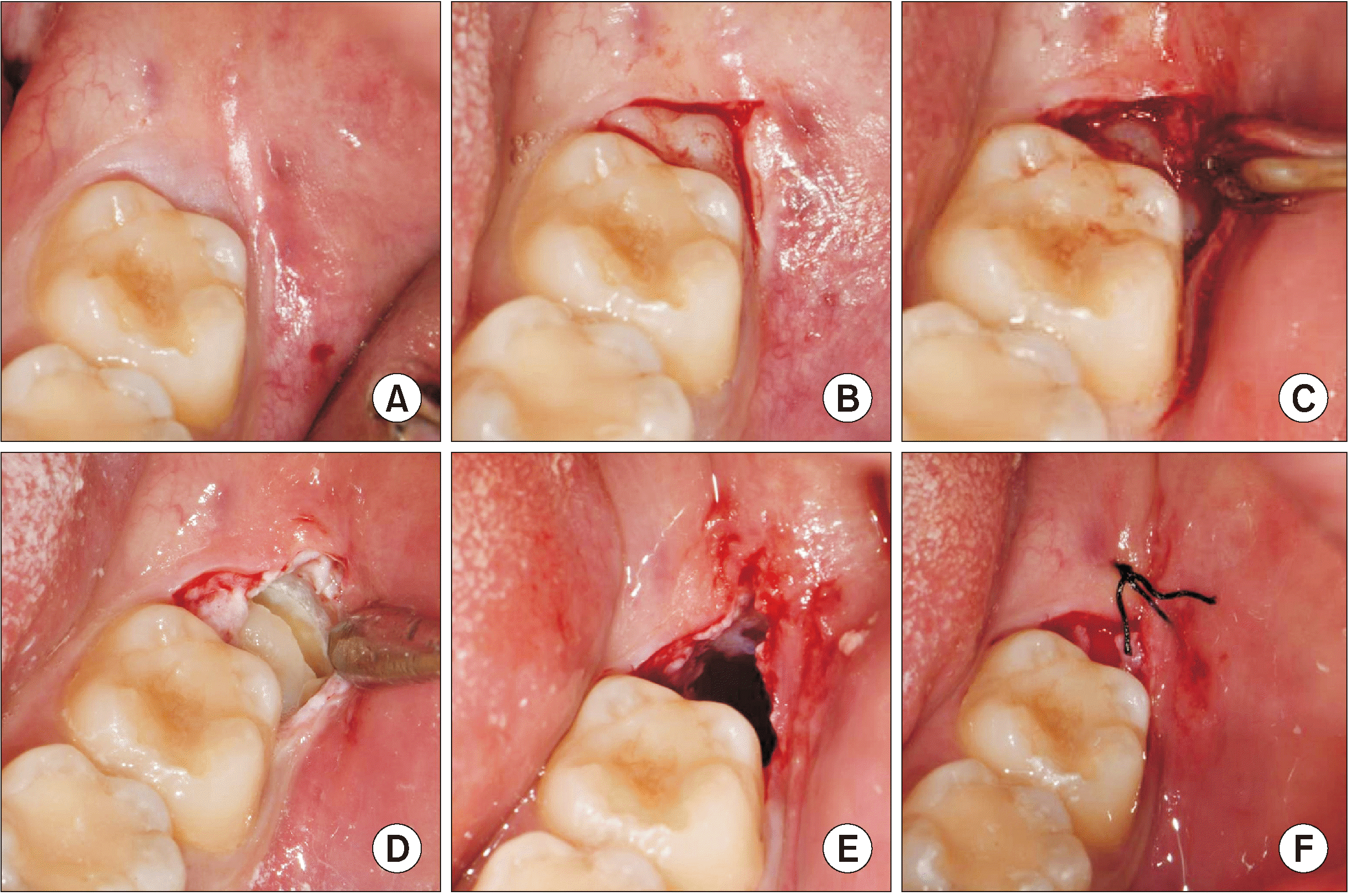 | Fig. 1Photographs of the surgical extractions with operculectomies. A. Preoperative photograph of the patient in Fig. 2. B. Incision for the operculectomy. C. After operculectomy, the impacted mandibular third molar was exposed. D. Odontomy of the third molar was performed. E. Extraction of the third molar was completed. F. Postoperative photograph after suturing with collagen plug packing.
|
The pathologic conditions associated with impacted third molars were defined as caries, pericoronitis, dentigerous cysts, deformed roots, cuspal contact below the CEJ of the adjacent molar, and root resorption of the adjacent tooth.
Extraction time was calculated from the start of the incision to the last suture, based on digital photographs.
1. Difficulty score and index
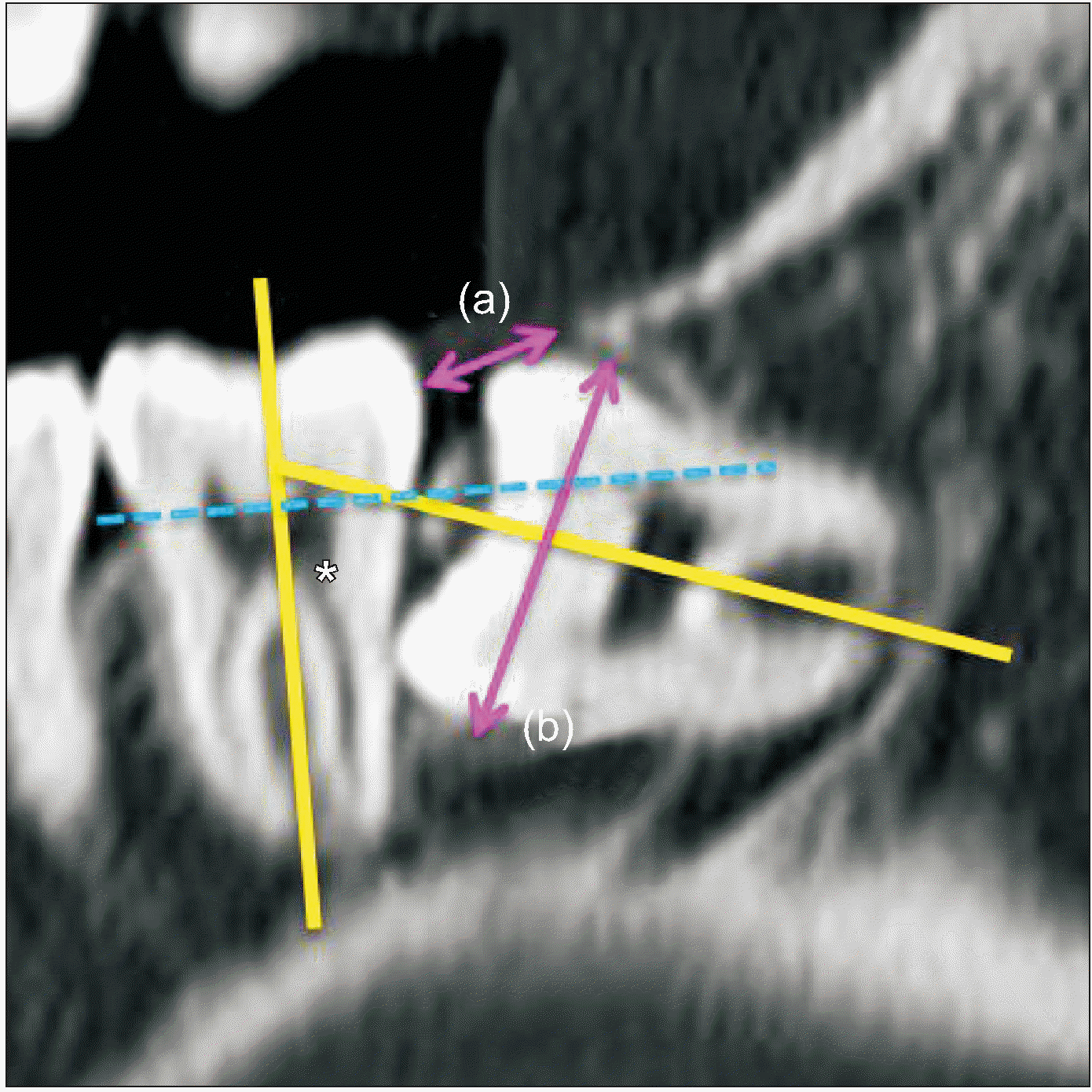
We reformatted the CT data to a panoramic view based on the mandibular arch (middle of the mandibular teeth and inferior mandibular canal) with an axial cut using platform software (SOMARIS/7; Siemens) and selected a single image cut that included the middle of the extracted third molar.
We followed the most recently published (2019) difficulty index5. Briefly, impacted third molars were classified and scored (difficulty score) based on spatial relationship (1-5 points), depth (1-4 points), and ramus relationship (1-3 points).(Fig. 2)
Spatial relationship was subcategorized based on the angle between the long axis of the third molar and the adjacent second molar as follows: (1) mesioangular (11° to 79°), (2) horizontal (80° to 100°), (3) vertical (–10° to 10°), (4) distoangular (–11° to –79°), or (5) reverse where the crown of the third molar was more root-oriented than horizontal.
Depth was subcategorized and scored as levels A (1), B (2), C (3), or D (4). Level A was defined as a condition when more than half of the third molar crown was above the CEJ of the adjacent second molar. Level B was defined when less than half of the third molar crown above the CEJ of the adjacent second molar. When the entire the third molar crown was positioned below the CEJ of the adjacent second molar, it was defined as either level C or level D. Level C was defined as a condition when more than half of the third molar crown was positioned superior to the mid-level of the adjacent second molar root. Level D was defined when the third molar crown level was inferior to that mentioned above.
The available ramus relationship/space was sub-classified and scored as class I (1), II (2), or III (3). Eruption space was defined as the ratio of distance between the distal side of the second molar to the ascending ramus (a) and the diameter of the third molar (b), as shown in Fig. 2. An eruption space (a/b) larger than two-thirds the distance was defined as class I, between one-third and two-thirds was class II, and smaller than one-third was class III.
The difficulty index was based on total points as follows: I (3-4 points), II (5-7 points), III (8-10 points), and IV (11-12 points). In addition, the modified difficulty score was calculated by adding one point to the difficulty score if the third molar was associated with a pathologic condition. Two modified difficulty indices that considered the presence of pathologic conditions were defined as follows: the half-level up difficulty index (HDI) and the one-level up difficulty index (ODI) based on the difficulty index reported by Kim et al.5.
2. Statistical analysis
Extraction time was analyzed with an independent t-test for difference between anesthetic techniques (general or local anesthesia) and left- or right-side molar. One-way ANOVA was used to analyze the difference in extraction time between difficulty indices. Post-hoc analysis was performed using the Mann–Whitney U test, and the Bonferroni method was used to control for type 1 error. Pearson correlation coefficients were used to reveal linear relationships of extraction time with difficulty score and index and with modified difficulty score and indices (HDI and ODI). The data were analyzed using IBM SPSS Statistics (ver. 23.0; IBM, Armonk, NY, USA). A P-value less than 0.05 indicated significance.
Go to :
III. Results
A total of 65 patients (all males, 21.25±1.37 years) with 68 extractions was included, and the average extraction time was 17.48±6.56 minutes. Fifty-one extractions were performed under local anesthesia (18.04±6.56 minutes), while 17 were completed under general anesthesia (17.48±6.56 minutes). Thirty-four extractions were performed on the left side (18.17±6.58 minutes), and 34 were completed on the right side (16.55±7.11 minutes). Extraction time exhibited no statistical difference according to anesthetic technique or side.(Table 1)
Table 1
Demographic and clinical informations of the patients with impacted third molar
| Variable | Value | P-value1 |
|---|---|---|
| No. of patients | 65 | |
| Age (yr) | 21.25±1.37 | |
| Sex | Male | |
| Extraction time (min) | ||
| Anesthesia | 0.439 | |
| Local (n=51) | 18.04±6.56 | |
| General (n=17) | 17.48±6.56 | |
| Location | 0.959 | |
| Left (n=34) | 18.17±6.58 | |
| Right (n=34) | 16.55±7.11 |
![]()
The mean extraction times according to difficulty score were 9.47±4.33, 15.08±5.40, 18.13±5.36, 17.28±4.20, 19.51±6.42, and 28.20±5.17 minutes from scores three to eight, respectively. The correlation between extraction time and difficulty score was significant (P<0.001), and the observed correlation coefficient (r) was 0.599, suggesting a moderate positive correlation (P<0.001). The mean extraction times according to modified difficulty scores were 8.59±4.10, 14.30±3.01, 15.41±4.59, 19.08±5.07, 17.18±6.06, 26.09±3.32, and 27.36±5.33 minutes from scores three to nine, respectively. The correlation between extraction time and modified difficulty score was significant (P<0.001). The observed correlation coefficient (r) was 0.690, suggesting a moderate positive correlation.
The association between extraction time and the difficulty indices are shown in Table 2. The mean extraction times according to difficulty index were 12.18±5.39, 18.28±5.34, and 28.20±5.17 minutes in I, II, and III, respectively. The correlation between extraction time and difficulty index was significant (P<0.001), and the observed correlation coefficient (r) was 0.584, suggesting a moderate positive correlation.(Fig. 3) Extraction time was significantly different between difficulty index grades I and II (P=0.001), I and III (P<0.001), and II and III (P<0.001).(Table 2)
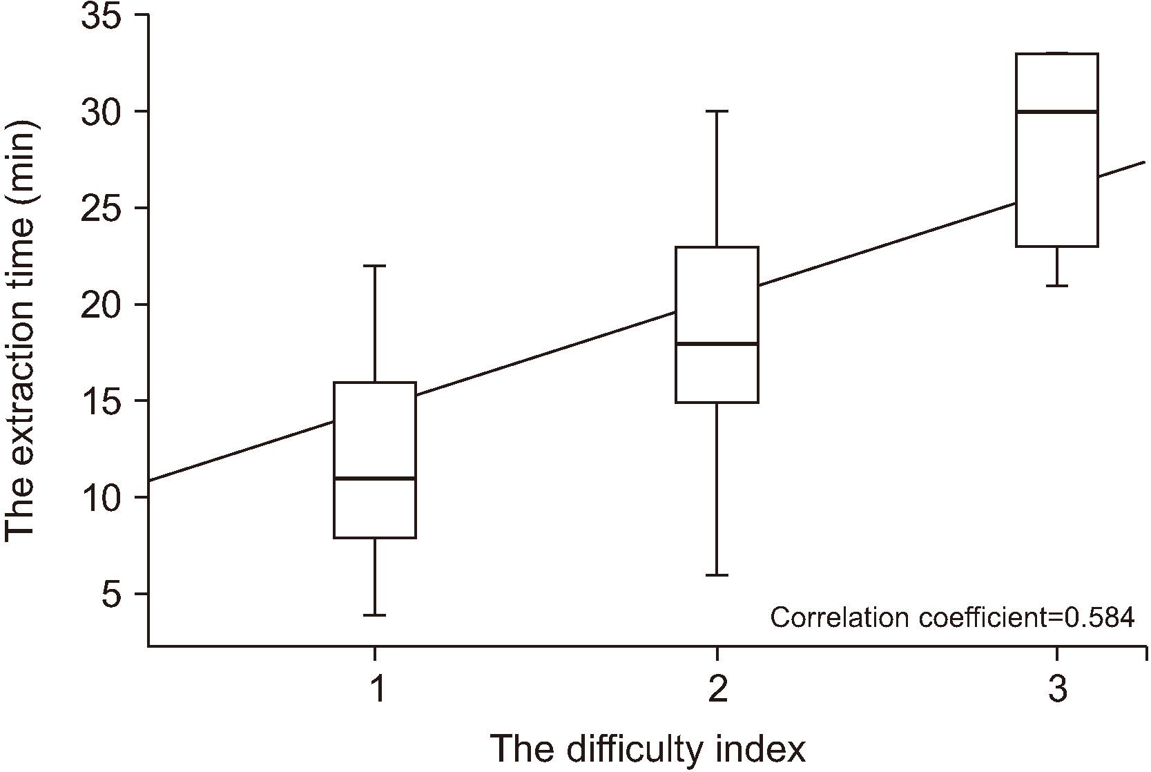 | Fig. 3The correlation between the extraction time and the difficulty index. The correlation coefficient was 0.584, suggesting a moderate positive correlation (P<0.001).
|
Table 2
Association between extraction time and difficulty indices of impacted third molars
| Grade | n | Extraction time (min) | P-value | Correlation coefficient (r) | |
|---|---|---|---|---|---|
| Difficulty index | |||||
| I | 17 | 12.18±5.39 | <0.0011 |
0.0012 (I vs II) <0.0012 (II vs III) |
0.599 |
| II | 45 | 18.28±5.34 | |||
| III | 6 | 28.20±5.17 | |||
| Half-up difficulty index | |||||
| 1 | 13 | 10.60±4.33 | <0.0011 |
0.3182 (1 vs 1.5) >0.9992 (1.5 vs 2) <0.0012 (2 vs 2.5) >0.9992 (2.5 vs 3.5) NA (2.5 vs 3) NA (3 vs 3.5) |
0.728 |
| 1.5 | 4 | 16.30±7.33 | |||
| 2 | 30 | 15.38±4.06 | |||
| 2.5 | 15 | 24.08±3.23 | |||
| 3 | 1 | 31.60 | |||
| 3.5 | 5 | 27.36±0.33 | |||
| One-up difficult index | |||||
| I | 13 | 10.60±4.33 | <0.0011 |
0.0102 (I vs II) <0.0012 (II vs III) >0.9992 (III vs IV) |
0.764 |
| II | 34 | 15.44±4.28 | |||
| III | 16 | 24.38±3.49 | |||
| IV | 5 | 27.36±5.33 | |||
![]()
The mean extraction times according to HDI were 10.60±4.33, 16.30±7.33, 15.38±4.06, 24.08±3.23, 31.60, and 27.36±0.33 minutes in one, one-half, two, two-half, three, and three-half, respectively, and those according to ODI were 10.60±4.33, 15.44±4.28, 24.38±3.49, and 27.36±5.33 minutes in I, II, III, and IV. The correlations between extraction time and HDI and ODI were significant (P<0.001). The observed correlation coefficients (r) were 0.728 and 0.764, respectively, suggesting strong positive correlations.(Fig. 4)
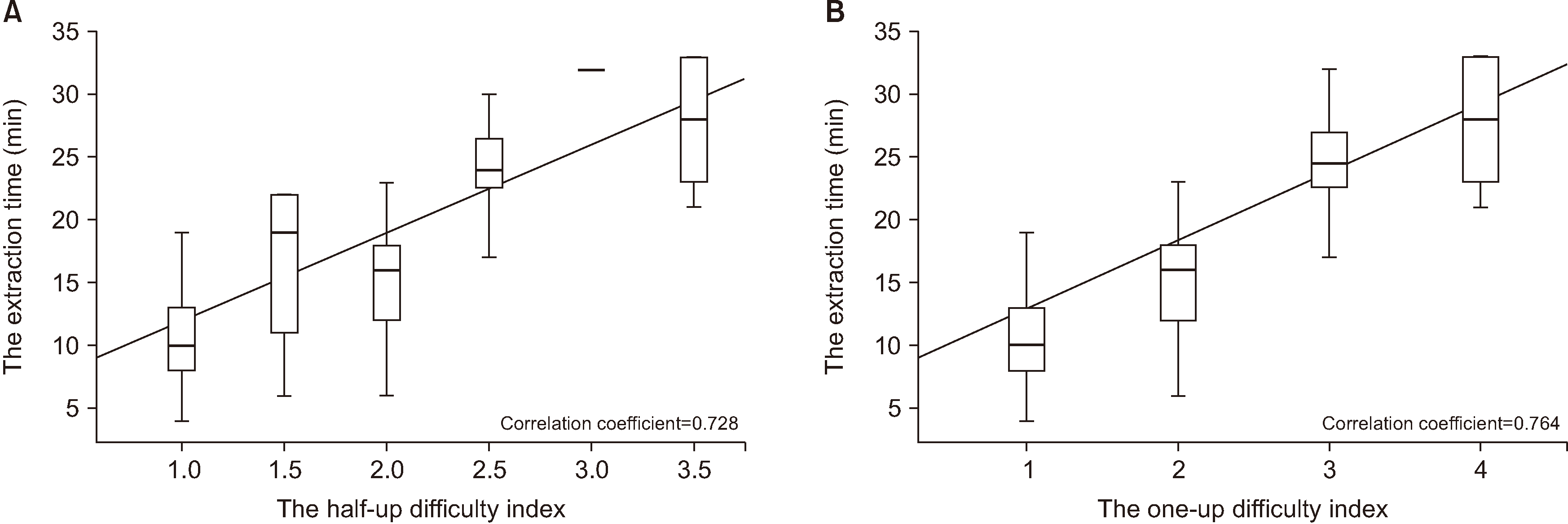 | Fig. 4The correlation between the extraction time and the modified difficulty indices. A. The correlation between the extraction time and the half-up difficulty index. The correlation coefficient was 0.728, suggesting a strong positive correlation (P<0.001). B. The correlation between the extraction time and the half-up difficulty index. The correlation coefficient was 0.764, suggesting a strong positive correlation (P<0.001).
|
Extraction time significantly differed among HDI grades (P<0.001), but post hoc analysis showed significance only between HDI grades two and two-half (P<0.001). Extraction time was significantly different among the ODI grades, ODI grades I and II (P=0.010), II and III (P<0.001), and II and IV (P<0.001).(Table 2)
Go to :
IV. Discussion
Two findings were revealed in this study. First, the difficulty index5 classified as grades I, II, and III based on CT data reformatted to a panoramic view was correlated with extraction time of impacted third molars other than grade IV. Second, the modified difficulty indices that considered the presence of pathologic conditions were strongly correlated with extraction time.
Prediction of surgical extraction difficulty is important for anticipating postoperative issues and planning treatment options such as general anesthesia in a controlled environment to prevent complications12,13. Extraction difficulties were evaluated based on position of the third molars using panoramic radiography1
-5. The position was associated with postoperative complications such as alveolar nerve injury6 and dry socket7. However, assessing the difficulty index with panoramic imaging has been controversial due to morphological variations and superimposition of adjacent structures9,14. A major concern in three-dimensional analysis using CT is poor reproducibility in setting the three-dimensional axis and selecting a single image cut15. The author evaluated the position with CT reformatted panoramic images. The correlation coefficients (r) of extraction time according to the recently proposed difficulty scores and index were 0.599 and 0.584, respectively, on the panoramic image5, moderate correlations (P<0.001).
Many studies on extraction of impacted third molars have focused on postoperative complications6,7,9,11,14. However, the presence of the following pathologic conditions associated with third molars could also be critical for intraoperative considerations: unfavorable fracture during odontomy in carious third molars, local anesthetic failure and prolonged bleeding during the management of inflammatory conditions, dentigerous cysts or curved roots, and poor accessibility for instrumentation due to cuspal contact between the third molar and the distal surface of the adjacent second molar. Therefore, we considered the presence of pathologic conditions as an independent factor to evaluate the difficulty of extractions. As a result, the coefficient (r) of extraction time was 0.690 with the modified difficulty score based on the presence of pathologic conditions, which was higher than that of the previous difficulty score and exhibited a strong positive correlation (P<0.001). The coefficients (r) further increased to 0.728 and 0.764 with HDI and ODI, respectively. Regarding the result, pathologic conditions could more strongly correlate with extraction time compared with the previous difficulty score by using radiography only. Furthermore, ODI exhibited a greater correlation with surgery time than did HDI.
The inter-grade of the previous difficulty index showed statistically significant differences in average extraction time, but the difference was almost twice as large between grades II and III (18.28 and 28.20 minutes; P<0.001) compared to the difference between grades I and II (12.18 and 18.28 minutes; P=0.001). However, the average extraction time of ODI seems to be subdivided between ODI grades I and II (10.60 and 15.44 minutes; P=0.010), II and III (15.44 and 24.38 minutes; P<0.001), and III and IV (24.38 and 27.36 minutes; P>0.999) compared with the previous difficulty index. ODI grade IV did not exhibit a statistical difference from ODI grade III in Bonferroni’s method, a finding that may be explained by the limitation of our retrospective study design that did not include a large number of high-grade extractions.
Surgery time is crucial for patients and clinicians and allows them to determine the difficulty of extraction. As impacted third molars and their surrounding structures vary in anatomy, various surgical techniques have been used. However, few studies have evaluated difficulties based on surgery time. Although we used a unified surgical technique (operculectomy), the operculum could develop after resorption of the bone overlying the eruption space. As a result, we could not include extremely difficult third molars (difficulty index IV) that were fully embedded in bone5. Impacted third molars with difficulty index IV have different histologic aspects compared to other impacted teeth and can be removed with alternative treatment strategies. Further studies with prospective designs and larger numbers of patients are needed to evaluate extremely difficult third molars associated with pathologic conditions.
Go to :
V. Conclusion
Extraction time of third molar surgery is strongly correlated with modified difficulty indices that take into consideration the pathologic condition compared to the previous difficulty index. With regard to the clinical situation, the difficulty index of surgical extractions should include an assessment of the pathologic conditions associated with third molars.
Go to :
 XML Download