Introduction
Listeria is widely distributed in the natural environment and food. In most cases, Listeria infection is caused by eating contaminated food. Listeriosis is usually diagnosed when Listeria monocytogenes grows in body fluids such as the blood and cerebrospinal fluid.
It is known to not cause infections in healthy adults with normal immune systems but can cause severe infections in immunocompromised patients, elderly people, pregnant women, and newborn babies following transmission from the placenta or birth canal [
1]. During pregnancy, listeriosis usually only presents with fever and flu-like symptoms, such as fatigue and myalgia. However, the complication of infections during pregnancy include miscarriage, stillbirth, premature delivery, or life-threatening neonatal sepsis and fetal loss [
2].
The incidence of infections in pregnant women is approximately 100 times more than that in the general population. In 1,651 cases reported in 2009 to 2011, the Centers for Disease Control and Prevention found that 14% were in pregnant women [
3]. The frequency of
Listeria infection in the US is 0.7 per 100,000 babies per year; the frequency in Europe is lower and the first case in Korea was identified in 1973. Congenital infections leading to prenatal meningitis and sepsis, which are considered representative clinical infections in reports on foreign countries, are unexpectedly rare in Korea according to domestic reports. Since the announcement of four cases of neonatal meningitis and sepsis in 1982, only one case of infection of the placenta in relation to the stillbirth of the fetus, one case of prenatal myeloma, and one case of septicemia in newborn babies who died prematurely birth were reported [
1,
4].
Recently, the incidence of listeriosis has increased in many countries [
5]. However, the diagnosis of listeriosis in pregnant women is difficult because the symptoms are often ambiguous.
We would like to report a case of pregnancy-associated listeriosis, which is not common in Korea relative to other countries, and share the proper approach and treatment of pregnant woman.
Go to :

Case
A 35-year-old woman who was 25 weeks and 1 day pregnant with no specific problem during antenatal care was admitted. The patient had eaten grilled clams 10 days before visiting the hospital and took acetaminophen due to a slight fever and headache. Therefore, she visited the obstetrics and gynecology department, which provided prenatal care due to the chills and fever over 38℃. The patient showed signs of improvement after the administration of an antipyretic drug and hydration. On the day of hospitalization, the patient was injected with ceftriaxone and atosiban acetate for her high fever and uterine contractions; however, the contractions of the uterus continued. Consequently, she was transferred to our hospital.
No specific findings were made during the initial examination with blood pressure 103/53 mmHg, body temperature 36.7℃, pulse 97/min, and respiration 20 breaths/min. Her consciousness was clear and there were no headache, dizziness, or premature rupture of membrane; however, uterine contractions continued at intervals of 4 to 5 minutes. Breathing sounds were normal, tonsil enlargement was not observed in the upper respiratory tract, and there was no abdominal tenderness nor rebound tenderness; however, the cervix was 2 cm dilated, 75% effaced.
The cervical length on ultrasound conducted at the private gynecology clinic was 3 cm with a U-shaped cervical funnel. Abdominal ultrasonography confirmed a fetal echo of 140 times/min. The fetus presentation was vertex, the amniotic fluid index was 15.21 cm, and the expected weight was 848 g at 25 weeks and 4 days. The placenta was located on the posterior of the uterus and no other abnormalities of the fetus were seen.
The patient was transferred to our emergency room and under suspicion of chorioamnionitis, a vaginal culture was performed. White blood cells at the time of hospitalization were 35,120/mm3 (neutrophils, 93.3%; lymphocytes, 3.8%; monocytes, 2.6%; eosinophils, 0%; basophils, 0.3%), 8.9 g/dL hemoglobin, 299,000/μL platelets, and chemical tests showed an increase in C-reactive protein (CRP) at 12.58 mg/dL. Due to continued contraction of the uterus, opened cervix, and vertex fetal presentation was vertex, we expected normal delivery. Magnesium sulfate administration was started for fetal neuroprotection. The patient delivered her baby vaginally and the amniotic fluid was meconium-stained.
From the time of hospitalization, the antibiotics administered were ceftriaxone 2 g once and metronidazole 500 mg three times a day. Because L. monocytogenes was detected in the blood culture of the newborn, a blood culture was conducted on the mother for three days after delivery but no pathogens were grown. No bacteria were detected in the vaginal culture.
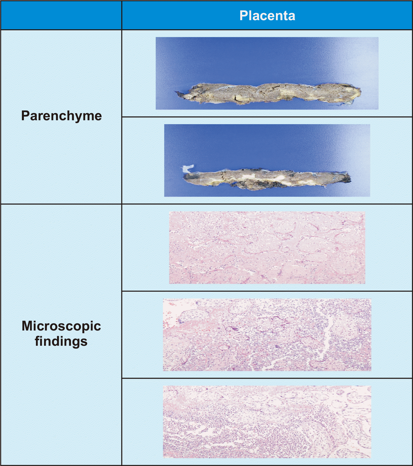
However, the final placental pathology test diagnosed acute suppurative inflammation and an infarct. The cut surface in the placenta shows multiple, variable-sized, yellowish nodules in the placental parenchyma. Microscopic findings show intervillous abscesses with large numbers of neutrophils (
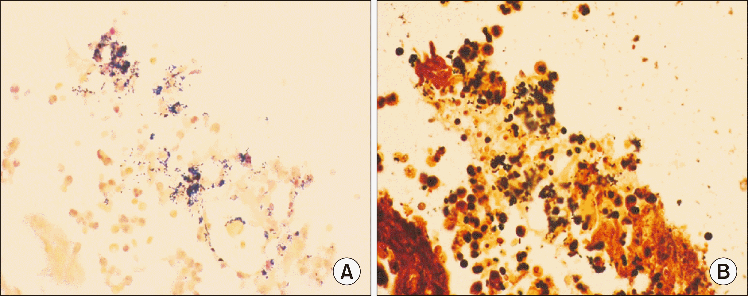
Fig. 1). Gram and Warthin-Starry stains of placental tissue revealed rod-shaped bacilli (
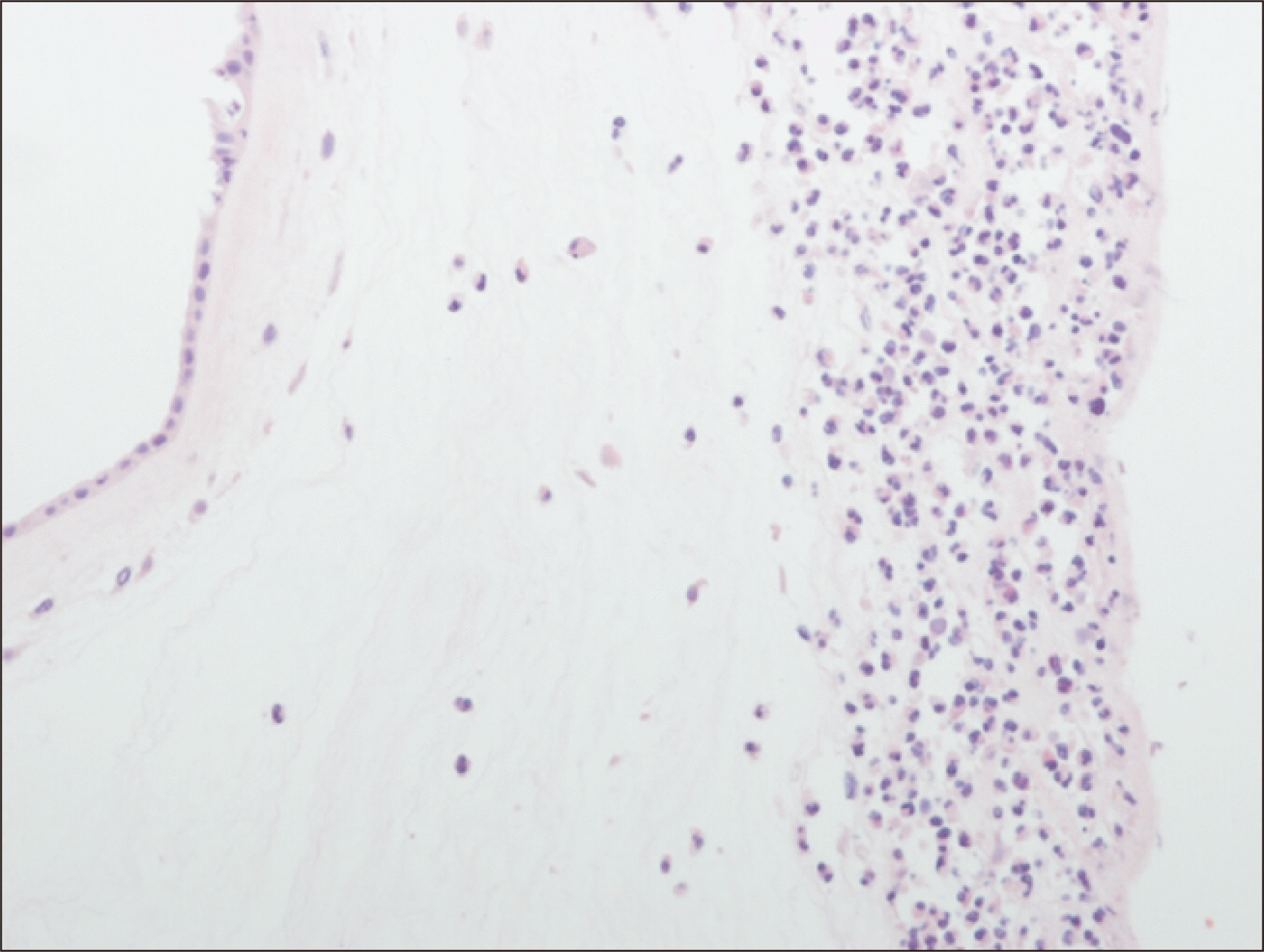
Fig. 2). Amniotic membrane pathology testing led to a diagnosis of acute and chronic chorioamnionitis (
Fig. 3). Antibiotics treatment was continued after delivery for 2 weeks. After then white blood cell and CRP blood levels are normalized and there is no fever, so she is being followed up after discharge.
 | Fig. 1Placental parenchyma and microscopic findings (H&E, ×100). The cut surface of the placenta shows multiple, variable-sized, yellowish nodules in placenta parenchyme. Microscopic findings show intervillous abscesses with large numbers of neutrophils. 
|
 | Fig. 2Gram and Warthin-Starry stain of placental tissue (×400). Gram and Warthin-Starry stain of placental tissue shows rod-shaped bacilli. (A) Gram stain. (B) Warthin-Starry stain. 
|
 | Fig. 3Microscopic amniotic membrane findings (H&E, ×200). Microscopic examination revealed acute and chronic chorioamnionitis. 
|
The newborn’s Apgar score at birth was 4 points at 1 minute and 4 points at 5 minutes. The baby had a weight of 860 g (50th to 75th percentile), height of 31 cm (10th to 50th percentile), head circumference of 22.2 cm, chest circumference of 20.0 cm, abdominal circumference of 18.5 cm, blood pressure of 38/20 mmHg (mean blood pressure 25 mmHg), pulse rate of 140 times/min, and body temperature of 35.0℃. The infant’s whole body was pale and breathing and activity were impaired. A blood test conducted at birth revealed a white blood cell count of 2,800/μL (reference range [RR], 9,000–30,000/μL), 10.1 g/dL hemoglobin (RR, 13.5–22.5 g/dL), 42,000/μL platelets (RR, 150,000–450,000/μL), and chemical tests showed an increase of CRP at 3.66 mg/dL (RR, 0-0.5 mg/dL) aspartate aminotransferase/alanine aminotransferase 42/5 IU/L (RR, 0-33/0-33 IU/L), partial thromboplastin time 2.07 international normalized ratio (RR, 0.9–1.20 international normalized ratio), activated partial thromboplastin time 111.9 sec (RR, 20.6–33.0 sec), fibrin degradation product 21.0 μg/mL (RR, 0–5 μg/mL), and D-dimer 6.81 mg/L (RR, 0–0.59 mg/L). Diffuse haziness was present in both lungs with air-bronchogram and suspected respiratory distress syndrome was observed on chest X-ray. As a result, intubation was performed, surfactant administered and the ventilator was applied.
A patent ductus arteriosus was identified on echocardiography at 2 days after birth and treated using ibuprofen. Persistent pulmonary hypertension was noted on the echocardiogram and and treated with sildenafil citrate. Based on the diagnosis of sepsis and disseminated intravascular coagulation, the treatment included the transfusion of red blood cells and platelets, and administration of hydrocortisone, antithrombin III. Ampicillin was used form 1 day after birth, and due to the growth of gram positive bacilli on the blood culture, cefotaxime and vacomycin was added from 2 days after birth. On the second day of hospitalization, the newborn’s blood pressure and oxygen saturation decreased, and emergency brain ultrasonography found stage 3–4 intraventricular hemorrhage. Despite conservative treatment with hydration, inotropics, antibiotics, and ventilators, sepsis, pulmonary hypertension, metabolic acidosis, hypoglycemia, heart failure, and septic shock worsened. The patient died on the second day of life despite treatment. L. monocytogenes was detected in the blood culture and gastric aspirate culture.
Go to :

Discussion
There are 10 distinct species of
Listeria, a bacterial genus that causes listeriosis, and
L. monocytogenesis pathogenic for humans [
6]. Numerous experts think that human
Listeria infections are foodborne and
L. monocytogeneshas been recovered from several foods, such as raw and undercooked seafood, eggs, meat, and poultry [
7]. However, due to the variable incubation period, it is hard to identify the food that was the source of the infection.
The incidence of listeriosis during pregnancy is 12 per 100,000, compared with 0.7 per 100,000 in the general population. Although rare in pregnancy, a mother with
Listeria infection may not show any outward symptoms and the unborn child may be severely affected [
8].
According to a review of the Korean literature, since infection through bacteremic pregnant women is rare, a case of infection detected in the placenta related to stillbirth, a case of fetal distress from a pregnant woman with bacteremia, and a case of neonatal sepsis-related death due to preterm birth are the only published case reports of neonatal meningitis and sepsis since in 1982 [
1].
In addition, according to statistics from National Health Insurance Sharing Service, the year that had the highest number of listeriosis incidence between 2008 and 2014 was 2014. In 2014, there were 33 patients with listeriosis of the nation's population (0.06 per 100,000), 6 of whom were reproductive age women. So the proportion of pregnant women is thought to be smaller.
Listeriosis can cause serious consequences, but its accurate diagnosis is difficult because its symptoms are non-specific, e.g., flu-like symptoms, and a standard diagnostic tool has not yet been identified. However, patients with fever and other symptoms are recommended to undergo blood culture or cerebrospinal fluid tapping depending on their symptoms. If the patient has given birth, placental culture must also be performed [
9,
10].
Due to the rarity of listeriosis, there are no prospective
in vivo studies of antibiotic regimens; however, the standard treatment of choice for listeriosis includes IV ampicillin and gentamicin for 14 to 21 days for patients without a penicillin allergy. Trimethoprim-sulfamethoxazole is a suitable alternative in persons allergic to penicillin [
10,
11]. On the other hand, the infection heals spontaneously in pregnant women who deliver infants during early-onset listeriosis [
12].
The limitation in this case was that we did not conduct a blood culture test on the mother who was transferred in mid-term pregnancy due to a high fever and preterm labor. Moreover, there was a lack of history taken from the mother. Therefore, the lesson that can be learned from this case is that history taking and physical examination are crucial for discriminating fevers without a clear focus and that blood cultures must be performed on mothers with a fever before using antibiotics, especially if they have symptoms such as cough, sore throat, or diarrhea [
12]. Although listeriosis can have serious consequences, it can be prevented by methods such as avoiding raw food during pregnancy, thus making thorough education of mothers important.
Go to :


 PDF
PDF Citation
 Print
Print


 XML Download