

 PDF
PDF Citation
 Print
Print


INTRODUCTION
Globally, the prevalence of diabetes in adults was estimated to be 8.8% in 2015 (415 million people) and is predicted to rise to 10.4% in 2040.1) In the USA, economic costs of diabetes increased by 26% from 2012 to 2017 due to the increased prevalence of diabetes and the increased cost per person with diabetes.2) Also, diabetic patients have a 30 times greater lifetime risk of undergoing an amputation than nondiabetic patients.3) After major amputation, including belowknee amputation, thirty-day postoperative mortality rates can range from 4% to 22%,4) and the 5-year mortality rate following amputation is estimated at 39%∼68%.5) As there is increased energy expenditure with higher amputation levels, amputation should be performed at the lowest possible level.6)
Conventional soft tissue coverage methods in the foot can range from primary healing with or without negative pressure therapy,7) secondary closure with a skin graft,8) dermal analog,9) regional flap10,11) and free flap.12) The method of the coverage is usually chosen due to the condition of the underlying wound bed, the size of patients’ defect and vascular status, or even the surgeon’s preference. But primary healing with negative pressure therapy requires treatment over a long period to fill the defect. Also, the application of negative pressure therapy with skin graft & dermal analog is inappropriate when the tendon and bones are exposed.13) Free flap operations are technically challenging, and many patients are not suitable for it because of the comorbidity. The easiest way to cover large skin defects is to shorten the bones, but sometimes more amputation is needed to cover the wound.
Since Emmett14) first introduced fillet toe flap for the coverage of foot skin defect in 1976, filleted flaps of the amputated toes or rays, or “spare parts” concept,15) has been used widely for first ray defect,16,17) 2nd ray defect,18) 5th ray defect,19,20) and transmetatarsal amputation.21,22) This strategy allows minimal bone resection and harvesting of flaps without additional donor-site morbidity. Fillet flap is a beneficial reconstruction strategy for major defects,15) and has the advantage of having the same skin and sensory characteristics as that part surrounding the defect.13) The published results contain small series with short follow-up; however, little data has been published on the potential risks.
This study aims to report our fillet flap results for the reconstruction of the forefoot and midfoot defects with the diabetic foot amputation.
MATERIALS AND METHODS
Patients and methods
The Institutional Review Board in Hallym University Sacred Heart Hospital (2020-08-009) approved this study, and the patient consent was exempted. A retrospective chart review was conducted. All patients in an outpatient clinic or emergency room who required fillet flap surgery of the foot due to diabetic complications by a single surgeon were included in the study. All defects were below the Chopart joint level, and in each case, reconstruction was performed by the same orthopedic surgeon. The patient was excluded if the patient lost to follow-up, had the possibility of amputation proximal to the tarsal bone level at the time of the first admission, and if the surgical method is not a fillet flap. We excluded patients who stopped follow-up even though the wound did not heal. Comprehensive clinical data were collected retrospectively and included sex, age, side, symptom duration, comorbidity (include diabetes mellitus [DM] and chronic kidney disease [CKD]), smoking history, height, weight, body mass index (BMI), ankle-brachial index (ABI), laboratory data (include white blood cell count [WBC], lymphocyte count, creatinine, albumin, the base level of C-reactive protein [CRP], CRP just before the operation, hemoglobin A1c percentage [HbA1c]), history of the previous operation, cultured pathogen, operation time, hospital day, complications. We defined ‘symptom duration’ as the period from the day the patient stated that symptoms first occurred to the day of surgery. Detailed data is described in Supplementary Table 1. We counted CKD history when there was a history of CKD diagnosis at the time of admission or when CKD was diagnosed by consultation with an internal medicine after hospitalization. Smoking history was identified by questionnaire and nursing information survey. We defined ‘base level of CRP’ as the first CRP performed by the patient with the symptom, and ‘CRP just before the operation’ as the CRP performed by the patient on the day or one day before surgery. HbA1c percentages were recorded if documented in the hospital chart within 30 days of the index amputation. The primary endpoint was clean wound without discharge and defined as the date of initial healing; initial healing days were calculated. The final endpoint was complete wound healing without any protection or dressing, defined as the date to complete healing; complete healing days were calculated.
Surgical technique
Our surgical technique’s theme was to resect enough bone and utilize as much of the abundant tissue of the foot as possible to minimize the wound tension and compensate for length loss caused by bone resection.
Preoperatively, all patients were treated with culturedirected antibiotic therapy according to the department of infectious disease medicine recommendations. The procedure is performed under local ankle block, spinal or general anesthesia. The patient is positioned supine, the leg is prepared and draped distal to the thigh. The author usually does not use a tourniquet while operating on diabetic feet to evaluate vascular patency. A bump is used to elevate the leg to minimize blood loss. After en bloc excision of the necrotic and ulcerated lesion, wounds were serially debrided until healthy, viable tissue was visible. The filleted flap is prepared by subperiosteal separation of the skin and subcutaneous tissue of the toe or metatarsal. The effort was made to leave the healthy skin intact without disturbing it and to keep intact toes. The bony stump was smoothed to remove any sharp margins and avoid injury to the fillet flap. Once the flap designing according to the defect pattern was completed, the margins of the flap have fitted the margins of the wound and were determined to be free of tension on the pedicle. The wound is then closed with nylon sutures, in a non-tight manner with or without a silastic drain. In the case of a relatively poor wound with discharge, complete wound closure was performed with a 2-stage operation. A slight compressive dressing is then applied. The patient is instructed for nonweight bearing ambulation until the wound has healed properly. If the flap had tension, a partial stitch out was done. During the follow-up period, we examined each patient to determine flap survival and to detect postoperative complications. Preoperative and postoperative X-rays were used to calculate the discrepancy between the preoperative and postoperative foot length.
RESULTS
Between March 2013 and November 2017, 16 cases of fillet flap surgery were operated. Among them, one patient expired due to medical condition and one patient did not come to follow-up. After exclusion, fourteen cases (13 male and one female) were included in the case series (Fig. 1). The patients’ mean age was 60.3 years (range, 52∼75 years; standard deviation, 5.8 years). The obtained data were summarized in Table 1. Necrotic and infected wound was in 2 toes, nine forefeet (1 medial, two central, and six lateral), and three midfoot. The comorbidities include two smoking and ten chronic kidney disease.
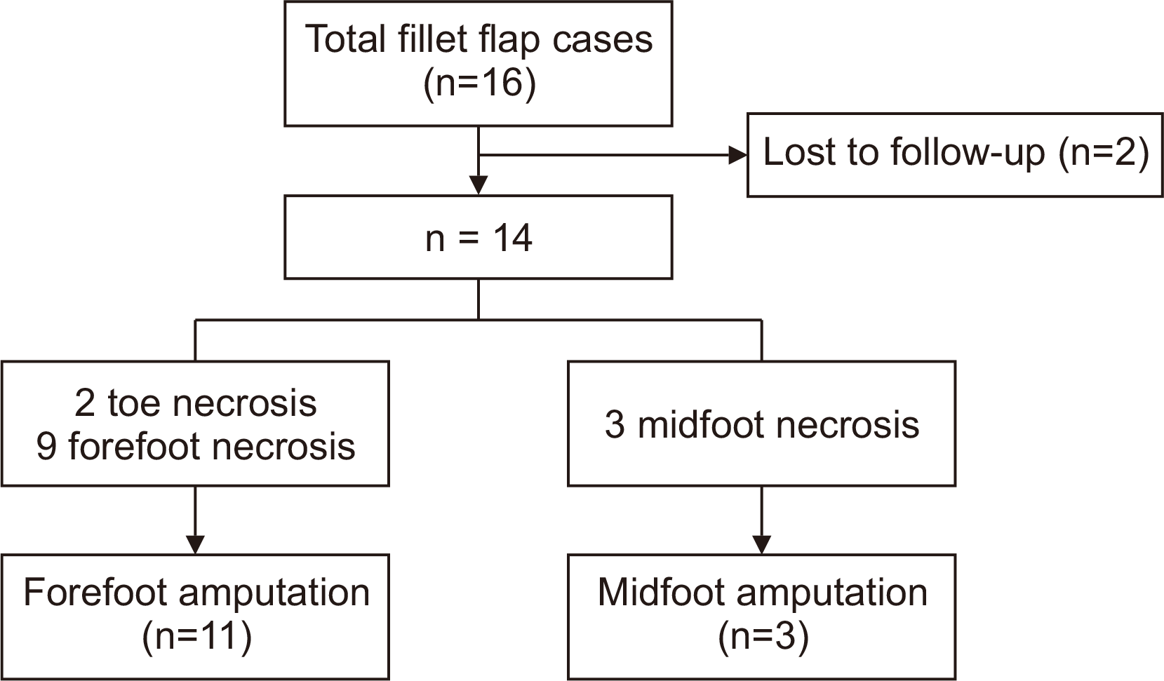
Fig. 1
Study patient’s exclusion/inclusion criteria. This figure illustrates the exclusion and inclusion criteria applied to the fillet flap surgery.
![]()
Table 1
Demographics and Baseline Data
![]()
Postoperative data are shown in Table 2. Eleven forefoot amputation and three midfoot amputations with fillet flap were performed. By the fillet flap, amputation size was reduced as much as possible.
Table 2
Outcome Analysis (n=14)
![]()
Three patients had revision surgery for partial necrosis of flap among forefoot necrosis, and two patients had an additional amputation. Wound revision or skin graft was performed at 2 to 8 weeks postoperatively. No additional operation was needed for those patients.
For the midfoot necrosis patients, two patients had an additional amputation. One patient had transmetatarsal amputation at four months postoperatively for wound necrosis. Another patient showed necrosis along the fascia of the medial calf and had below-knee amputations at two weeks postoperatively.
We further describe three cases according to their complication.
Case without complication: case no. 10
A 55-year-old male patient visited the hospital because of left forefoot necrosis. Previously, he had 3rd, 4th metatarsal ray amputation, and wound closure in our hospital three months ago. The defect extended to the lateral aspect of 2nd toe, and its size was 2 cm×4 cm (Fig. 2A). Preoperatively, osteomyelitic change was seen in the proximal phalanx neck of 2nd toe on plain radiograph. Debridement was performed, and the proximal phalanx was amputated because it showed osteomyelitis change (Fig. 2B). If a primary closure using nearby tissue is to be attempted, then the need for 2nd to 5th metatarsal shortening procedure becomes inevitable because the necrotic tissue exists at the same level as that of the proximal phalanx (Fig. 2B). The fillet flap was designed to match the defect site’s size and dissected, placed at the defect site and sutured. The occlusive dressing was performed. After 3 months, flap wound healed without any complication.
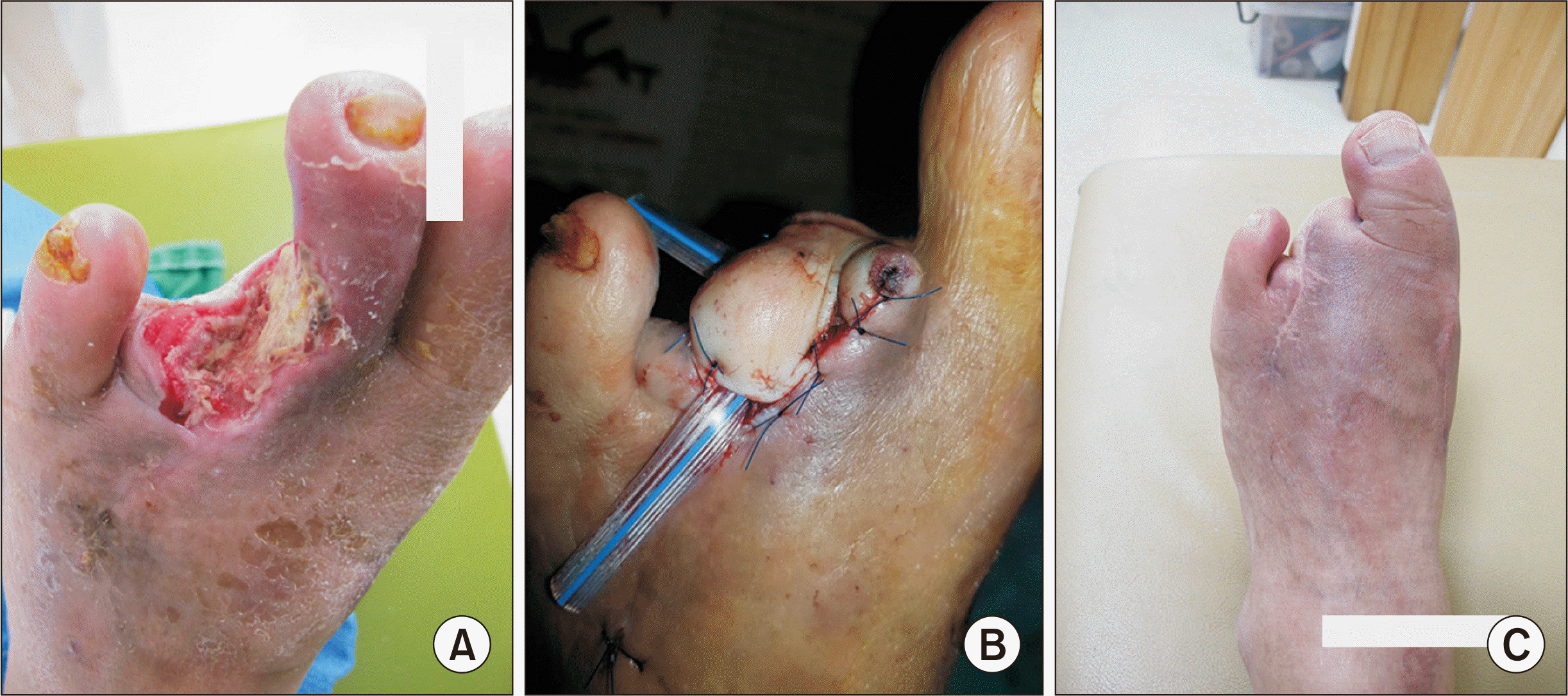
Case with partial necrosis of flap: case no. 1
A 63-year-old female patient visited the hospital for her right foot skin defect and 3rd toe necrosis (Fig. 3A). Osteomyelitis on the 2nd and 3rd metatarsal head was suspected with magnetic resonance imaging (MRI), and the excision of the metatarsal head was performed. Skin defect was seen on the dorsal aspect of the 2nd and 3rd metatarsal and extended to the Lisfranc joint (Fig. 3B). Partial amputation of 3rd metatarsal (distal 2/3) was performed for closure, and a fillet flap was done for skin defect. This procedure could prevent additional amputation and preserved forefoot. At postoperative two weeks, necrosis of the 2nd toe flap was seen (Fig. 3C). We excised the necrotic flap, and the wound was closed. After one month from the last operation, the wound healed without complication (Fig. 3D).
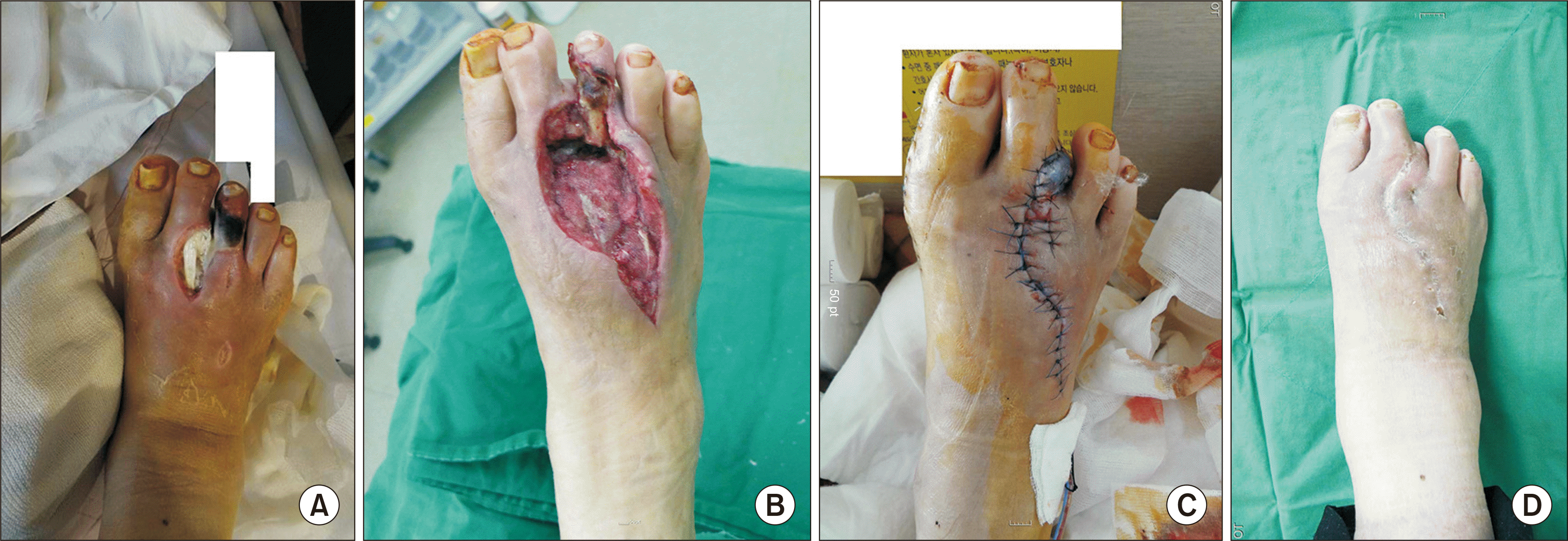
Fig. 3
Case no. 1. In a 63-year-old female patient, a diabetic foot with a chronic infection on the right forefoot. (A) Preoperative skin defect on the dorsal aspect of second metatarsal and necrosis of third toe. (B) Skin defect on the dorsal aspect of second and third metatarsal after excision of the metatarsal head. (C) Postoperative coverage with fillet flap of third toe, along with partial necrosis of flap. (D) Complete wound healing at one month postoperative follow-up.
![]()
Case with additional amputation: case no. 12
A 63-year-old male patient visited the hospital for his right 4th toe necrosis (Fig. 4A). After the amputation of 4th toe, further necrosis along the 4th metatarsal was seen (Fig. 4B). The soft-tissue gas and contrast enhancement were found on MRI, and transmetatarsal amputation with fillet flap was performed (Fig. 4C). However, the patient complained of pain on the lower leg, and a color change on the medial calf was seen. We strongly suspected necrotizing fasciitis. After fasciotomy, necrosis along the fascia was seen, and debridement was performed. As the wound worsened rapidly, below-knee amputation was performed ten days after amputation. After about three months from the last operation, the wound healed without complication and prosthesis was applied.
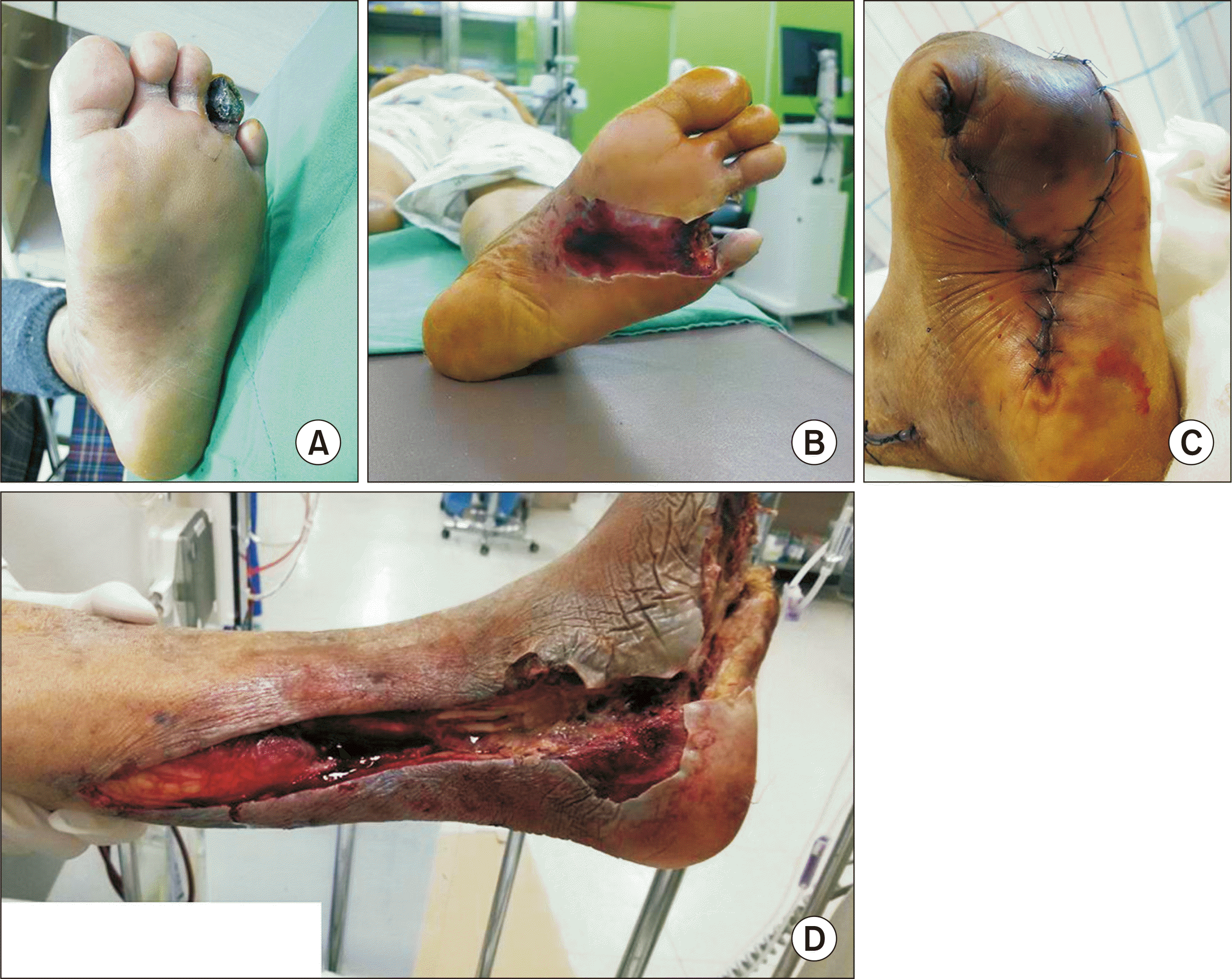
Fig. 4
Case no. 12. In a 63-year-old male patient, a diabetic foot with right fourth toe necrosis. (A) Preoperative necrosis of fourth toe. (B) Further necrosis along fourth metatarsal after fourth toe amputation. (C) Postoperative coverage with transmetatarsal amputation and fillet flap. (D) Necrosis along the medial calf at ten days after amputation.
![]()
DISCUSSION
The skin defect of the necrotic foot frequently requires flap surgery and presents an extremely difficult reconstructive challenge because of their vascular insufficiency. However, flap surgery can be lengthy and is often not a suitable option for older patients with comorbidities. It also requires considerable microvascular surgery experience, and hence its availability is usually limited to larger medical centers.
The fillet flap technique has several advantages over traditional amputation and flap surgery, including minimal bone resection and flap harmony with surrounding soft tissue. Since the probability of reamputation after ray amputation is remarkably high,23,24) it is essential to avoid ray amputation as much as possible. Moreover, fillet flap can be harvested quickly without donor-site morbidity and does not require microvascular surgery. This idea is in line with a recent report; selected patients may benefit from “conservative” bone resections that preserve soft tissue and assist healing by primary and secondary intention.25) Published literature show the use of fillet flaps in various areas including finger,15) toe,14) first ray defect,16,17) 2nd ray defect,18) 5th ray defect,19,20) transmetatarsal amputation,21,22) knee,26) and even shoulder15); it is called by various names including fillet flap, rotational flap, and angiosome flap.
To compare the results of surgery with conventional surgery, we studied only the lesions of the forefoot. Fillet flap for coverage for the closure of diabetic forefoot skin defect first reported a long time ago,27) but not much paper has yet been published. Kuntscher et al.15) reported 11-foot cases with “pedicled limb fillet flaps” in various causes, including burn, trauma, tumor, and diabetic gangrene. For diabetic foot, few cases were reported from several works of literature.16,17,19,20,28)To our knowledge, this paper has the highest number of diabetic patients among the fillet flap papers. Also, followup time was missing in some patients28) or in all patients,15) sex,28,29) and laterality15) were not reported, and description of the previous surgery was omitted16,29) in some literature.
Of those reported, the recipient site’s common complications included wound dehiscence, recurrent infection, sepsis, partial flap necrosis and further amputation. Kuntscher et al.15) reported an overall complication rate of 18% and major complications such as flap loss, flap revision, or severe infection at 7.5%. In this study, flap complications included three flap necrosis and four revision amputations. All flap necrosis cases recovered without additional surgery. Although there were four cases of amputation, it was difficult to analyze the effect on amputation because the total number of patients was small. Our initial healing 70.6 days (10 weeks) and complete healing days of 4 months are between 6 weeks and sex months of previous reports.16,20) However, variations in the study designs, the study populations, and different treatment methods could explain the conflicting results of prior studies. Although this procedure required sacrificing of an adjacent toe in some cases, this axial pattern local flap was a nearly ideal replacement for the excised tissue, including excellent matches regarding the color, texture, and durability of the skin18) and prevented additional longer amputations. Also, the well-vascularized soft tissue provides padding over bony prominences and contributes to the elimination of infection.19) The mean length of the shortening of the foot was 31.0 mm (range, 12.4∼67.8 mm) in this study. Considering the skin defect area, this shortening could not be long, and the overall length of the foot did not decrease. Moreover, the fillet flap could obtain wound healing with a smaller range of cuts than the conventional ray amputation option. As shown in Supplementary Table 2, extensive amputation was prevented. Although the advantage of partial foot amputation over transtibial amputation was reported,30) advantage of partial forefoot amputation over midfoot amputation is not clear. Further studies are warranted to evaluate this approach.
Our study had some limitations. This study is a small case series, and there is no control group. Also, we might select patients suitable for the fillet flap technique.16) In our results, the results were better in the forefoot than in midfoot. Randomized controlled trials comparing patient selection criteria and different flap coverage methods for partial ray amputations in diabetic patients are needed.20) Although we have described comprehensive clinical data that may affect the diabetic foot, some data are missing. Ours include chronic kidney disease, smoking, ABI, and WBC, which were associated with poor outcomes.23) However, our data is absent of pulses, delayed capillary filling, toe-brachial index. We are currently conducting tests for neuropathy, but unfortunately, the tests were not routinely performed in the past. Nonetheless, this series contributes valuable information. To the best of our knowledge, ours is the largest study of its kind to date.
CONCLUSION
The fillet flap facilitates the restoration of normal foot contour and allows salvage of the metatarsal or toe. Symptom duration and size of the defect are among the risk factors and must be considered for successful fillet flap. This technique can avoid additional morbidity and facilitate limb salvage in diabetic foot infection.
SUPPLEMENTARY MATERIALS
Supplementary materials can be found via https://doi.org/10.14193/jkfas.2020.24.4.148
 XML Download