Journal List > J Korean Foot Ankle Soc > v.24(4) > 1145869

중족부에 발생한 다발성 악성 말초 신경초 종양 1예: 증례 보고

Abstract

Malignant peripheral nerve sheath tumors (MPNSTs) usually arise in soft tissues; they are rarely found in the bone. This paper reports a case of MPNST in the foot and ankle joint involving the distal tibia, talus, calcaneus, navicular, medial intermediate, and lateral cuneiform, cuboid, and 2nd to 4th metatarsal bone. Palliative treatment was performed. The authors encountered a patient with intraosseous MPNST of the midfoot who presented with nonspecific clinical and radiologic findings. This case shows that a high index of suspicion and a histopathology examination, including immunohistochemistry, will be necessary for an accurate diagnosis.

말초 신경초 종양은 연부조직에 주로 발생한다. 또한 골에 발생하는 말초 신경초 종양은 대부분 양성이며 원발성 골종양의 0.2% 미만을 차지할 정도로 매우 드물다.1,2) 골의 악성 말초 신경초 종양은 드문 형태의 악성 종양이며 대부분 인접한 연부조직으로부터 이차적으로 발생하게 된다.1) 골에 생긴 원발성 악성 말초 신경초 종양이 보고된 경우는 드물다.3) 대부분 하악골(약 50%), 상악골1,4-6) 또는 척추4,7) 에 발생하며 사지에 발생하는 경우는 더욱 드물다. 사지 중에서도 상지의 골에 발생하는 경우가 많으며 하지의 골에 발생하는 경우는 극히 드물다.2) 본 저자들은 족부족관절에 다발성으로 발생한 악성 말초 신경초 종양을 경험하여 이를 보고하고자 한다. 본 증례는 본원 임상연구 윤리위원회의 승인을 받았다.

증례 보고

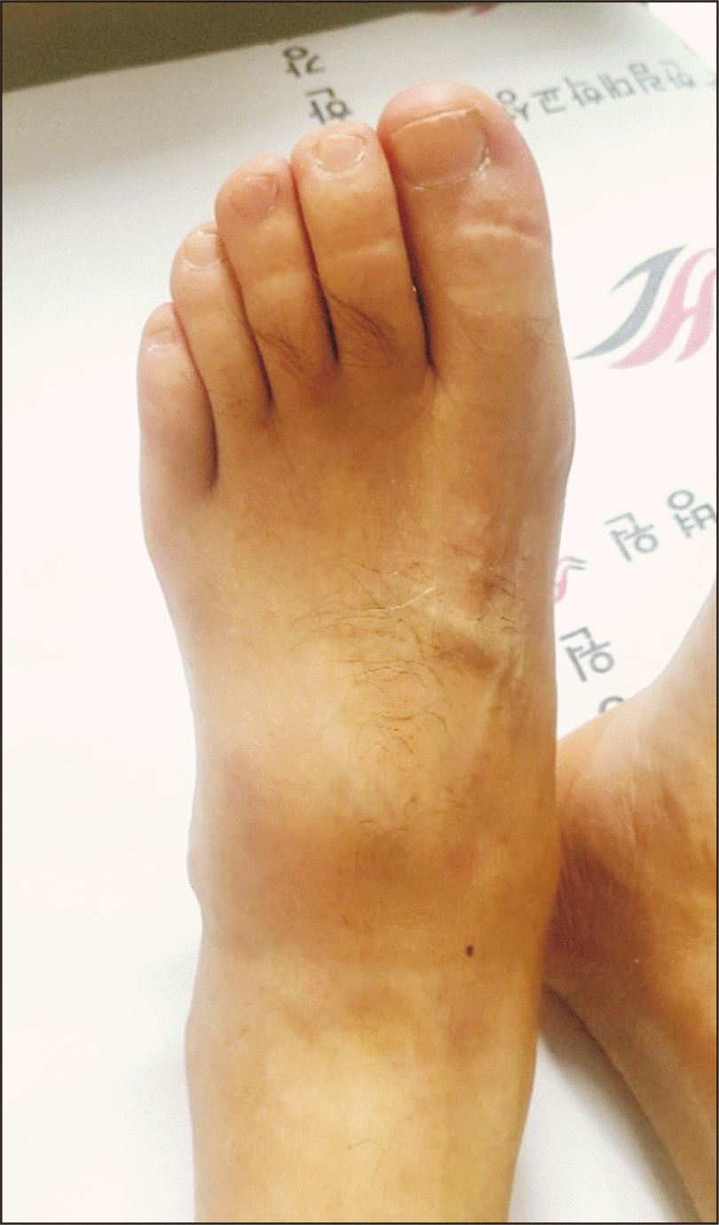
64세 남자환자가 내원 3달 전부터 시작된 발등의 종창과 통증을 주소로 내원하였다. 환자는 당뇨 및 고지혈증으로 약물 복용 중이었으며 가족력상 특이 소견은 없었다. 1년 전 족부 외상의 과거력이 있었으나 당시 특이 소견은 보이지 않았다. 3개월 전부터는 통증 및 종창이 시작되어 타원에서 치료를 받았으나 호전이 없어 본원으로 전원되었다. 종창은 점차 진행되었으며 본원 내원 당시 좌측 발등에 4 cm×3 cm 정도의 다소 단단하고 잘 움직이지 않는 종물이 만져졌다. 열감, 통증 및 압통을 호소하고 있었으나 보행은 가능한 상태였으며 말초의 운동 및 감각은 모두 정상이었다(Fig. 1).
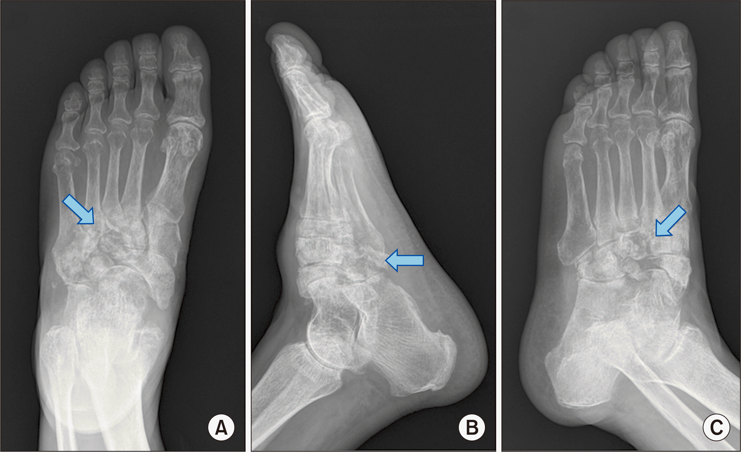
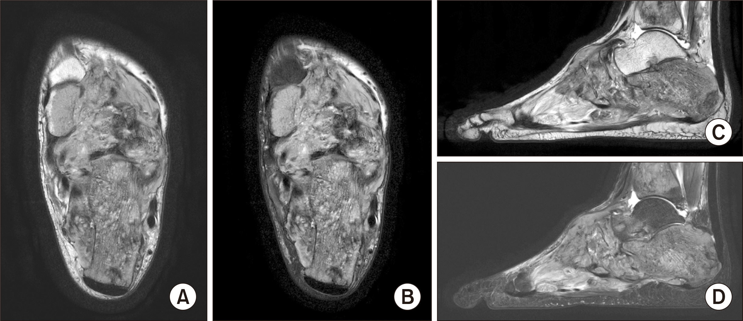
내원하여 시행한 단순 방사선 사진에서 좌측의 경골 원위부, 거골, 종골, 주상골, 세 개의 설상골, 입방골, 2∼4번째 중족골의 골용해 소견을 보이고 있었다(Fig. 2). 자기공명영상 사진에서 경골 원위부, 거골, 종골, 주상골, 세 개의 설상골, 입방골, 2∼4번째 중족골에 다발성의 골내강을 포함하는 비균일적 조영증강을 보이고 있었으며 주변부로의 종괴 형성을 보이고 있었다(Fig. 3). 이와 같은 소견으로 골종양 또는 주변 농양을 형성하고 있는 골수염에 대한 감별 진단이 필요하였고 조직학적 확진 및 치료를 위하여 절제 생검술을 시행하기로 하였다.
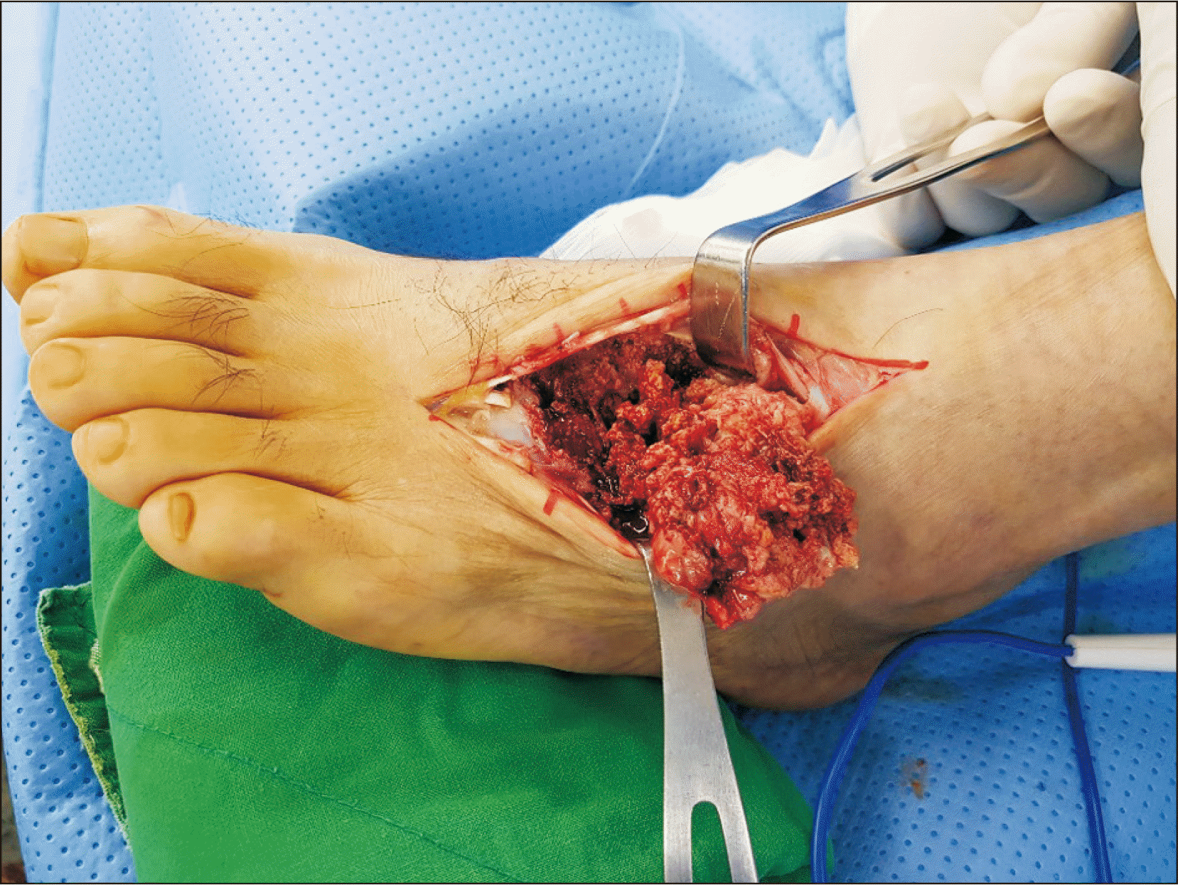
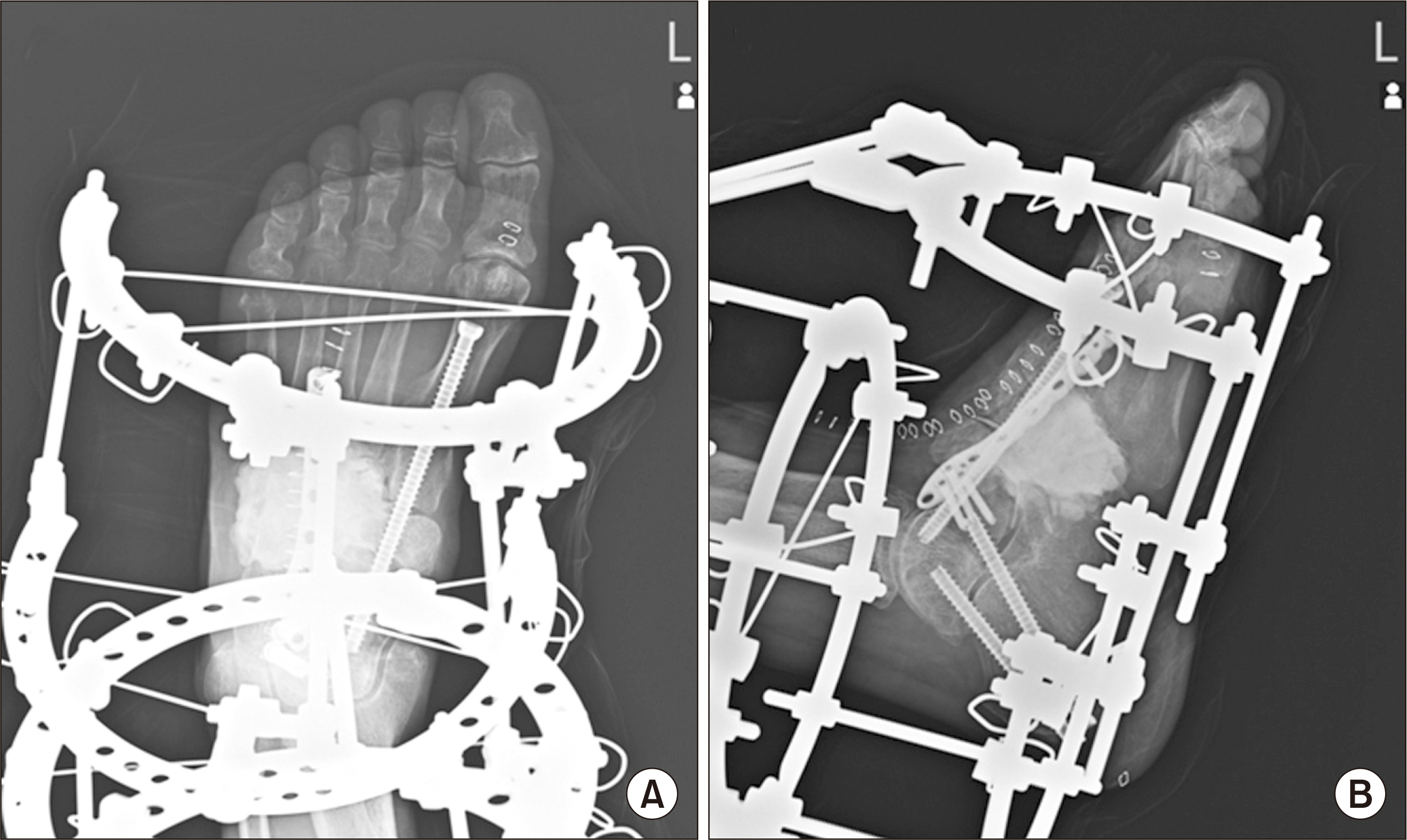
수술은 족배부 부위에 약 10 cm 가량 종절개를 시행하였으며 골의 형태가 명확하지 않고 주변부의 농양은 관찰되지 않아 수술 중 동결절편 조직검사를 시행하였다(Fig. 4). 검사결과 다수의 고등급 악성 세포 소견을 보였다. 이에 악성 종양으로 생각되어 추후 조직검사 확인 후 이차 수술을 계획하기로 하고 완화 치료로 종양에 대한 절제 술을 시행하였다. 제거된 골 및 종양의 범위가 넓은 상태로, 발의 모양 유지 및 보행을 위하여 손실 부위에 시멘트를 이용해 공간을 채웠으며 체중부하를 가능하게 하기위해 금속판과 유관나사를 이용한 고정술 및 일리자로프 외고정기를 이용한 고정을 하였다(Fig. 5).
병리학적으로 방추형의 종양세포로 구성되어 있었고 핵의 비정 형성과 다형성이 관찰되었으며 세포 분화도는 4.5/10 high-power field로 보였다. 면역조직화학적 염색상 S-100 단백질에 강한 양상 소견이 관찰되었다(Fig. 6). 이와 같은 소견을 바탕으로 Federation Nationale des Centres de Lutte Contre Le Cancer (FNCLCC) grade 3의 악성 말초 신경초 종양으로 확진하였다.
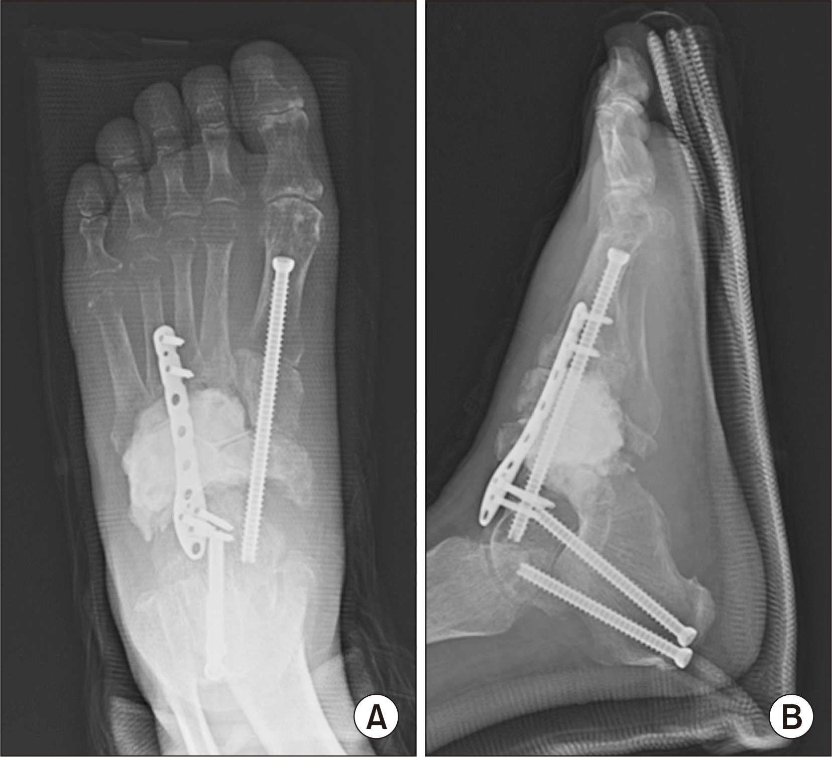
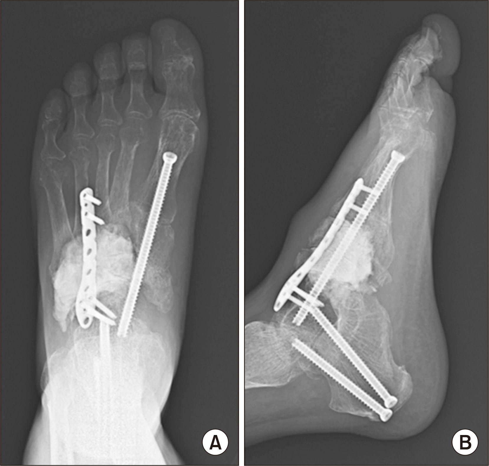
이후 원칙적으로 골병변을 제거하기 위해 하퇴 절단술을 계획하였으나 수술 후 시행한 양전자 방출 단층촬영(positron emission tomography computed tomography, PET-CT) 검사상 폐전이가 확인되었다. 이로 인해 생존율이 낮을 것이라 판단되었고 환자 및 보호자와 상의 후 추가적인 수술적 치료는 시행하지 않고 보조 항암 화학요법(adjuvant chemotherapy)을 진행하기로 하였다. 수술 후 6 주째 외고정기를 제거하였으며 6개월 추시에서 수술부위의 통증은 호소하고 있지 않는 상태로 보행이 가능하였다(Fig. 7, 8). 하지만 이후 진행한 흉부, 복부 및 하지 CT상 기존 폐 및 족부족관절 부위의 상태 악화와 함께 새롭게 췌장에도 의심되는 병변이 나타나기 시작 하며 점차 전신상태 악화로 보행은 어려운 상태가 되었다.

고 찰

악성 말초 신경초 종양은 말초신경의 Schwann 세포로부터 생기는 희귀한 질환으로 1949년 Stout8)에 의해 처음 보고되었다. 그러나 종양의 기원에 대한 의견이 분분하여 악성 신경초 종양, 신경섬유 육종 신경원성육종, 악성 신경섬유종 등의 병명이 혼용되고 있으며 신경섬유종증 제 1형(neurofibromatosis type I)을 동반하는 경우 가 많다.9)
골의 악성 말초 신경초 종양은 대부분 고분화 육종이다. 폐전이가 가장 흔히 일어나며 림프절로의 전이는 10% 미만으로 나타난다. 국소적 재발은 40%∼65%에서 발생하며 전이의 빈도는 40%∼68%로 나타난다.3)
수술 전 진단은 그 희귀성 및 영상학적 검사상의 비특이적 소견으로 인하여 종종 놓치는 경우가 있다. 단순 방사선 검사상 경계가 불분명한 용해성 병변을 나타나게 된다. 자기공명영상 역시 비특이적 소견을 보인다.3) T1증강영상에서는 비균일적인 증강과 중간∼저신 호강도를 보이며 T2증강영상에서는 고신호강도를 보이게 된다. 조영증강영상에서는 병변의 퇴화 정도에 따라 다양한 양상의 조영증강을 보이게 된다. 흉부 CT는 폐전이 여부를 감별하는 데 도움이 될 수 있다. 골피질의 파괴가 없고 주변 연부조직으로의 확장이 없다면 골에 생긴 원발성 악성 말초 신경초 종양을 나타낼 수 있다. 그러나 이는 양성과 악성, 골수염 등의 감별진단이 필요하며 영상학적 검사만으로는 확진을 내릴 수 없다. 따라서 의심이 될 때는 면역화학염색을 포함한 조직학적 검사가 수행되어야 한다.
일반적인 치료는 다른 악성 골종양들과 마찬가지로 광범위 절제 술 후 필요 시 방사선 또는 화학적 항암요법을 시행한다. 하지만 방사선 또는 화학적 항암요법에 대해서는 여러 논란이 있다.10) 방사선 치료 후에도 상태가 악화 시에는 예후가 좋지 않다고 알려져 있다.
본 증례에서는 족부족관절 부위에서 발생한 다발성 악성 말초 신경초 종양에 대해서 보고하고자 한다. 이 질환의 진단은 희귀하고 비특이적인 임상 및 영상학적 소견을 보이고 있어 어렵다. 따라서 진단을 정확히 하기위해서는 면역조직화학을 포함한 조직 병리학적 검사를 반드시 시행하여야 한다. 본 증례와 같이 족부 및 족관절 부위의 다양한 골을 침범한 경우 원칙적 치료로는 하퇴부 절단술과 같은 광범위 절제술을 시행하여야 한다. 다만 환자의 생존율 및 수술부위 범 위 등을 종합하여 고려하는 것이 필요하다. 본 증례에서처럼 보행을 위해 족부의 형태를 유지하고 기능을 보존하는 완화치료도 좋은 치료의 하나가 될 수 있을 것으로 생각되며 이는 다른 족부족관절 내에 발생하는 악성 종양에서도 비슷하게 적용될 수 있을 것으로 생각된 다.

Notes

Financial support: None.

Conflict of interest: None.

REFERENCES

1. Terry DG, Sauser DD, Gordon MD. 1998; Intraosseous malignant peripheral nerve sheath tumor in a patient with neurofibromatosis. Skeletal Ra-diol. 27:346–9. doi: 10.1007/s002560050395. DOI: 10.1007/s002560050395. PMID: 9677654.
crossref
2. Lesic A, Bumbasirevic M, Atkinson HD, Maksimovic R, Sopta J, Atanackovic M. 2006; Malignant intraosseous peripheral nerve sheath tumour of the proximal femur: a case report. J Orthop Surg (Hong Kong). 14:84–9. doi: 10.1177/230949900601400119. DOI: 10.1177/230949900601400119. PMID: 16598095.
crossref
3. Kendi TK, Erakar A, Yildiz HY, Saglik Y, Erekul S. 2004; Intraosseous malig-nant peripheral nerve sheath tumor with local recurrence, lung metas-tases and death. Skeletal Radiol. 33:223–5. doi: 10.1007/s00256-003-0678-1. DOI: 10.1007/s00256-003-0678-1. PMID: 14758514.
crossref
4. Sham ME, Ghorpade , Shetty A, Hari S, Vinay . 2010; Malignant peripheral nerve cell tumour. J Maxillofac Oral Surg. 9:68–71. doi: 10.1007/s12663-010-0019-6. DOI: 10.1007/s12663-010-0019-6. PMID: 23139572. PMCID: PMC3453695.
crossref
5. Ma ZW, Ward R, Hoda SA. 1996; Intraosseous malignant peripheral nerve sheath tumor. Arch Pathol Lab Med. 120:517–8.
6. Dunnick NR. 2000; Image interpretation session: 1999. Intraosseous ma-lignant peripheral nerve sheath tumor (malignant schwannoma) in a patient with neurofibromatosis. Radiographics. 20:271–3. doi: 10.1148/radiographics.20.1.g00ja27257. DOI: 10.1148/radiographics.20.1.g00ja27257.
7. Moon SJ, Lee JK, Seo BR, Kim JH, Kim SH, Lee KH, et al. 2008; An intraos-seous malignant peripheral nerve sheath tumor of the cervical spine: a case report and review of the literature. Spine. 33:E712–6. doi: 10.1097/BRS.0b013e31817e6995. DOI: 10.1097/BRS.0b013e31817e6995. PMID: 18758353.
8. Stout AP. 1949; Tumors of the peripheral nervous system. J Mo State Med Assoc. 46:255–9.
9. Moon MS, Kim HJ, Lee DS, Kim RK, Seo EJ. 1983; Malignant schwan-noma (a case report). J Korean Orthop Assoc. 18:1029–32. doi: 10.4055/jkoa.1983.18.5.1029. DOI: 10.4055/jkoa.1983.18.5.1029.
crossref
10. Muthusamy S, Conway SA, Pitcher JD, Temple HT. 2017; Primary intraos-seous malignant peripheral nerve sheath tumor of the medial cunei-form: a case report and review of the literature. J Foot Ankle Surg. 56:129–34. doi: 10.1053/j.jfas.2016.05.013. DOI: 10.1053/j.jfas.2016.05.013. PMID: 27449524.

Fig. 1
View showing the mass about 4 cm×3 cm over the left dorsum of foot.
JKFAS-24-156-f1.tif
Fig. 2
Anteroposterior (A), lateral (B), and oblique (C) radiographs show multifocal osteolytic lesions involving distal tibia, talus, calcaneus, navicular, medial, intermediate and lateral cuneiform, cuboid and 2nd to 4th metatarsal bones (arrows).
JKFAS-24-156-f2.tif
Fig. 3
Axial T2-weighted (A), fat suppres-sion (B), and sagittal T2-weighted (C), fat suppression (D) magnetic resonance imag-ings demonstrate multiple marrow replacing lesions with heterogeneous enhancement involving distal tibia, talus, calcaneus, navicular, medial, intermediate and lateral cuneiform, cuboid and 2nd to 4th metatarsal bones. It also shows multifocal extraosseous extension with mass formation.
JKFAS-24-156-f3.tif
Fig. 4
Bone morphology was not clear from the operation, and no abscess was observed around the mass.
JKFAS-24-156-f4.tif
Fig. 5
Anteroposterior (A), lateral (B) radiographs showing the bone cement was filled to fill the space of the removed bone. To maintain the shape of the foot and to walk, fixation using a metal plate and cannulated screws and fixation using an Ilizarov external fixator were performed.
JKFAS-24-156-f5.tif
Fig. 6
(A) Low-power view shows an invasive solid mass (deep violet color part), which is infiltrating to surrounding skeletal muscle and fat tissue (H&E stain, ×10). (B) One area of the tumor shows an entrapped normal peripheral nerve among the sarcoma cells. This area shows that the morphology of the sarcoma spindle tumor cells are similar to normal nerve spindle cells (H&E stain, ×200). (C) At other areas, most spindle tumor cells show prominently irregular arrangement and severe pleomorphic histology (H&E stain, ×200). (D) On immunohistochemical study, the tumor cells are positive for S-100 protein (neural origin marker). The brown-colored cells means positive immunoreactivity (S-100 immunostain, ×200). (E) On immunohistochemical study, other markers other than S-100 protein show no brown coloration, which means negative immunoreactivity (HMB-45 immunostain, ×200).
JKFAS-24-156-f6.tif
Fig. 7
(A, B) The external fixator was removed 6 weeks after surgery.
JKFAS-24-156-f7.tif
Fig. 8
(A, B) At the 6 months follow-up point no radiologic evidence of recurrence and the bone cement and metal fixation at the surgical site were well maintained.
JKFAS-24-156-f8.tif
TOOLS
Similar articles