

 PDF
PDF Citation
 Print
Print


INTRODUCTION
Craniofacial cleft is a rare congenital malformation with diverse spectrum of severity, causing significant impairment of function, facial aesthetics, and psychosocial problems for both the patients and their family.1-5 Since it can occur in both the soft and hard tissues of the craniofacial region, thorough clinical examination and radiological evaluation are necessary to assess the degree and path of the craniofacial clefts.6
Although the precise etiology of craniofacial cleft is unknown, the normal embryogenesis process in the craniofacial region might be disrupted by genetic and environmental predisposing factors during the first 12 weeks of gestation.7-13 The incidence of craniofacial clefts has been reported to be 1.4–6.0 per 100,000 live births.11,14 The reasons for this wide range of estimate might be due to the rare occurrence of the craniofacial clefts and the lack of standard methods for data collection.15
The repairing of a craniofacial cleft needs multistage surgical treatment with pre-surgical orthopedic correction, wide tissue mobilization, and/or a bone graft.16-18 In addition, some patients with craniofacial clefts exhibited other anomalies in the dentition, eye, nerve, and respiratory system. Therefore, it is necessary to establish a multidisciplinary approach with a well-experienced cooperative team that includes plastic surgeons, neurosurgeons, ophthalmologists, otolaryngologists, orthopedic surgeons, orthodontists, and psychiatrists to restore function, improve aesthetics, and resolve psychosocial issues.13,19,20
Most of the previous case reports or clinical studies have investigated the clinical characteristics of a specific type of craniofacial cleft in Korea21-30 and other countries.13,31-36 Furthermore, there have been no clinical demographic studies on the distribution and phenotype of all patients with craniofacial cleft in Korea to date. Therefore, the purpose of this retrospective study was to investigate the distribution, side involvement, phenotype, and associated anomalies of Korean patients with craniofacial clefts.
MATERIALS AND METHODS
The design of this study was a retrospective one. The initial samples were patients with craniofacial cleft, who visited the Multi-disciplinary Clinic, Department of Plastic and Reconstructive Surgery, Seoul National University Children's Hospital and the Department of Orthodontics, Seoul National University Dental Hospital. The inclusion criteria were as follows: (1) patients whose ethnic background was Korean, (2) patients who were diagnosed with craniofacial clefts, (3) patients who were treated and/or followed-up from 1998 to 2018, and (4) patients whose charts, clinical photographs, cephalometric and panoramic radiographs, and/or three-dimensional computed tomography scans were available. Patients who were not treated or followed up longitudinally were excluded.
As a result, the final cohort consisted of 38 Korean patients with craniofacial clefts (17 males and 21 females; mean age at the first consultation: 5.34 ± 3.03 years). The frequency and distribution of Tessier cleft types, sex, side involvement, phenotype, and associated anomalies were investigated. This study was reviewed and approved by the Institutional Review Board of the Seoul National University Dental Hospital (ERI19030).
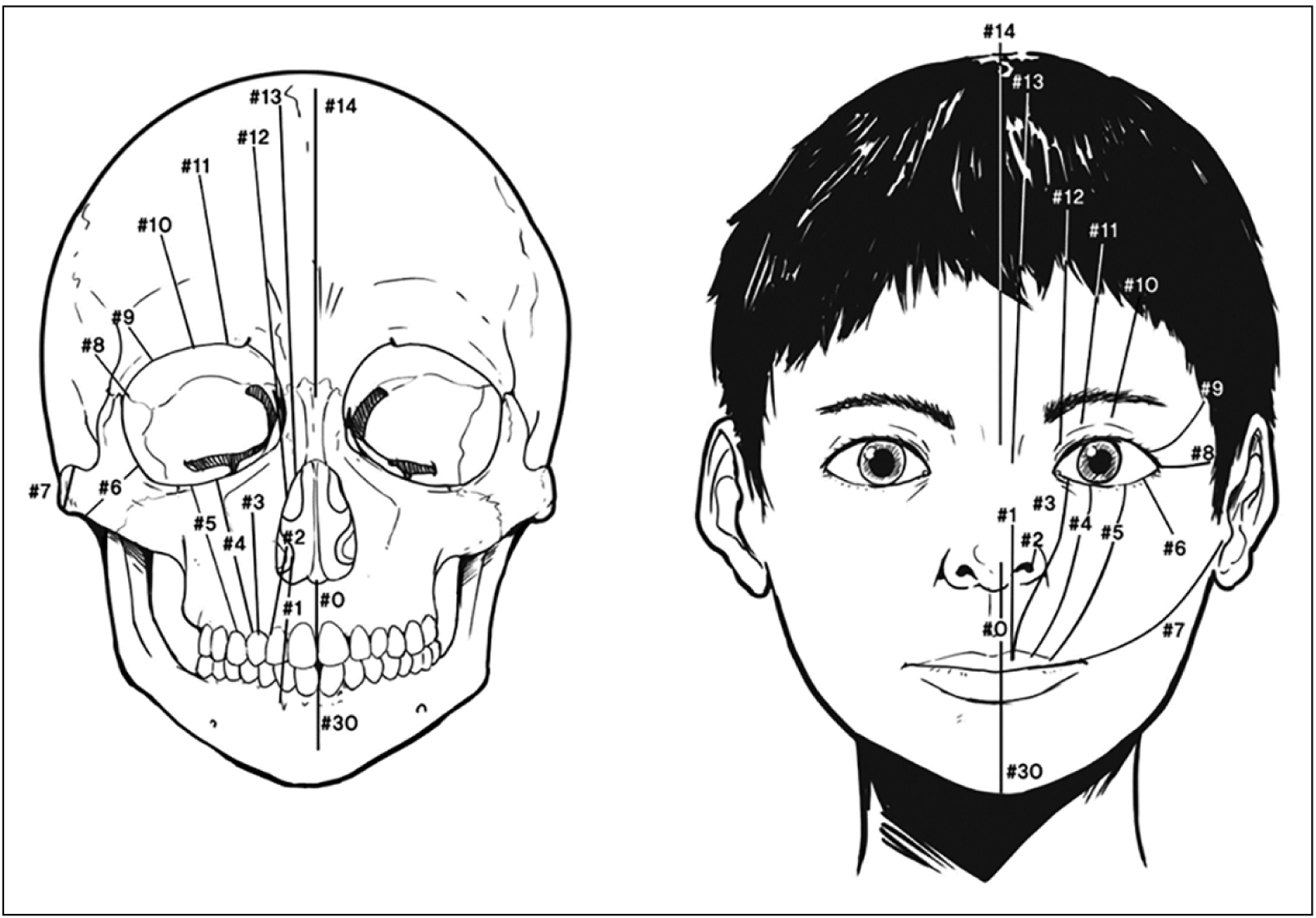
Using the orbit as the primary reference structure, Tessier37 numbered craniofacial clefts from #0 to #14 in a counterclockwise fashion, extending through the lips, nostrils, maxilla, eyelids, and eye brows (Figure 1). The median cleft of the lower jaw was designated as #30 cleft. Its subgroup was divided into midline clefts (#0, #14, and #30), paramedian clefts (#1, #2, #12, and #13), orbital clefts (#3, #4, #5, #9, #10, and #11), and lateral clefts (#6, #7, and #8).
Statistical analysis was performed using descriptive statistics, chi-square goodness of fit test, and Fisher’s exact test using SPSS software (ver. 12.0; SPSS Inc., Chicago, IL, USA). A p-value of less than 0.05 was considered a significant difference.
RESULTS
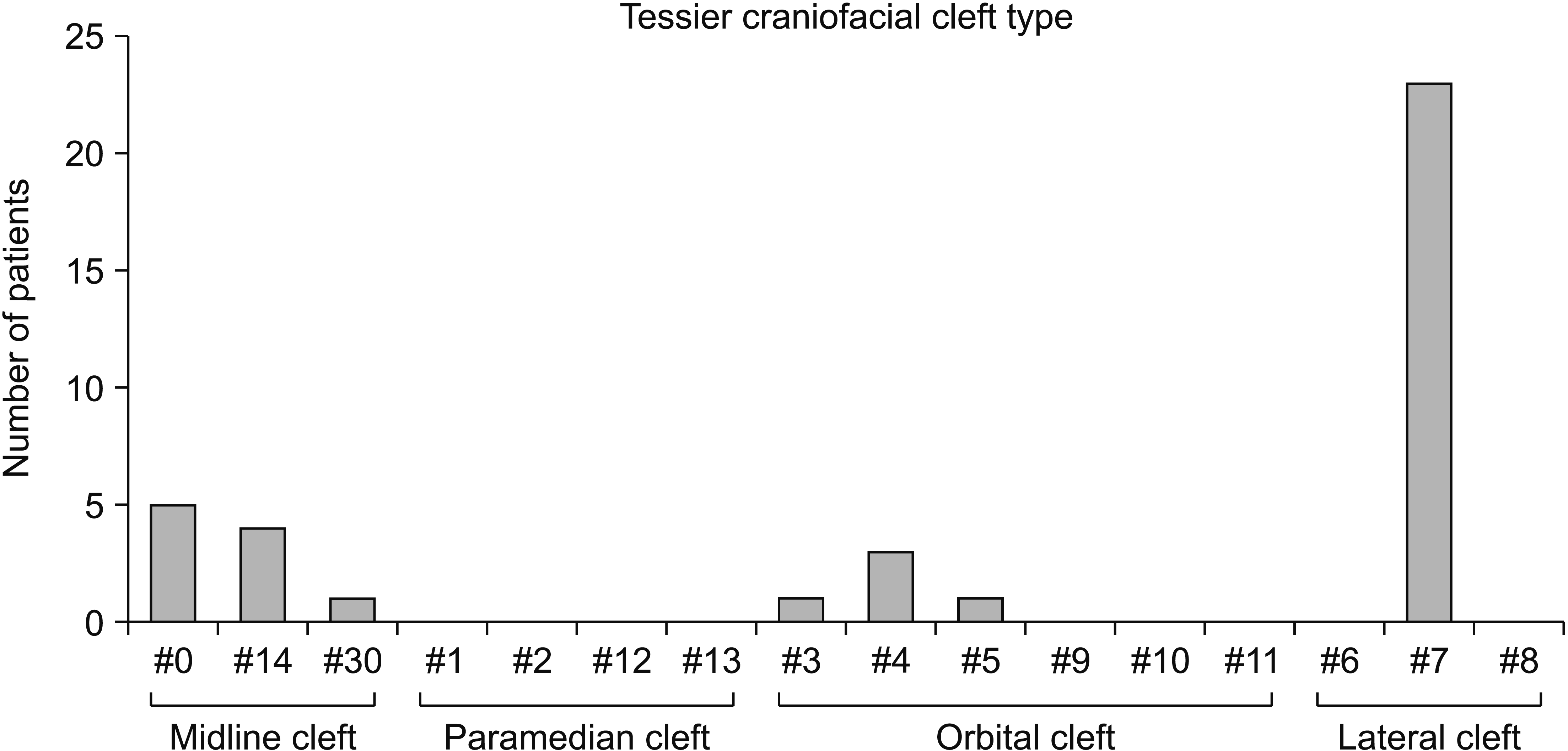
Composition of patients with craniofacial cleft (Figure 2)
The subjects included 10 patients with midline cleft (26.3%; n = 5 for #0 cleft, n = 4 for #14 cleft, and n = 1 for #30 cleft), 5 patients with orbital cleft (13.2%; n = 1 for #3 cleft, n = 3 for #4 cleft, n = 1 for #5 cleft), and 23 patients with lateral cleft (60.5%; n = 23 for #7 cleft).
Prevalence and distribution of Tessier cleft type (Figure 2)
The most common type was #7 cleft (n = 23), followed by #0 cleft (n = 5), #14 cleft (n = 4), #4 cleft (n = 3), and #3, #5 and #30 clefts (all n = 1). However, all paramedian clefts (#1, #2, #12, and #13 clefts) and a portion of orbital clefts (#9, #10, and #11 clefts) and lateral clefts (#6 and #8 clefts) were not detected.
Prevalence and distribution of sex in patients with craniofacial cleft (Table 1)
There was no difference in the prevalence of craniofacial clefts between male and female patients (total: n = 17 [44.7%] vs. n = 21 [55.3%]; midline cleft: n = 4 [40.0%] vs. n = 6 [60.0%]; orbital cleft: n = 3 [60.0%] vs. n = 2 [40.0%]; lateral cleft: n = 10 [43.5%] vs. n = 13 [56.5%]; all p > 0.05).
Comparison of side involvement in patients with craniofacial cleft (Table 2)
In patients with orbital cleft (#3, #4, and #5 clefts), there were no significant differences between the prevalence of unilateral and bilateral types (n = 2 [50.0%] vs. n = 2 [50.0%]; p > 0.05).
In patients with lateral cleft (all #7 clefts), the unilateral type was more prevalent than the bilateral type (n = 20 [87.0%] vs. n = 3 [13.0%]; p < 0.001). However, there was no significant difference between the affected side involvement of the right and left sides in the unilateral type (n = 13 [65.0%] vs. n = 7 [35.0%]; p > 0.05).
Association with hemifacial microsomia and side involvement in patients with #7 cleft (Table 3)
The association between hemifacial microsomia (HFM) and #7 cleft could not be stated in this study (yes: n = 16 [69.6%] vs. no: n = 7 [30.4%]; p > 0.05). However, in #7 cleft cases that had HFM, there was a significant match in the side involvement of #7 cleft and HFM (n = 14/16 [87.5%]; p < 0.01).
Phenotype and associated anomalies in patients with midline cleft (Figure 3)
Patients with #0 cleft (n = 5) exhibited nasal deformity (n = 3), bony defect in the premaxilla (n = 3), missing teeth in the premaxilla (n = 2), midline cleft lip (n = 3), microphthalmia (n = 2), congenital cataract (n = 1), optic nerve hypoplasia (n = 1), corpus callosum agenesis (n = 2), encephalocele (n = 3), hemangioma of neck (n = 1), duplex kidney (n = 1), pulmonary stenosis (n = 1), and funnel chest deformity (n = 1).
Patients with #14 cleft (n = 4) had unilateral coronal synostosis (n = 1), unilateral cleft lip and palate (UCLP) (n = 2), hyperterorbitism (n = 4), congenital ptosis with amblyopia (n = 1), strabismus (n = 2), and nasal deformity (n = 2). A patient with #30 cleft (n = 1) demonstrated tongue tie and missing mandibular left central incisor.
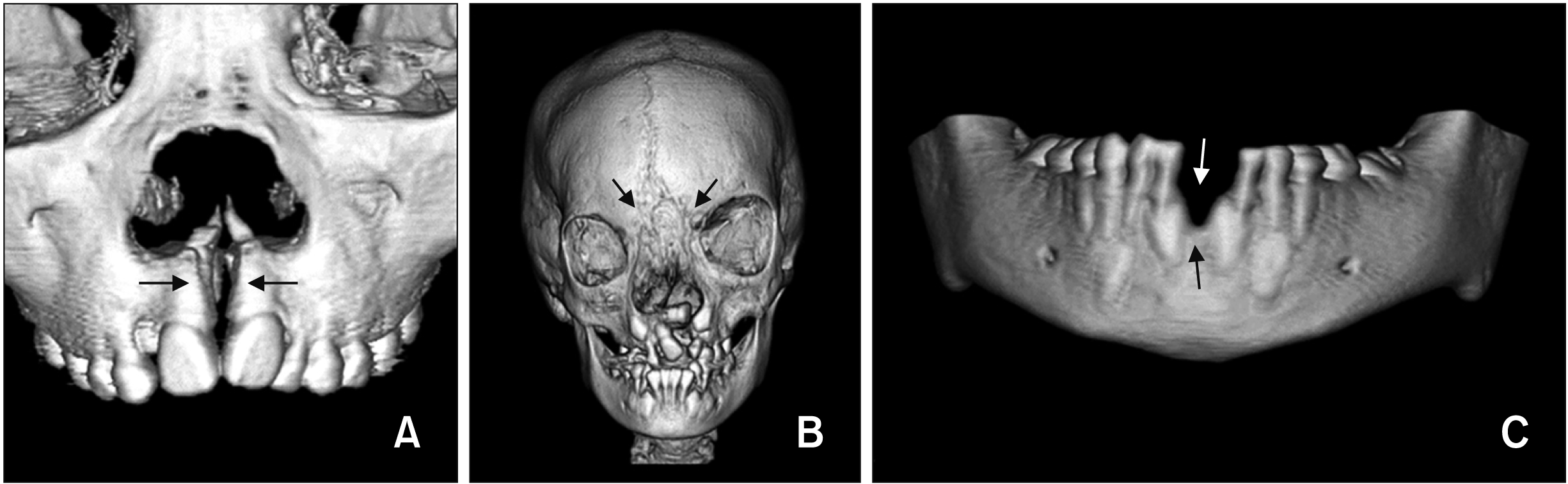
Phenotype and associated anomalies in patients with orbital cleft (Figure 4)
A patient with #3 cleft (n = 1) was the unilateral type (right side) and had bilateral cleft lip and alveolus (BCLA). All patients with #4 cleft (n = 3) were the bilateral type, including a combination of #3 and #4 clefts (n = 1), and had multiple teeth missing (n = 1). A patient with #5 cleft (n = 1) was a unilateral type (right side) and had a posterior openbite.
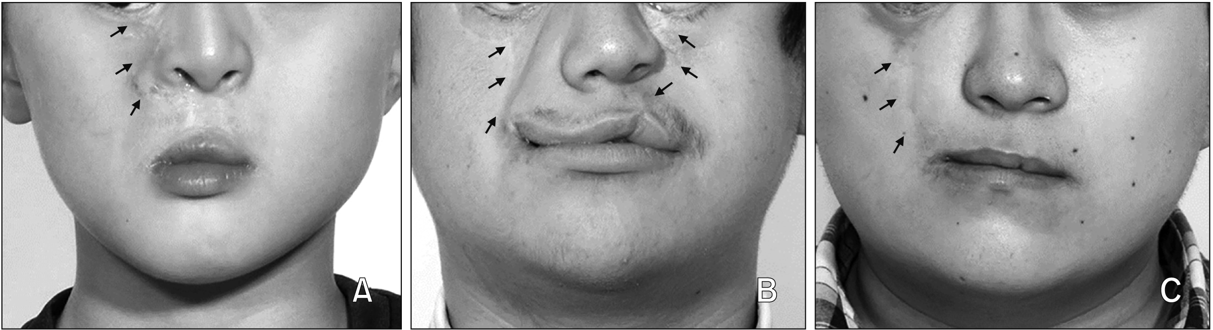
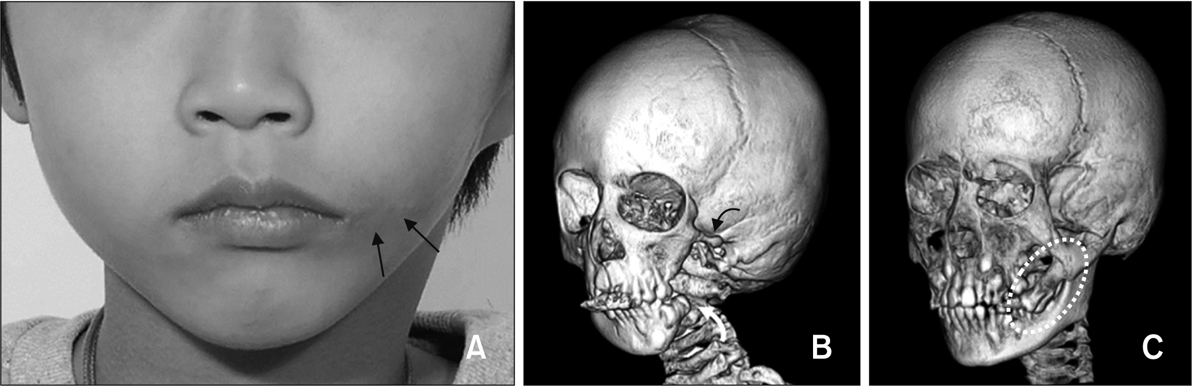
Phenotype and associated anomalies in patients with lateral cleft (Figure 5)
Among the patients with #7 cleft (n = 23), 20 patients were the unilateral type (87.0%; 13 right side and 7 left side) and three were the bilateral type (13.0%). A total of 16 patients were associated with HFM (69.6%; 12 unilateral HFM [Type I, n = 1; Type IIa, n = 5; Type IIb, n = 4; Type III, n = 2] and 4 bilateral HFM). In addition, they were associated with Goldenhar syndrome (n = 4, with internal organ anomalies, spinal anomalies and other skeletal anomalies), Klippel-Feil syndrome (n = 1, fusion of spines), and UCLP (n = 1). Seven patients were the isolated type (30.4%). Although six patients had missing teeth, two exhibited the posterior accessory bony process and supernumerary teeth.
DISCUSSION
Frequency and distribution of Tessier cleft types
Since it was difficult to find previous studies that investigated the frequency and distribution of whole craniofacial cleft types using PubMed, Scopus and others, it is necessary to indirectly compare the results of this study with those of previous studies. In the present study, the most common type was #7 cleft (n = 23, 60.5%, Figure 1), followed by #0 cleft (n = 5, 13.2%, Figure 2) and #14 cleft (n = 4, 10.5%, Figure 2). These findings were similar to those reported by da Silva Freitas et al.36 and Kalantar-Hormozi et al.13 (Table 4). The findings that #3, #5, and #30 clefts were found in only one patient (all n = 1, 2.6%; Figure 2) and #1, #2, #6, #8, #9, #10, #11, #12, #13 were not found (all n = 0, Figure 2) were similar to the results of Mishra and Purwar,35 who reported that #3 cleft was extremely rare (Table 4), and da Silva Freitas et al.,36 who indicated that #5 cleft was an extremely rare craniofacial cleft (Table 4). However, although Alonso et al.33 and Mishra and Purwar35 reported #4 cleft as the rarest in their studies, the present study exhibited that #4 cleft was the fourth common cleft in this study (n = 3 [7.9%], Figure 2). These discrepancies might be due to the difference in the ethnic background, difficulties in visiting the hospitals, and differences in skills of the hospitals in the respective countries.
Frequency and distribution of sex
In the present study, there was no difference between the prevalence of craniofacial clefts in male and female patients (total, midline cleft, orbital cleft, lateral cleft; all p > 0.05; Table 1). This was different from the results of Kalantar-Hormozi et al.,13 who reported that girls are more commonly affected than boys. These discrepancies might also be due to the difference in ethnic background, and parents’ attitude towards children’s facial appearance according to sex in the respective countries.
Comparison of side involvement in patients with craniofacial cleft
In patients with orbital cleft (#3, #4, and #5 clefts), there was no statistical difference between the prevalence of the unilateral and bilateral types (50.0% vs. 50.0%; p > 0.05; Table 2). However, this result can be attributed to the small sample size; therefore, it is necessary to analyze larger samples to validate the findings.
In patients with lateral cleft (all #7 clefts), the unilateral type was more prevalent than the bilateral type (87.0% vs. 13.0%, p < 0.001; Table 2). This finding was similar to the results of Kim et al.21 (n = 15 [93.7%] vs. n = 1 [6.3%]). Our results revealed that there was no significant difference between the affected side involvement of the right and left sides in the unilateral type (65.0% vs. 35.0%, p > 0.05; Table 2), which was also similar to the results of Kim et al.21 (n = 9 [60.0%] vs. n = 6 [40.0%]).
Association with hemifacial microsomia and side involvement in patients with #7 cleft
In the present study, the association between HFM and #7 cleft was not significant (yes, 69.6% vs. no, 30.4%; p > 0.05; Table 3). However, the percentage of positive association in this study (69.6%) was higher than that reported in previous studies (Kim et al.21 [n = 3/16, 18.8%] and Ryu et al.30 [n = 4/11, 36.4%]). Since these results were obtained from small sample groups, it is necessary to increase the sample size to obtain a more meaningful outcome.
In patients with #7 cleft who had HFM, there was a significant match in terms of the side involvement of #7 cleft and HFM (87.5%, p < 0.01; Table 3), indicating that these two anomalies might result from the same disturbance during the craniofacial embryogenesis process.
Posterior accessory bony process in patients with #7 cleft
Among patients with #7 cleft, 2 patients exhibited a posterior accessory bony process and supernumerary teeth (Figure 5). Woods et al.38 reported that 55.0% of patients with #7 cleft exhibited bony abnormalities including simple clefting of the maxillary molar region, 39.0% showed maxillary duplication, and 6.0% showed intermaxillary fusion. According to Cheung et al.,39 the pathogenesis of the posterior accessory bony process in patients with lateral cleft might be due to a compensatory mesenchymal growth from the temporal region rather than the duplication of the maxilla.
Associated anomalies with craniofacial cleft
In the present study, we found that most craniofacial clefts were associated with intra- and extra-craniofacial anomalies. Hence, there is a need of a multidisciplinary approach with a well-experienced cooperative team to tackle this issue. Although they are sporadic, orthodontists need to understand the clinical characteristics of craniofacial cleft and other anomalies associated with it.
Limitation of this study and suggestions for future studies
The present study attempted to provide an overview of the distribution, side involvement, phenotype, and associated anomalies of Korean patients with all types of craniofacial clefts. Despite the potential limitations due to the use of specific single university hospital-based data, we obtained meaningful basic data on the above-mentioned clinical characteristics of Korean patients with craniofacial cleft, who were diagnosed, treated, and/or followed-up over the last 20 years (1998–2018). The results of this study may provide basic data to establish a guideline for the use of health care for orthodontic treatment and surgical intervention of patients with craniofacial cleft in Korea in the near future. However, further studies are necessary to investigate the etiology of craniofacial cleft and the mechanism of its association with other anomalies during embryological development and to perform nationwide multicenter studies with systematic statistical analyses for orthodontic and surgical treatment of patients with craniofacial cleft.
CONCLUSION
Among the types of craniofacial clefts, #7 cleft was the most commonly found.
In patients with #7 cleft, the unilateral type was more prevalent than the bilateral type.
There was a high degree of match in the side involvement between #7 cleft and HFM.
Due to the diverse associated craniofacial anomalies in patients with craniofacial cleft, a multidisciplinary approach involving a well-experienced cooperative team is mandatory for individualized diagnosis and treatment planning of these patients.
 XML Download