

 PDF
PDF Citation
 Print
Print


INTRODUCTION
The Kidney Disease: Improving Global Outcomes (KDIGO) Clinical Practice Guidelines for Acute Kidney Injury (AKI) defines AKI according to the serum creatinine (S-Cr) concentration [1]. However, the role of S-Cr as a kidney function marker is limited because its concentration is influenced by multiple non-kidney factors, including age, sex, muscle mass and metabolism, dietary habits, medications, and hydration status. Moreover, the half-life of S-Cr increases from four hrs to 24–72 hours, if the baseline glomerular filtration rate (GFR) falls [2–5]. Therefore, a true decrement in GFR may not be appropriately reflected by the S-Cr concentration in patients with sepsis, liver disease, and/or muscle wasting [2–4]. Baseline S-Cr concentrations are already high in patients with chronic kidney disease (CKD), regardless of an AKI event, leading to difficulty in discriminating AKI. Thus, there is a need for a reliable AKI biomarker that is sensitive, specific, and easily measured to predict early changes following kidney injury without the need for exogenous substance administration [6]. Early AKI prediction is especially crucial to prevent AKI development or progression in high-risk patients who are older, have sepsis, are affected by multiorgan failure, have preexisting chronic comorbidities, and/or are taking nephrotoxic drugs, which can be a cause of AKI [7].
As an alternative to S-Cr, neutrophil gelatinase-associated lipocalin (NGAL) is a useful biomarker for AKI prediction [8–12]. Plasma NGAL (PNGAL) and urine NGAL (UNGAL) were previously measured using separate conventional immunoassays [13–17]. Recently developed assays enable the measurement of both PNGAL and UNGAL using the same reagent in routine chemistry analyzers. However, there is still no consensus on the type of specimen (blood or urine) that is most appropriate for NGAL assays in the initial work-up for AKI prediction; PNGAL concentrations might be influenced by extra-kidney factors and that elevated UNGAL concentrations provided a more specific estimation of kidney tubular damage [18]. Blood collection is routinely performed to test several laboratory parameters; the same specimens could be used for PNGAL tests [18]. By contrast, urine collection is difficult in incontinent patients, requiring bladder catheterization, which is an invasive procedure. Despite the need for a comparison between PNGAL and UNGAL for the AKI prediction in high-risk patients, few studies have evaluated the diagnostic performances of PNGAL and UNGAL tests simultaneously in patients without CKD, admitted to an intensive care unit (ICU) or a critical care setting and in oliguric, critically ill patients without CKD [19–21].
Therefore, there is a need to explore PNGAL and UNGAL-based approaches for AKI prediction and attempt to reach a consensus regarding the most appropriate assay. We evaluated the AKI prediction powers of PNGAL and UNGAL in AKI high-risk patients with and without CKD. We also assessed whether PNGAL and UNGAL measurements add value to S-Cr measurement for predicting AKI in AKI high-risk patients. To our knowledge, this is the first study to investigate PNGAL and UNGAL measurements simultaneously for predicting AKI in AKI high-risk patients mainly based on patients visiting the emergency department, especially with CKD.
Go to : 
MATERIALS AND METHODS
Subjects and specimens
We enrolled patients for whom the NGAL assay was recommended for suspected AKI at Konkuk University Medical Center, Seoul, Korea, between October 2016 and April 2017. In total, 200 patients were enrolled in this retrospective study. After excluding 49 patients without S-Cr data to determine AKI and/or CKD status, a total of 151 AKI high-risk patients were included (140 from the emergency department, nine from the obstetrics and gynecology department, two from the internal medicine department, and one from the neurosurgery department). High risk for AKI was determined based on the presence of one or more AKI risk factors, such as old age (>65 years old), sepsis, chronic preexisting chronic comorbidities (e.g., diabetes mellitus, hypertension, chronic heart disease, and CKD), and intake of nephrotoxic drugs (e.g., antibiotics, iodinated contrast agents, nonsteroidal anti-inflammatory drugs, and anticancer agents) [7, 22]. AKI was defined based on the KDIGO clinical practice guidelines [1]; the initial and serial S-Cr and estimated-GFR (eGFR) data of every patient were reviewed and applied. Urine output criteria were excluded. The patients with eGFR below 60 mL/min/1.73 m2 for more than 90 days were considered to have CKD. This study was approved by the Institutional Review Board (KUH1200071) of Konkuk University Medical Center. This registry study required neither study-specific blood or urine collection nor other interventions. Therefore, the requirement of written informed consent from the patients was exempted.
A total of 151 residual specimens (EDTA plasma and urine) were consecutively collected immediately after the onset of suspected clinical manifestations of AKI. The specimens were divided into small aliquots to avoid repeated freezing and thawing and were stored at −70°C until use. Frozen specimens were thawed at room temperature (23°C to 24°C) and gently mixed just before biomarker measurement. The PNGAL and UNGAL concentrations were measured between March and June 2017.
Assays
PNGAL and UNGAL concentrations were simultaneously measured using a particle-enhanced turbidimetric immunoassay (The NGAL Test, Bioporto Diagnostics A/S, Hellerup, Denmark) with a TBA-C16000 instrument (Toshiba Co., Tokyo, Japan). The analytical measurement range of the NGAL test is 25–3,000 ng/mL for both plasma and urine. The total coefficient of variation (CV%) during the study period was <3.3% for controls (from The NGAL Test Control Kit ST003CA), urine, and plasma specimens according to the manufacturer.
Statistical analysis
Data are expressed as median (interquartile range) or number (percentage). Areas under the receiver operating characteristic curves (AuROCs) and their 95% confidence intervals (CIs) were analyzed to assess the diagnostic performances and optimal cut-off values of PNGAL and UNGAL to predict AKI in all patients and according to the CKD status. PNGAL and UNGAL concentrations were compared between groups created according to clinical events and outcomes, such as AKI, in-hospital mortality, ICU admission, and need for renal replacement therapy (RRT), using the Mann–Whitney U test. Univariate and multivariate logistic regression analyses were performed to assess the predictors of AKI development. Variables with univariate P<0.05 (age and kidney biomarkers) were entered into the model for multivariate logistic regression analysis. The MedCalc Statistical Software (version 17.4.4, MedCalc Software bvba, Ostend, Belgium) was used for statistical analyses. P<0.05 was considered statistically significant.
Go to :
RESULTS
Patient characteristics
The characteristics of the patients according to AKI risk factors, PNGAL and UNGAL concentrations, initial S-Cr concentration, and initial eGFR are summarized in Tables 1 and 2. All patients had at least one AKI risk factor, demonstrating the diversity of AKI high-risk factors in this population. The AKI and non-AKI groups showed statistically significant differences in the concentrations of PNGAL, UNGAL, and initial S-Cr, and in initial eGFR (P<0.05 for all, Table 2). The same factors differed for patients with and without AKI when stratified by CKD status, except that patients with CKD (N=16) did not differ in PNGAL concentration from those without CKD (Table 2).
Table 1
Distribution of the study population according to AKI risk factors (N=151)
| Susceptibility factor | N (%) |
|---|---|
| Female | 80 (53.0) |
|
|
|
| Black | 0 (0) |
|
|
|
| CKD | 16 (10.6) |
|
|
|
| Chronic diseases | |
| Heart | 39 (25.8) |
| Lung | 66 (43.7) |
| Liver | 11 (7.3) |
| Diabetes mellitus | 37 (24.5) |
| Hypertension | 75 (49.7) |
| Cancer | 40 (26.5) |
| Anemia | 90 (59.6) |
|
|
|
| > 65 yr old | 103 (68.2) |
|
|
|
| Exposure | |
|
|
|
| Sepsis | 36 (23.8) |
|
|
|
| Critical illness* | 151 (100) |
|
|
|
| Circulatory shock | 14 (9.3) |
|
|
|
| Burns | 0 (0) |
|
|
|
| Trauma | 6 (4.0) |
|
|
|
| Cardiac surgery (especially with CPB) | 11 (7.3)/3 (2.0) |
|
|
|
| Major noncardiac surgery | 44 (29.1) |
|
|
|
| Nephrotoxic drugs | |
| Antibiotics | 30 (20.0) |
| Radiocontrast agents | 3 (2.0) |
| NSAIDs | 7 (4.6) |
| Anticancer drugs | 25 (16.6) |
|
|
|
| Pesticides | 1 (0.7) |
|
|
|
| Overall number of risk factors per patient | |
|
|
|
| 1 | 1 (0.7) |
|
|
|
| 2 | 6 (4.0) |
|
|
|
| 3 | 14 (9.3) |
|
|
|
| 4 | 20 (13.2) |
|
|
|
| 5 | 19 (12.6) |
|
|
|
| 6 | 34 (22.5) |
|
|
|
| 7 | 27 (17.9) |
|
|
|
| 8 | 16 (10.6) |
|
|
|
| 9 | 7 (4.6) |
|
|
|
| 10 | 6 (4.0) |
|
|
|
| 11 | 1 (0.7) |
![]()
Table 2
Distribution of PNGAL, UNGAL, initial S-Cr, and initial eGFR in the study population
| Variable | All patients (N=151) | All patients (N=151) | AKI high-risk CKD (N=16) | AKI high-risk non-CKD (N=135) | ||||||
|---|---|---|---|---|---|---|---|---|---|---|
|
|
|
|
||||||||
| AKI (N=58) | Non-AKI (N=93) | P* | AKI (N=9) | Non-AKI (N=7) | P* | AKI (N=49) | Non-AKI (N=86) | P* | ||
| PNGAL (ng/mL) | 216.3 (137.8–395.1) | 392.6 (206.3 –587.5) | 159.4 (117.7–262.8) | <0.001 | 621.0 (485.1–820.8) | 355.0 (307.6–494.9) | 0.081 | 338.3 (195.1–485.5) | 155.3 (116.0–239.2) | <0.001 |
|
|
||||||||||
| UNGAL (ng/mL) | 90.5 (28.2–429.1) | 250.2 (75.3–770.6) | 55.6 (19.8–216.3) | <0.001 | 877.4 (537.9–1,973.0) | 69.9 (9.2–143.9) | 0.004 | 196.6 (64.9–591.2) | 53.6 (21.0–227.0) | 0.002 |
|
|
||||||||||
| Initial S-Cr (μmol/L) | 79.6 (64.5–114.0) | 117.6 (84.0–157.4) | 70.7 (57.5–85.8) | <0.001 | 302.3 (226.3–508.3) | 114.0 (102.6–125.5) | 0.004 | 98.13 (80.5–132.6) | 69.0 (53.9–79.6) | <0.001 |
|
|
||||||||||
| Initial eGFR based on the MDRD study equation (mL·min−1·1.73 m−2) | 71 (49–90) | 47 (34–67) | 87 (70–90) | <0.001 | 13 (10–25) | 47 (36–51) | 0.005 | 51 (39–70) | 89 (73–90) | <0.001 |
![]()
Multivariate logistic regression analysis of risk factors to predict AKI development
Four models were analyzed by multivariate logistic regression (Table 3). In Model 0 (age and initial S-Cr concentration) and Model 1 (age, initial S-Cr, and UNGAL), only initial S-Cr concentration was a significant predictor of AKI. In Model 2 (age, initial S-Cr, and PNGAL) and Model 3 (age, initial S-Cr, PNGAL, and UNGAL), initial S-Cr and PNGAL were significant predictors of AKI. The R2 value was the highest overall for Model 3. Therefore, initial S-Cr (P<0.001) and PNGAL (P=0.013) concentrations, but not the UNGAL concentration, were considered independent predictors of AKI development in AKI high-risk patients, and the combination of these two factors increased the predictive power of the model.
Table 3
Multivariate logistic regression analyses of risk factors for AKI prediction in AKI high-risk patients (N=151)
![]()
Performance of PNGAL and UNGAL to predict AKI development and the effect of CKD
The predictive value of PNGAL and UNGAL concentrations for AKI in AKI high-risk patients is shown in Fig. 1 based on the AuROC values. For all patients, the AuROC for PNGAL was higher than that for UNGAL; there was moderate sensitivity and high specificity for a PNGAL cut-off concentration of 306.1 ng/mL and moderate sensitivity and specificity for a UNGAL cut-off concentration of 90.5 ng/mL (Fig. 1A). However, the AuROC value for UNGAL was higher than that for PNGAL in patients with CKD; there was moderate sensitivity and good specificity for a PNGAL cut-off concentration of 504.4 ng/mL and high sensitivity and 100% specificity for a UNGAL cut-off concentration of 477.4 ng/mL (Fig. 1B). The AuROC value in patients without CKD was higher for PNGAL than for UNGAL; there was moderate sensitivity and high specificity for a PNGAL cut-off concentration of 271.0 ng/mL and moderate sensitivity and specificity for a UNGAL cut-off concentration of 90.5 ng/mL (Fig. 1C).
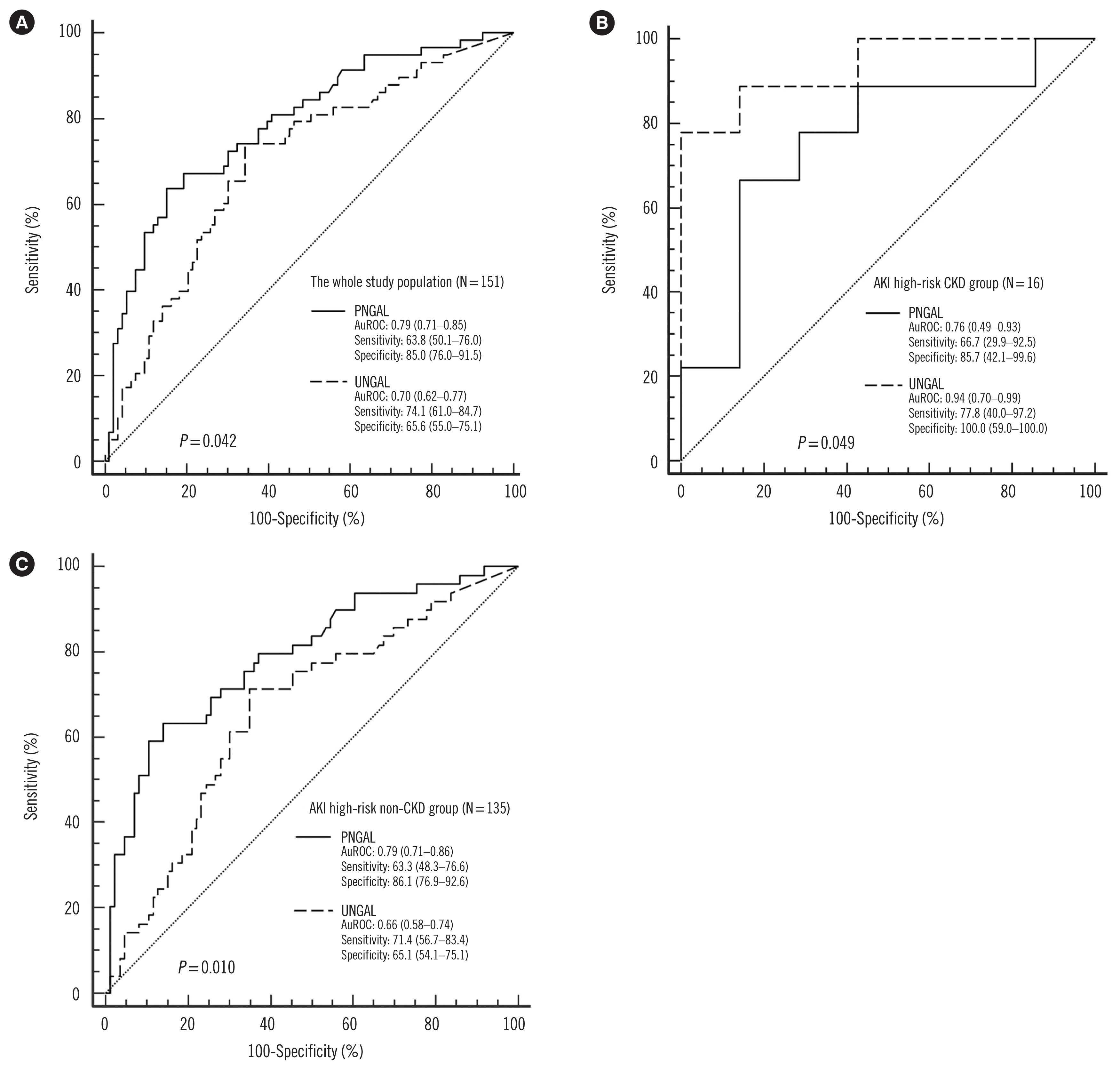 | Fig. 1Comparison of AuROCs for PNGAL and UNGAL for AKI prediction in AKI high-risk patients. (A) All patients (N=151), (B) AKI high-risk CKD group (N=16), (C) AKI high-risk non-CKD group (N=135).
Abbreviations: AKI, acute kidney injury; AuROC, area under the receiver operating characteristic curve for the prediction of AKI; CKD, chronic kidney disease; PNGAL, plasma neutrophil gelatinase-associated lipocalin; UNGAL, urine neutrophil gelatinase-associated lipocalin.
|
The AuROC value of PNGAL was significantly higher than that of UNGAL (P=0.042; Fig. 1A) in all patients, whereas in the CKD group (N=16), AuROC value of UNGAL was significantly higher than that of PNGAL (P=0.049; Fig. 1B). In the AKI high-risk non-CKD group (N=135), PNGAL showed a significantly higher AuROC value than UNGAL (P=0.010; Fig. 1C).
Comparison of PNGAL and UNGAL concentrations according to clinical outcomes
PNGAL and UNGAL concentrations differed significantly according to clinical outcomes, such as in-hospital mortality, ICU admission, and RRT requirement (Table 4). Specifically, PNGAL concentrations were higher in patients who died, were admitted to the ICU, and received RRT, and UNGAL concentrations were higher in patients who were admitted to the ICU and/or received RRT.
Table 4
Comparison of PNGAL and UNGAL concentrations according to clinical outcomes
| In-hospital mortality | ICU admission | RRT requirement | |||||||
|---|---|---|---|---|---|---|---|---|---|
|
|
|
|
|||||||
| −† (N=137) | +† (N=14) | P* | −† (N=110) | +† (N=41) | P* | −† (N=147) | +† (N=4) | P* | |
| PNGAL (ng/mL) | 206.3 (133.9–351.5) | 436.6 (152.5–724.7) | 0.023 | 184.9 (128.3–306.1) | 387.4 (170.2–513.5) | <0.001 | 211.7 (134.7–360.4) | 801.5 (543.3–1,071.4) | 0.003 |
|
|
|||||||||
| UNGAL (ng/mL) | 87.0 (26.1–376.6) | 340.5 (82.6–779.8) | 0.064 | 76.4 (21.8–315.8) | 153.3 (51.2–771.9) | 0.020 | 87.0 (27.7–382.7) | 2,008.7 (1,004.3–3,327.3) | 0.003 |
![]()
Go to :
DISCUSSION
The main purpose of this study was to evaluate the presence of AKI at admission and to determine the predictive power for AKI development based on S-Cr, PNGAL, and UNGAL concentration measurements in suspected patients. In almost all patients with AKI, except for six (52/58), the condition developed on admission. Soto, et al. [23] reported that PNGAL is an accurate biomarker for the AKI prediction in patients admitted to the emergency department and proposed a three-grade classification of AKI risk based on PNGAL concentration.
The incidence of AKI in this study (58/151, 38.4%) was higher than that previously reported [19, 20]. Tecson, et al. [19] reported an AKI incidence of 13.5% in ICU patients, and Egal, et al. [20] reported an AKI incidence of 34.7% in oliguric critically ill patients. Differences in AKI incidence across study populations could be caused by differences in the constitutions of the patients (AKI risk factors, such as susceptibilities and/or exposures) and/or the clinical judgments of experienced clinicians regarding AKI evaluation. PNGAL, UNGAL, and initial S-Cr concentrations, and initial eGFR all differed significantly between the AKI and non-AKI groups; however, the difference of PNGAL concentration was no longer significant in patients with CKD. This result confirms a strong association of PNGAL and UNGAL concentrations with AKI.
PNGAL and initial S-Cr concentrations emerged as independent predictors of AKI development in AKI high-risk patients based on multivariate regression analysis. However, UNGAL concentration did not show a significant regression coefficient. These findings indicate that AKI was largely determined by the S-Cr concentration, which was already high in the AKI patients due to the characteristics of this population, such as admission to the emergency department and presentation with symptoms that categorized them as AKI high-risk patients. However, the R2 value increased in the model combining PNGAL and UNGAL concentrations with initial S-Cr concentration, reflecting the additive value of NGAL and S-Cr for the AKI prediction.
PNGAL showed similar AuROCs in the AKI high-risk CKD group and AKI high-risk non-CKD group. UNGAL showed a higher AuROC in the AKI high-risk CKD group. In CKD patients, the elimination of NGAL by the kidney is severely deteriorated, while basal PNGAL concentrations tend to be maintained at high concentrations. This leads to a difficulty in discriminating AKI in CKD patients based on PNGAL concentration. Corbacıoglu, et al. [24] reported that PNGAL concentrations were higher in AKI patients than in CKD patients. However, in clinical practice, the use of PNGAL concentrations to distinguish between AKI and CKD is limited [24]. Smith, et al. [25] reported that using the UNGAL-to-urine creatinine ratio in addition to conventional-cardiovascular and kidney risk factors may improve the prediction of disease progression in elderly Caucasian pre-dialysis CKD patients with low-grade proteinuria. Therefore, PNGAL concentration evaluation may be useful as a first step in the AKI prediction in AKI high-risk patients without CKD, whereas UNGAL concentration evaluation may be useful as a first step in the AKI prediction in AKI high-risk patients with CKD. This study provides several cut-off values for PNGAL and UNGAL for these patients and subgroups. The median UNGAL concentration in the AKI high-risk CKD group was significantly higher in AKI patients than in the non-AKI ones. Although the median PNGAL concentration was higher in the AKI group than in the non-AKI group, the difference was not statistically significant.
PNGAL and UNGAL concentrations could also reflect patient outcomes in AKI high-risk patients. Several studies identified both PNGAL and UNGAL concentrations as predictors of mortality or diagnostic markers for AKI in critically ill patients or patients who underwent cardiac surgery with cardiopulmonary bypass [26–29]. However, these studies excluded CKD patients at the patient selection step or concluded that the discriminative performance of both PNGAL and UNGAL concentrations was reduced by preexisting CKD. By contrast, we included CKD patients as a subgroup in this study and identified that UNGAL concentration evaluation may be useful as a first step for AKI prediction in AKI high-risk patients with CKD.
The present study has several limitations. The definition for AKI was extended because S-Cr records in the seven days before UNGAL measurement were not available for most patients. In addition, urine output criteria were excluded in the determination of AKI. The median initial S-Cr concentration in the AKI group was high, at 117.6 μmol/L, because kidney injury had already occurred in most patients who visited the emergency department. In addition, 16 out of the 151 patients had CKD. Therefore, further studies should be performed with a larger number or various populations of patients. Finally, we used eGFR values for the determination of AKI and CKD instead of measured GFR values. GFR measurement remains a significant confirmatory test for reduced eGFR [30]. However, all GFR measurement methods are imprecise and could be biased against true GFR [30]. Standardization and calibration among methods, with the development of more accurate eGFR equations may provide helpful data for both clinical work and research [30].
In conclusion, PNGAL may serve as a useful biomarker for AKI prediction in AKI high-risk patients. However, in AKI high-risk patients with CKD, UNGAL showed better AKI prediction power than PNGAL. This approach could be beneficial for the timely and optimized management of AKI in AKI high-risk patients with or without CKD. Our findings may guide in selecting the most appropriate specimens for NGAL assays based on the presence of CKD in AKI high-risk patients. The current trend in NGAL studies is focused on evaluating the potential role of NGAL in specific study populations, such as patients with decompensated cirrhosis, diabetic kidney disease, heart failure with or without left ventricular assisted device implantation; infants with congenital urinary tract obstruction; and patients with abdominal aortic aneurysm [31–35]. Further studies are needed to investigate the role of NGAL as a biomarker of AKI in specific patient groups.
Go to :
 XML Download