

 PDF
PDF Citation
 Print
Print


INTRODUCTION
Parasitic infections can cause anemia, malnutrition, and other impairments in physical and cognitive development, especially in children [1, 2]. Giardia duodenalis is one of the main etiological agents of diarrhea worldwide, accounting for approximately 28.2 million cases of diarrhea each year due to food contamination [3]. Protozoa transmission is considered a public health problem in developing countries, and since 2004, Giardia has been included in the WHO’s “Neglected Diseases Initiative” group [4].
G. duodenalis infection shows a broad clinical spectrum, ranging from asymptomatic cases to acute or chronic diarrhea, abdominal pain, nausea and vomiting, dehydration, and weight loss [5, 6]. Children, especially those that attend childcare centers, are considered a high-risk group for G. duodenalis infection and its consequences, including impairment in physical and cognitive development [5, 6]. The laboratory diagnosis of G. duodenalis is conventionally performed by microscopic identification of cysts and/or trophozoites in feces [7]. However, microscopic identification has limited sensitivity due to the intermittent elimination of G. duodenalis cysts in feces and requires trained technicians for accurate diagnosis [4, 5]. Coproantigen tests based on ELISA or immunochromatography were also developed for detecting parasite proteins in feces and are considered more sensitive than microscopy-based methods [8–11]. In addition, the detection of antibodies against G. duodenalis in sera by ELISA or immunofluorescence can also be useful for diagnosis and seroepidemiological studies in large communities [12, 13]. High levels of specific antibodies against Giardia have been detected in populations from Mexico [12], the Caribbean [13], the United States [14], and Venezuela [15]. Although the detection of specific serum IgG antibodies cannot distinguish past from current infections, this approach nevertheless provides information on the overall exposure of a population. Studies suggest that the presence of serum or salivary anti-Giardia IgA indicates recent infections by G. duodenalis [15, 16]. However, the results are controversial, and some reports have shown that neither IgA nor IgG can differentiate between past and current infection [17, 18]. These debatable reports indicate the need for more studies to assess the efficacy of serology in G. duodenalis diagnosis as well to investigate its performance in pediatric population from endemic areas for intestinal parasitic infections. Commercially produced ELISA kits are not promptly available for detecting serum antibodies to Giardia infection. Therefore, the main objective of this study was to compare the diagnostic potential of an in house-ELISA for detecting specific antibodies in sera with the current G. duodenalis infection determined by microscopy and/or the presence of parasite antigens in the feces of children from Salvador, Bahia, Brazil.
Go to : 
MATERIALS AND METHODS
Study design and population
This cross-sectional study was conducted on children undergoing routine laboratory examinations at the Clinical Analysis Laboratory of Pharmacy College of the Federal University of Bahia (N=287) and those attending daycare centers (N=187) located in the same city district of Salvador, Bahia, Brazil. Overall, the children’s ages ranged from 0–14 years, with those from daycares mostly 2–7 years old.
The Ethics Committee of Nursing School, Federal University of Bahia, Brazil, approved the study (project approval No. 907.867). Children whose parents agreed to participate in the study and signed an informed consent form were enrolled during the study period. Children over eight years old were informed about the research and they signed a consent form. All parasitological tests results were sent to the children’s parents.
The children were selected by convenience sampling from January 2015 to January 2016. Fecal and serum samples were collected from all participating children. At least two fecal samples were submitted for the diagnosis of G. duodenalis. Part of the feces was used for the parasitological examination, and the other was frozen at −20°C for up to six months before testing for Giardia coproantigen. Tubes containing polymer gel for serum separation were centrifuged for 10 minutes at 1,620×g, and sera were frozen at −20°C until use. Standardization of the in-house ELISA and study of seroprevalence of anti-Giardia IgG and IgA in children sera were performed in 2017.
Diagnosis of intestinal parasites in fecal samples
Stool samples were subjected to the following parasitological tests: (a) sedimentation by centrifugation in water [19]; (b) zinc sulfate (density of solution 1.18 g/mL) centrifugal flotation [20]; and (c) modified Ziehl-Neelsen staining [21]. Two slides were examined for each test. In addition to these parasitological tests, an ELISA kit (RIDASCREEN® Giardia, R-Biopharm AG, Germany) was used for detecting the Giardia coproantigens.
In-house ELISA for detection of anti-Giardia IgG and IgA
G. duodenalis soluble antigen preparation
G. duodenalis trophozoites (strain WB) were axenically cultured in TYI-S-33 medium supplemented with serum and bovine bile (Keister’s Modified TYIS33 Giardia Medium - ATCC Medium 2695; Virginia, USA), according to the procedure described by Keister [22]. The trophozoites were washed three times at 4°C, 720×g for 10 minutes in sterile phosphate-buffered saline (PBS), pH 7.2. The suspension was then sonicated in an ice bath (15 cycles of 60 seconds at 90 Hz). Protease inhibitors [0.05 mmol/L EDTA, 1 mmol/L phenylmethanesulfonyl fluoride, 0.05 mmol/L tosyl-L-phenylalanine chloromethyl ketone/tosyl-L-lysine chloromethyl ketone, and 1 μg/mL leupeptin] were added to the antigen extract and centrifuged at 4,500×g for 30 minutes at 4°C. The supernatant with soluble antigen was analyzed for protein content according to the method of Lowry, et al. [23], divided into aliquots, and stored at −20°C until use.
Standardization of in-house ELISA
The indirect ELISA was standardized using 94 serum samples obtained from 30 G. duodenalis monoinfected children, 30 non-parasitized children, and 34 children who were infected with other intestinal parasites, including Endolimax nana, Entamoeba coli, Iodamoeba butschilli, Blastocystis hominis, Trichuris trichiura, Ascaris lumbricoides, Enterobius vermicularis, and Strongyloides stercoralis. These sera were different from the main samples tested to determine the seroprevalence of anti-Giardia IgG and IgA in children, and previously selected from children who routinely attended at the Clinical Analysis Laboratory of Pharmacy College, Federal University of Bahia. All fecal samples were examined at the Parasitology Laboratory as described in the previous section “Diagnosis of intestinal parasites in fecal samples” and, according to their parasitological results, they were chosen to integrate the set of control sera. G. duodenalis-negative samples were selected by parasitological examination of three fecal samples and by a coproantigen test, as mentioned above.
ELISA for anti-Giardia IgG and IgA detection
Microplate wells (Corning Costar polystyrene EIA/RIA plates, Corning) were coated with 20 μg/mL of G. duodenalis antigen in 0.06 mol/L carbonate-bicarbonate buffer (pH 9.6), incubated overnight at 4°C, and washed three times with PBS containing 0.05% Tween-20 (PBS-T). The plates were blocked with 200 μL of PBS-T containing 5% w/v skim milk (PBS-T-milk) for 1 hour at 37°C. After blocking, the wells were washed five times with PBS-T, filled with 100 μL of serum samples diluted at 1/100 (IgG) or 1/25 (IgA) diluted in PBS-T-milk, and incubated in duplicate for 1 hour at room temperature (RT; ~27°C). After another washing step, 100 μL of horseradish peroxidase-conjugated anti-human IgG (Sigma-Aldrich, St. Louis, MO, USA) or IgA (Thermo Fisher Scientific) antibodies diluted 1/1,000 in PBS-T-milk were added, followed by 1-hour incubation at 37°C. The plates were washed three times with PBS-T and twice with pure PBS. The reaction was visualized by adding 100 μL of substrate (0.051 mol/L citrate-phosphate buffer, pH 5.0, containing 0.004 mol/L p-phenylenediamine and 0.040% hydrogen peroxide), and the plate was incubated for 20 minutes in the dark at RT. Twenty microliters of 8 N sulfuric acid was added to stop the reaction, and the optical density (OD) was measured at 450–630 nm with a microplate reader (Awareness Technology, Palm City, FL, USA). The antibody levels were expressed as the ELISA index (EI), calculated as the ratio between the OD of each tested sample and that at the cut-off point; samples with an EI >1 were considered positive.
Statistical analysis
The data were analyzed using SPSS Statistics for Windows version 19 (IBM Corp., Armonk, NY, USA), and statistical analyses were performed with GraphPad Prism 7 (GraphPad Software, Inc., San Diego, CA, USA). The ELISA OD cut-off, sensitivity and specificity values were determined by the receiver operating characteristic (ROC) curve. The agreement between ELISA and a parasitological diagnosis of G. duodenalis infection was assessed with the kappa index [24]. Analysis of variance followed by Dunn’s test was used to compare EI values among groups (infected with G. duodenalis, infected with other protozoa or helminths, and non-infected children). P<0.05 was considered statistically significant.
Go to :
RESULTS
Characterization of the population and frequency of enteroparasites
There was no significant difference among the children groups in relation to gender. Overall, 77.4% of children fell within the 2–10 years range; children under 2 years and 11–14 years of age were all from the group examined at the clinical laboratory (Table 1). There was a predominance of monoparasitism by protozoa, and G. duodenalis was the most predominant pathogenic parasite found (8.2%; N=39; Table 1).
Table 1
Characteristics of children and frequency of parasite infection
![]()
Detection of anti-Giardia IgG and IgA in sera
The sensitivity and specificity of ELISA were 80% and 90% for anti-Giardia IgG and 80% and 83.3% for IgA, respectively (Table 2). The OD cut-off value was 0.136 for IgG and 0.068 for IgA, which were used to calculate the Giardia antibody positivity rates in the study population.
Table 2
Sensitivity, specificity, and cut-off value of the in-house ELISA for detection of anti-Giardia IgG and IgA antibodies in sera
| ELISA* | Optical density cut-off value | Sensitivity (95% CI) | Specificity (95% CI) |
|---|---|---|---|
| IgG | 0.136 | 80.0% (61.4–92.3%) | 90.0% (73.5–97.9%) |
| IgA | 0.068 | 80.0% (78.9–89.2%) | 83.3% (51.6–89.8%) |
![]()
The antibody reactivities were significantly higher in children infected with G. duodenalis (P<0.001) compared with those of the other groups (Fig. 1). The seropositivity for anti-Giardia IgG and IgA was 13.9% (N=66) and 23.6% (N=112), respectively (Table 3). Of the 39 children who had G. duodenalis in feces, 79.5% (N=31) and 71.8% (N=28) showed seropositivity for IgG and IgA, respectively.
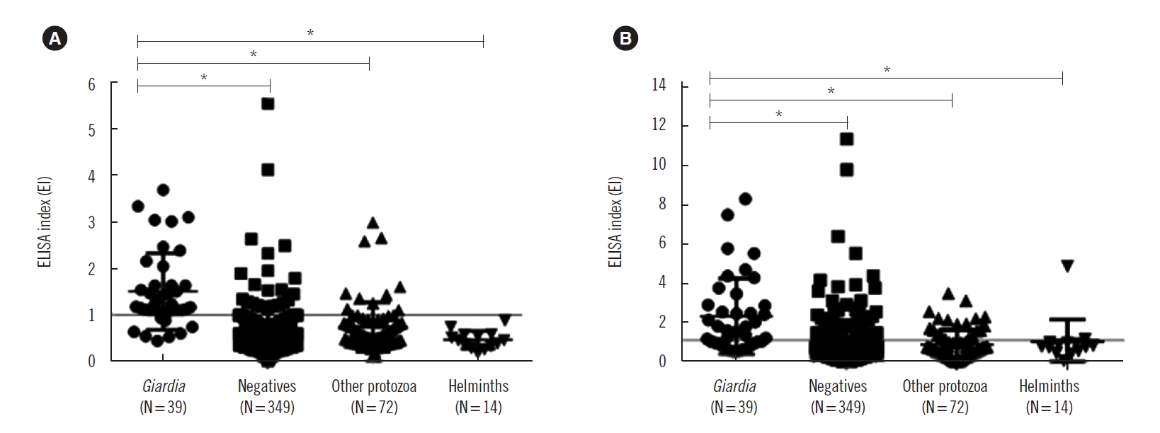 | Fig. 1Reactivities of anti-Giardia antibodies in sera of children according to their parasitological results. IgG (A) and IgA (B) levels of anti-Giardia in serum samples are expressed as the ELISA index (EI). Symbols represent G. duodenalis-infected children (●), negatives (■), and children infected with other protozoa (▲) or helminths (▼).
s*P<0.001 (ANOVA, Dunn test).
|
Table 3
Seropositivity of anti-Giardia IgG and IgA in children, according to the presence of parasitic infection
| Groups |
ELISA positivity for anti-G. duodenalis antibodies Total positives N/tested N (%) |
|||||
|---|---|---|---|---|---|---|
|
|
||||||
| IgG | IgA | |||||
|
|
|
|||||
| Clinical laboratory 36/287 (12.5) | Daycare centers 30/187 (16.0) | Total 66/474 (13.9) | Clinical laboratory 58/287 (20.2) | Daycare centers 54/187 (28.9) | Total 112/474 (23.6) | |
| Children with G. duodenalis | 18/23 (78.3)† | 13/16 (81.3)‡ | 31/39 (79.5)* | 17/23 (73.9)§ | 11/16 (68.8)|| | 28/39 (71.8)* |
|
|
||||||
| Children with other protozoa | 6/46 (13.0)† | 4/26 (15.4)‡ | 10/72 (13.9) | 10/46 (21.7)§ | 11/26 (42.3)|| | 21/72 (29.2) |
|
|
||||||
| Children with helminths | 0/11 (0.0) | 1/3 (33.3) | 1/14 (7.1) | 2/11 (18.2)§ | 0/3 (0.0) | 2/14 (14.3) |
|
|
||||||
| Non-parasitized children | 12/207 (5.8)† | 10/142 (7.0)‡ | 22/349 (6.3) | 31/207 (15)§ | 32/142 (22.5)|| | 63/349 (18.1) |
![]()
The agreement between the positivity rate of specific antibodies and the detection of G. duodenalis in feces was moderate for ELISA-IgG (kappa index [95% CI]=0.543 [0.422–0.664]) and mild for ELISA-IgA (kappa index [95% CI]=0.283 [0.162–0.404]) (Table 4). Of the 86 children infected with other enteroparasites, 10 (11.6%) and 21 (24.4%) showed reactivity to specific IgG and IgA, respectively. Giardia antibodies were more frequent in those infected by E. nana and E. coli among all parasite infections (Table 5).
Table 4
Comparison of IgG- and IgA-ELISA positivity rate with the diagnosis of G. duodenalis in feces
![]()
Table 5
Giardia duodenalis IgG- and IgA-ELISA positivity rate in sera of children infected with other parasites
![]()
Go to :
DISCUSSION
In this study, we found a positivity rate of 27.9% for one or more intestinal parasites in children from Salvador, Bahia. This corroborates with other studies undertaken in Brazil that reported frequencies varying between 5% and 50% [25, 26]. G. duodenalis infection is more common in the infant population than in adults, with especially high frequencies detected in daycare centers and schools [27]. The frequency of G. duodenalis in children from Salvador, Brazil, was reported to range from 13% to 18.4% [11, 28]. Although we observed a lower frequency of G. duodenalis infection in our study, it was the most common pathogenic parasite found, with no differences observed between children in daycare centers and those attending the clinical laboratory. The considerable frequency of G. duodenalis and other protozoa observed in this study may be explained, in part, by the small size of the cysts, which facilitates their passage through filters and enables their escape from the standard processes of water treatment, in addition to the poor hygiene habits and immune defense immaturity of children [29].
Experimental, clinical, and epidemiological observations indicate that G. duodenalis stimulates an immune response in the host [30, 31], and anti-Giardia antibodies have been detected in individuals with G. duodenalis infection [15, 16]. Based on these findings, serological tests were developed to detect specific IgG, IgM, and IgA in the serum of patients. According to Garcia [32], the detection of these antibodies in the serum is not yet suitable for the diagnosis of current G. duodenalis infection; however, it can serve as an important tool for epidemiological surveys to determine the extent of G. duodenalis exposure of a population.
In our study, the ELISA for anti-G. duodenalis IgG and IgA showed a sensitivity of 80% and a specificity of 90% and 83.3%, respectively. Previous studies have found sensitivities ranging from 64–97% and specificities from 84–85% in anti-Giardia IgG ELISA [12, 16]. These differences are possibly due to the different populations studied, stage of infection, or technical variations in the immunoassays. Our standardized in-house ELISA demonstrated a seroprevalence of anti-Giardia IgG and IgA of 13.9% (N=66) and 23.6% (N=112), respectively. The detection rate of both antibodies was slightly higher in children from daycare centers (IgG: 16%; IgA: 28.9%) than in those tested at the clinical laboratory (IgG: 12.5%; IgA: 20.2%), which may reflect higher exposure to the parasite in the former group.
High rates of anti-Giardia IgG have been reported in Mexico, where anti-Giardia IgG was found in 77% of the sera of lactating women [33] and in 55.3% of the general population [12]. Guimarães and Sogayar [34] found an anti-Giardia IgG seroprevalence of 63.3% in children at daycare centers of São Paulo, Brazil. Other studies have shown a reduction in the prevalence of G. duodenalis and other enteroparasitoses infections in developing countries due to improvements in hygienic-sanitary and educational conditions in these regions [27, 35], thus reducing exposure to these pathogens. This may explain the lower anti-Giardia IgG seroprevalence in our study.
Studies have suggested that anti-Giardia IgA antibody is a better indicator of current infection than IgG, since it is predominantly produced in the gastrointestinal tract [15, 16]. However, the seroprevalence of IgA in our study was higher than that of IgG (23.6% vs. 13.9%). Overall, 26.7% of the children did not show G. duodenalis in feces, but were positive for specific IgA in serum, which is higher than IgG (12.8%). Moreover, when we compared the detection of anti-Giardia IgG or IgA in the serum with the detection of cysts and/or of Giardia antigens in feces, the kappa index agreement was moderate for IgG and weak for IgA. This finding suggests that like IgG, IgA is not always associated with current infection. In fact, in endemic areas where exposure to G. duodenalis infection is frequent and early, the levels of both serum specific IgG and IgA remain high in adulthood, reflecting recurrent exposure to the parasite [18, 36]. Experimental studies have also shown that IgA antibodies remain elevated for a long period even after elimination of the parasite [37], corroborating the present results.
Cross-reactivity is a common problem in the detection of antibodies against parasitic antigens, especially in endemic areas. However, few studies have described the cross-reactivity in immunoassays for the detection of anti-Giardia antibodies. We found that 11.6% (10/86) and 24.4% (21/86) of the children infected by other enteroparasites showed reactivity for anti-Giardia IgG and IgA, respectively, which were mostly cases of infection with E. nana or E. coli. This antibody cross-reactivity may indicate the antigenic similarity between protozoa species, undiagnosed Giardia infection due to a low discharge of cysts or fecal antigens, and/or previous exposure to G. duodenalis (immunological memory). Therefore, one limitation of our study was the use of G. duodenalis crude antigens for the in-house ELISA, which may have produced false-positive results due to cross-reactions with other intestinal protozoa. These can occur either because of the high sensitivity of the assay or proximity of protozoan epitopes. Moreover, G. duodenalis and intestinal amoebae are usually co-endemic in several regions of Brazil. Thus, recombinant antigens would be advantageous in terms of greater specificity in comparison with crude preparations, although there is a consensus of the need for a mixture of recombinant antigens to improve sensitivity [38]. More studies are needed to explain these cross-reactions, including the absorption of sera with E. nana and E. coli antigens before testing in antibody assays.
In conclusion, the parasitological examination and high detection rate of anti-G. duodenalis IgG and IgA observed in this study suggest the high endemicity and early exposure to this protozoan in the children. It is advisable that clinicians should carefully interpret the results of antibody tests as alternative tools for parasite diagnosis, given that IgG and IgA may persist for long periods after parasite clearance, especially in patients from enteroparasite-endemic countries.
Go to :
 XML Download