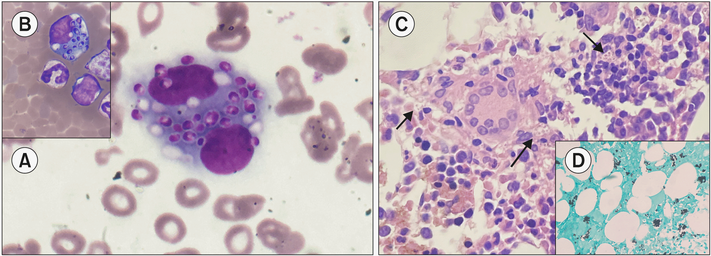
A 15-year-old female from Uttar Pradesh, India presented to pediatric emergency unit with bilateral joint pain, breathlessness, multiple necrotic skin lesions, and a six-month history of intermittent fever. On examination, she had multiple maculopapular lesions over her whole body and hepatosplenomegaly. Earlier investigations revealed pancytopenia. We considered a clinical diagnosis of lupus. Complete blood count showed hemoglobin 8.5 g/dL, total leucocyte count was 2,220/μL, platelets were 108,000/μL, and the absolute neutrophil count was 1,887/μL. The peripheral blood smear revealed occasional monocytic-macrophage lineage cells with engulfed yeast-like forms of Histoplasma capsulatum (A, MGG, ×100; B, ×40). Bone marrow biopsy showed increased plasma cells and histiocytes with many intracellular and extracellular forms of the fungus (arrow) (C, H&E stain, ×40). Occasional loose clusters of epithelioid histiocytes and foreign body giant cells were noted. Inset (D, ×40) shows fungus on the Gomori methenamine silver stain. The fungus was obtained from skin scraping. She was immediately started on amphotericin B and itraconazole. The immunological workup showed elevated IgG and a normal CD4:CD8 ratio. The workup for chronic granulomatous disease and HIV was negative. Despite dual antifungal therapy she succumbed to disseminated disease. Finding circulating fungal forms is a rare ominous event, especially in immunocompetent hosts.


 PDF
PDF Citation
 Print
Print


 XML Download