INTRODUCTION
Hemifacial microsomia (HFM) is a congenital anomaly of the facial structures, that leads to hypoplasia of the mandible, maxilla, zygoma, calvarial bone, external and middle ear, masticatory muscles, facial and trigeminal nerves, and overlying soft tissue, with a broad spectrum of phenotypic manifestations.
1-4 Most patients with HFM are treated using multi-disciplinary protocols to improve facial esthetics and rehabilitate the masticatory and respiratory functions.
5-14
A wide range of treatment modalities (Tx-Mods) exists for HFM based on the age of the patient and the severity of the phenotypic manifestation. Although unified consensus regarding the indications, optimal method, and optimal timing for the surgical treatment of HFM is still insufficient, the severity of mandibular deformity in HFM patients (
Figure 1) is one of the most important factors in determining the treatment method and predicting prognosis after treatment.
3,5,13-18
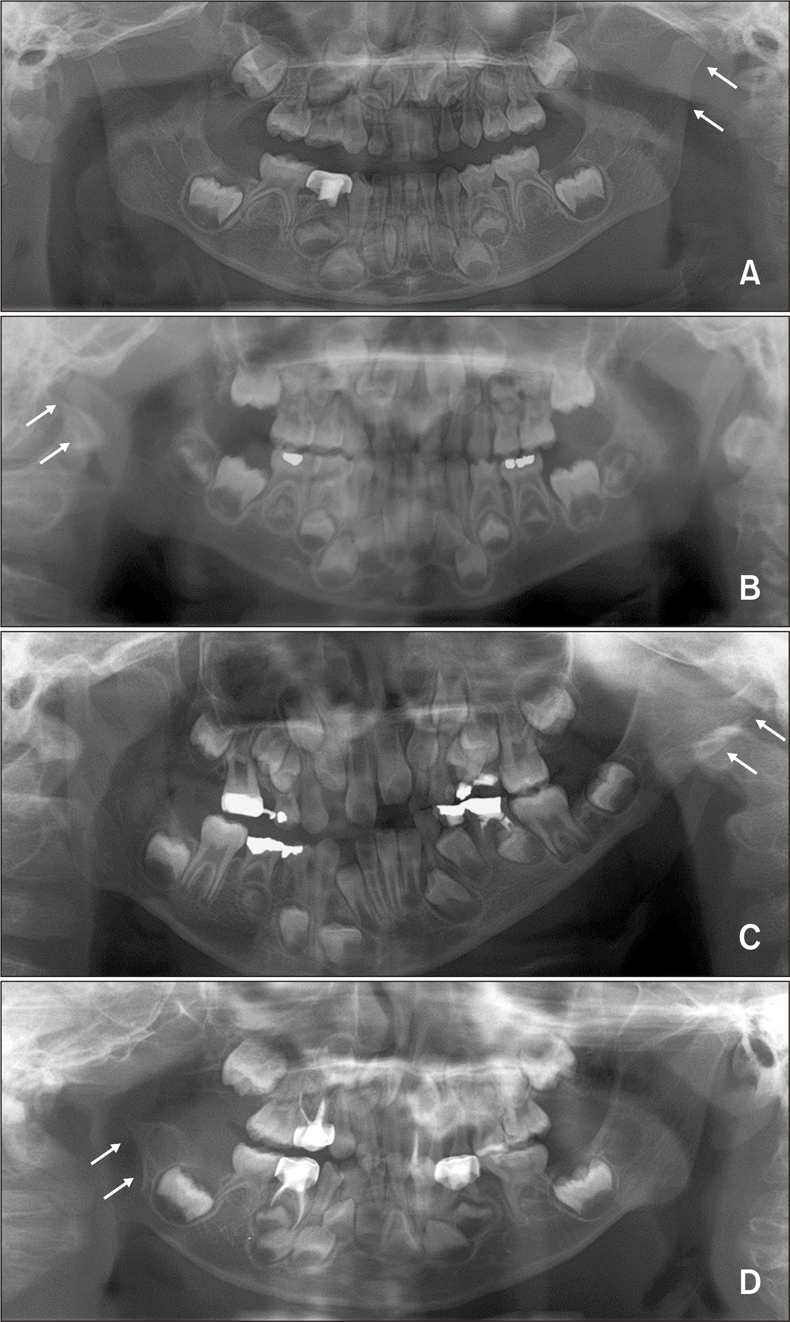 | Figure 1The Pruzansky–Kaban classification for hemifacial microsomia. A, Type I, the ramus/condyle complex has a normal shape but small size. B, Type IIa, the ramus/condyle complex is hypoplastic and abnormally shaped although the glenoid fossa is placed at the right position and the temporomandibular joint is functional. C, Type IIb, the glenoid fossa is placed at the inferiorly, medially, and anteriorly altered position with a severely hypoplastic ramus/condyle complex. D, Type III, complete absence of the ramus/condyle complex and the glenoid fossa. Arrow indicates the involvement side. 
|
There are two aspects to consider in surgical correction of HFM patients. First, the long-term outcome of surgical treatment is dependent on the severity of the mandibular deformity and facial asymmetry, rather than the type of surgical treatment (i.e., HFM patients with Pruzansky–Kaban type I and IIa exhibited stable results compared to those with Pruzansky–Kaban type IIb and III).
13,16 Second, surgical correction of moderate-to-severe facial asymmetry in HFM patients should be postponed till higher skeletal maturity is achieved, to reduce the number of surgical interventions and burden of care for HFM patients and their caregivers.
13,16,19,20
Pluijmers et al.,
18 in their retrospective chart review study, reported meaningful clinical data on the type and frequency of surgical interventions from three different major hospitals. However, their study did not consider the time-frame for which HFM patients were treated or followed-up under the same treatment protocol and the age of patients which would provide information about their growth stage.
18 Furthermore, there are no statistical studies on the type and frequency of orthodontic treatment for HFM patients. Therefore, the purpose of this retrospective study was to investigate the Tx-Mods for Korean patients with unilateral hemifacial microsomia (UHFM), including the type and frequency of orthodontic and surgical Tx-Mods, the number of patients who underwent surgical procedures, and the number of surgeries that each patient underwent, according to Pruzansky–Kaban types and growth stages, using strict criteria for the time period and the age of patients.
Go to :

MATERIALS AND METHODS
Subjects
The samples consisted of 82 Korean patients with UHFM (43 male and 39 female; 37 with right-sided involvement and 45 with left-sided involvement; mean age at the initial stage, 7.60 ± 4.68 years), who were treated or followed-up at the Multi-disciplinary Clinic, Department of Pediatric Plastic and Reconstructive Surgery of Seoul National University Children's Hospital and the Department of Oral and Maxillofacial Surgery and Department of Orthodontics of Seoul National University Dental Hospital (SNUDH) between 1998 and 2008.
The inclusion criteria were as follows: (1) patients who were born before 2005; (2) patients whose clinical chart, photographs, cephalograms, and orthopantomograms were available; and (3) patients whose skeletodental growth pattern could be evaluated using a longitudinal record. The initial stage referred to the time when the patient visited the clinic for the first time and the final stage indicated the time when the patient’s latest treatment was accomplished.
The patients were divided into four groups, based on the Pruzansky–Kaban classification (type I, n = 29; type IIa, n = 17; type IIb, n = 32; and type III, n = 4). This study was reviewed and approved by the Institutional Review Board of the SNUDH (ERI19043).
Types of treatment modalities
The orthodontic and surgical Tx-Mods for UHFM patients were classified into eight types according to the growth stage of patients and the degree of invasiveness and complexity of treatment modality: Tx-Mod-1, growth observation due to mild facial asymmetry; Tx-Mod-2, unilateral functional appliance treatment; Tx-Mod-3, fixed orthodontic treatment; Tx-Mod-4, growth observation due to a definite need for surgical intervention; Tx-Mod-5, unilateral mandibular or bimaxillary distraction osteogenesis (DO); Tx-Mod-6, fixation of the maxilla using LeFort I osteotomy for correcting the cant of the maxillary occlusal plane, and unilateral mandibular DO on the affected side and sagittal split ramus osteotomy (SSRO) on the unaffected side (Max-LeFort-I-Man-DO/SSRO);
10 Tx-Mod-7, orthognathic surgery including two-jaw surgery; and Tx-Mod-8, costochondral grafting with/without orthognathic surgery. If different Tx-Mods were consecutively used or combined at the final stage, the most invasive one was counted.
Tx-Mod-2 and Tx-Mod-3 were scored differently because of two reasons: First, Tx-Mod-2 is applied to patients before and during their adolescent period, while Tx-Mod-3 is applied to adolescent and adult patients. Second, Tx-Mod-2 is usually applied to Pruznasky–Kaban type I and IIa patients only; while some adult patients received Tx-Mod-3 for pre- and post-operative orthodontic treatment in case of two-jaw surgery.
Tx-Mod-6 and Tx-Mod-7 were scored differently because of two reasons: First, Tx-Mod-6 is applied to adolescent patients, while Tx-Mod-7 is applied to adult patients. Second, since fixation of the maxilla after Le Fort I osteotomy in Tx-Mod-6 is performed just after the pubertal growth peak, it can correct the cant of the maxillary occlusal plane as well as reduce the further growth restriction of the maxilla in the affected side. In addition, vertical lengthening of the affected side by mandibular DO can help maintain the corrected maxillary occlusal plane cant and correct the chin point deviation. By using this approach, clinicians can reduce the possibility of major operation to correct the facial asymmetry and severe maxillary occlusal plane cant. However, without proper management of the problems mentioned above in the adolescent period, two-jaw surgery might be challenging in terms of the amount of surgical movement, and the degree of correction of facial asymmetry and maxillary occlusal plane cant.
Variables
The type of treatment modality, the number of patients who underwent surgical procedures, and the number of surgical procedures that each patient underwent were investigated.
Statistical analysis
Non-parametric statistical analysis including Fisher’s exact test, Kruskal–Wallis test with Bonferroni correction, and mixed model analysis with Bonferroni method was adopted to overcome the unequal sample sizes between the groups using SPSS ver. 12.0 (SPSS Inc., Chicago, IL, USA). A p-value of less than 0.05 was considered statistically significant.
Go to :
RESULTS
Demographic data (Table 1)
There were no significant differences with respect to sex, side involvement, and mean age at the initial stage among the Pruzansky–Kaban type groups (all p > 0.05).
Table 1
Demographic data of unilateral hemifacial microsomia patients
|
Variable |
Total
(n = 82) |
Pruzansky–Kaban type |
p-value |
|
Type I
(n = 29) |
Type IIa
(n = 17) |
Type IIb
(n = 32) |
Type III
(n = 4) |
|
Distribution of sex (male:female) |
43:39 |
14:15 |
9:8 |
18:14 |
2:2 |
0.939*
|
|
Side involvement (right:left) |
37:45 |
14:15 |
9:8 |
12:20 |
2:2 |
0.719*
|
|
Mean age at the first consultation (yr) |
7.60 ± 4.68 |
7.11 ± 4.22 |
5.45 ± 2.72 |
8.63 ± 5.78 |
7.14 ± 1.81 |
0.138†
|

Distribution of the type of treatment modality Pruzansky–Kaban type I group (Table 2)
At the initial stage, growth observation (Tx-Mod-1 and Tx-Mod-4) was performed in 27.6% and 10.3% of the patients, respectively. At the final stage, growth observation (Tx-Mod-1) was performed in 41.3% of the patients.
The majority of type I patients was treated with a unilateral functional appliance at the initial stage (Tx-Mod-2, 55.2%), and with fixed orthodontic treatment and orthognathic surgery at the final stage (Tx-Mod-3, 34.5%; Tx-Mod-7, 20.7%).
Table 2
Distribution of the type of treatment modality in the Pruzansky–Kaban type I group
Previous
surgical
treatment
history |
Pruzansky–Kaban type I (n = 29) |
|
|
Tx-Mod |
|
|
Tx-Mod performed at the initial stage |
Tx-Mod performed at the final stage |
|
n = 0 (0%) |
Growth
observation |
Tx-Mod-1
Growth observation (n = 8, 27.6%) |
Tx-Mod-1
Growth observation (n = 7, 24.1%) |
|
|
|
Tx-Mod-3
Fixed-Ortho-Tx (n = 1, 3.4%) |
|
|
Tx-Mod-4
Growth observation (n = 3, 10.3%) |
Tx-Mod-7
Fixed-Ortho-Tx + two-jaw surgery (n = 3, 10.3%) |
|
Orthodontic
treatment |
Tx-Mod-2
Growth modification using unilateral
functional appliance (Unilat-Fx-App)
(n = 16, 55.2%) |
Tx-Mod-1
Growth observation (n = 5, 17.2%) |
|
|
|
Tx-Mod-3
Fixed-Ortho-Tx (n = 8, 27.6%) |
|
|
|
Tx-Mod-5
Fixed-Ortho-Tx + Unilat-Man-DO (n = 1, 3.4%) |
|
|
|
Tx-Mod-7
Fixed-Ortho-Tx + growth observation
+ two-jaw surgery (n = 2, 6.9%) |
|
|
Tx-Mod-3
Fixed orthodontic treatment using
brackets and wires (Fixed-Ortho-Tx)
(n = 1, 3.4%) |
Tx-Mod-3
Fixed-Ortho-Tx (n = 1, 3.4%) |
|
Surgical
treatment |
Tx-Mod-5
Unilateral mandibular distraction
osteogenesis (Unilat-Man-DO)
(n = 1, 3.4%) |
Tx-Mod-7
Fixed-Ortho-Tx + two-jaw surgery
(n = 1, 3.4%) |
Pruzansky–Kaban type IIa group (Table 3)
At the initial stage, growth observation (Tx-Mod-1 and Tx-Mod-4) was performed in 47.1% and 17.6% of the patients, respectively. At the final stage, growth observation (Tx-Mod-1) was performed in 47.1% of the patients.
At the initial stage, 35.3% of type IIa patients were treated with unilateral mandibular DO and unilateral functional appliance (Tx-Mod-5, 23.5%; Tx-Mod-2, 11.8%). However, at the final stage, 41.2% of the patients were treated with orthognathic surgery (Tx-Mod-7).
Table 3
Distribution of the type of treatment modality in the Pruzansky–Kaban type IIa group
|
Previous surgical treatment history |
Pruzansky–Kaban type IIa (n = 17) |
|
|
Tx-Mod |
|
|
Tx-Mod performed at the initial stage |
Tx-Mod performed at the final stage |
|
n = 0 (0%) |
Growth
observation |
Tx-Mod-1
Growth observation (n = 8, 47.1%) |
Tx-Mod-1
Growth observation (n = 8, 47.1%) |
|
|
Tx-Mod-4
Growth observation (n = 3, 17.6%) |
Tx-Mod-7
Fixed-Ortho-Tx + growth observation
+ two-jaw surgery (n = 3, 17.6%) |
|
Orthodontic
treatment |
Tx-Mod-2
Growth modification using
Unilat-Fx-App (n = 2, 11.8%) |
Tx-Mod-2
Growth modification using
Unilat-Fx-App (n = 1, 5.9%) |
|
|
|
Tx-Mod-7
Fixed-Ortho-Tx + Unilat-Man-DO + growth
observation + two-jaw surgery (n = 1, 5.9%) |
|
Surgical
treatment |
Tx-Mod-5
Unilat-Man-DO (n = 4, 23.5%) |
Tx-Mod-3
Growth observation + Fixed-Ortho-Tx
(n = 1, 5.9%) |
|
|
|
Tx-Mod-7
Growth observation + Fixed-Ortho-Tx +
two-jaw surgery (n = 3, 17.6%) |
Pruzansky–Kaban type IIb group (Table 4)
Among type IIb patients, 21.9% had a history of surgical treatment (5 patients with unilateral mandibular DO, 1 patient with unilateral maxillary DO, and 1 patient with Max-LeFort-I-Man-DO/SSRO).
At the initial stage, growth observation (Tx-Mod-1 and Tx-Mod-4) was performed in 12.5% and 28.1% of the patients, respectively. At the final stage, growth observation (Tx-Mod-1) was performed in 12.5% of the patients.
At the initial stage, 50.1% of type IIb patients were treated with orthodontic treatment (fixed orthodontic treatment, Tx-Mod-3, 31.3% and unilateral functional appliance treatment, Tx-Mod-2, 18.8%). However, surgical treatment including unilateral mandibular or bimaxillary DO (Tx-Mod-5, 6.2%) and Max-LeFort-I-Man-DO/bilateral SSRO (BSSRO) (Tx-Mod-6, 3.1%) was performed in only 9.3% of the patients.
At the final stage, surgical treatment was the most frequently used treatment modality (84.4%) including orthognathic surgery (Tx-Mod-7, 59.4%), Max-LeFort-I-Man-DO/BSSRO (Tx-Mod-6, 12.5%), unilateral mandibular DO (Tx-Mod-5, 9.4%), and costochondral grafting with/without orthognathic surgery (Tx-Mod-8, 3.1%). However, orthodontic treatment (Tx-Mod-3) was used in only 3.1% of the patients.
Table 4
Distribution of the type of treatment modality in the Pruzansky–Kaban type IIb group
Previous surgical
treatment history
(n = 7 of 32, 21.9%) |
Pruzansky–Kaban type IIb (n = 32) |
|
|
Tx-Mod |
|
|
Tx-Mod performed at the initial stage |
Tx-Mod performed at the final stage |
Unilateral mandibular DO
(n = 5, 71.4%)
Unilateral maxillary DO
(n = 1, 14.3%)
Max-LeFort-I-Man-DO/
BSSRO (n = 1, 14.3%) |
Growth
observation |
Tx-Mod-1
Growth observation
(n = 4, 12.5%) |
Tx-Mod-1
Growth observation (n = 4, 12.5%) |
|
|
Tx-Mod-4
Growth observation
(n = 9, 28.1%) |
Tx-Mod-3
Fixed-Ortho-Tx (n = 1, 3.1%) |
|
|
|
Tx-Mod-5
Unilat-Man-DO + Fixed-Ortho-Tx (n = 1, 3.1%) |
|
|
|
Tx-Mod-6
Max-LeFort-I-Man-DO/BSSRO
+ Fixed-Ortho-Tx (n = 2, 6.3%) |
|
|
|
Tx-Mod-7
Fixed-Ortho-Tx + two-jaw surgery (n = 5, 15.6%) |
|
Orthodontic
treatment |
Tx-Mod-2
Growth modification using
Unilat-Fx-App (n = 6, 18.8%) |
Tx-Mod-5
Fixed-Ortho-Tx + Unilat-Man-DO (n = 1, 3.1%) |
|
|
|
Tx-Mod-6
Fixed-Ortho-Tx + Unilat-Man-DO + growth
observation + Max-LeFort-I-Man-DO/BSSRO
(n = 1, 3.1%) |
|
|
|
Tx-Mod-7
Fixed-Ortho-Tx + Unilat-Bimax-DO + growth
observation + two-jaw surgery (n = 1, 3.1%) |
|
|
|
Tx-Mod-7
Fixed-Ortho-Tx + growth observation
+ two-jaw surgery (n = 3, 9.4%) |
|
|
Tx-Mod-3
Fixed-Ortho-Tx
(n = 10, 31.3%) |
Tx-Mod-5
Unilat-Man-DO (n = 1, 3.1%) |
|
|
|
Tx-Mod-6
Max-LeFort-I-Man-DO/BSSRO (n = 1, 3.1%) |
|
|
|
Tx-Mod-7
Unilat-Man-DO + growth observation
+ two-jaw surgery (n = 1, 3.1%) |
|
|
|
Tx-Mod-7
Two-jaw surgery (n = 6, 18.8%) |
|
|
|
Tx-Mod-8
Unilat-Man-DO + growth observation + two-jaw
surgery + costochondral graft (n = 1, 3.1%) |
|
Surgical
treatment |
Tx-Mod-5
Unilat-Man-DO (n = 1, 3.1%) |
Tx-Mod-7
Growth observation + Fixed-Ortho-Tx
+ two-jaw surgery (n = 3, 9.4%) |
|
|
Tx-Mod-5
Unilateral bimaxillary DO
(Unilat-Bimax-DO) (n = 1, 3.1%) |
|
|
|
Tx-Mod-6
Max-LeFort-I-Man-DO/BSSRO
(n = 1, 3.1%) |
|
Pruzansky–Kaban type III group (Table 5)
Half of type III patients had a history of surgical treatment (1 patient with unilateral mandibular DO and 1 patient with costochondral grafting).
At the initial stage, only growth observation (Tx-Mod-4, 100%) was used. At the final stage, only costochondral grafting with/without orthognathic surgery was used (Tx-Mod-8, 100%).
Table 5
Distribution of the type of treatment modality in the Pruzansky–Kaban type III group
Previous surgical
treatment history
(n = 2 of 4, 50%) |
Pruzansky–Kaban type III (n = 4) |
|
|
Tx-Mod |
|
|
Tx-Mod performed at the initial stage |
Tx-Mod performed at the final stage |
Unilateral mandibular DO
(n = 1, 50%)
Costochondral graft
(n = 1, 50%) |
Tx-Mod-4
Growth observation (n = 4, 100%) |
Tx-Mod-8
Two-jaw surgery + costochondral grafting (n = 1, 25%) |
|
|
Tx-Mod-8
Fixed-Ortho-Tx + costochondral grafting
+ growth observation + two-jaw surgery (n = 1, 25%) |
|
|
Tx-Mod-8
Fixed-Ortho-Tx + costochondral grafting
+ growth observation (n = 1, 25%) |
|
|
Tx-Mod-8
Fixed-Ortho-Tx + two-jaw surgery
+ costochondral grafting (n = 1, 25%) |
Comparison of the treatment modalities according to the Pruzansky–Kaban type and treatment stage (Table 6)
The more invasive and complex Tx-Mods were used for Pruzansky–Kaban types IIb and III compared to those for Pruzansky–Kaban types I and IIa ([I, IIa] < [IIb, III], p < 0.001). When the initial and final stages were compared to determine if the patients had undergone different Tx-Mods, the degree of invasiveness and complexity in the treatment modality at the final stage was higher than that at the initial stage (initial < final, p < 0.001).
Table 6
Comparison of the degree of invasiveness of treatment modalities according to the Pruzansky–Kaban type and treatment stage
|
Pruzansky–Kaban type |
Treatment modality |
Pruzansky–Kaban type |
Treatment stage |
|
|
Initial stage |
Final stage |
|
I (n = 29) |
2.07 ± 1.03 |
3.07 ± 2.30 |
< 0.001***
(I, IIa) < (IIb, III) |
< 0.001***
Initial < final |
|
IIa (n = 17) |
2.59 ± 1.77 |
3.65 ± 2.94 |
|
IIb (n = 32) |
2.97 ± 1.10 |
5.84 ± 2.08 |
|
III (n = 4) |
4.00 ± 0.00 |
8.00 ± 0.00 |
Comparison of the number of patients who underwent surgical procedures (Table 7)
In total, 56.1% of the patients (n = 46 of 82) had surgical treatment (Tx-Mod-5, 6, 7, and 8) during the treatment period. The number of patients who had surgical procedures increased up to 4.2 times, with an increase in the severity of HFM (type I, n = 7 of 29 [24.1%]; type IIa, n = 8 of 17 [47.1%]; type IIb, n = 27 of 32 [84.4%]; type III, n = 4 of 4 [100%]; p < 0.001; Exp(B) = 4.242).
Table 7
Comparison of the number of patients who underwent surgical procedures among the Pruzansky–Kaban type groups
|
Pruzansky–Kaban type |
Patients who underwent surgical procedures |
|
|
Number |
Percentage |
Exp(B) |
p-value |
|
I |
7/29 |
24.1 |
4.242 |
< 0.001***
|
|
IIa |
8/17 |
47.1 |
|
IIb |
27/32 |
84.4 |
|
III |
4/4 |
100.0 |
Comparison of the mean number of surgeries that each patient underwent and the number of patients who underwent multiple surgical procedures (Table 8)
Despite a tendency of increase in the mean number of surgical procedures from type I to type III, there was no statistically significant difference among the Pruzansky–Kaban types (type I, n = 1.1; type IIa, n = 1.5; type IIb, n = 1.6; and type III, n = 2.3; p > 0.05).
No statistically significant difference was observed in the number of patients who had multiple surgical procedures among the Pruzansky–Kaban types, although type I patients showed a lower percentage compared to the other types (type I, n = 1 [14.3%]; type IIa, n = 4 [50.0%]; type IIb, n = 12 [44.4%]; and type III, n = 3 [75.0%]; p > 0.05).
Table 8
Comparison of the number of surgeries that each patient underwent and the number of patients who underwent multiple surgical procedures among the Pruzansky–Kaban type groups
|
Pruzansky–Kaban type |
Number of surgical procedures in patients who underwent surgery |
|
Number of patients who underwent multiple surgical procedures |
|
|
|
Mean |
SD |
p-value |
Number |
Percentage |
p-value |
|
I (n = 7) |
1.14 |
0.38 |
0.154*
|
|
1/7 |
14.3 |
0.267†
|
|
IIa (n = 8) |
1.50 |
0.54 |
|
4/8 |
50.0 |
|
IIb (n = 27) |
1.63 |
0.88 |
|
12/27 |
44.4 |
|
III (n = 4) |
2.25 |
0.96 |
|
3/4 |
75.0 |
Go to :
DISCUSSION
Demographic data
There were no significant differences in the distribution of sex and side involvement among the Pruzansky–Kaban type groups (
Table 1). These findings were similar to the results of previous studies, despite the difference in geographical and ethnic factors.
19,21,22
Distribution of the type of treatment modality Pruzansky–Kaban type I group (Table 2)
The finding that growth observation (Tx-Mod-1) was performed in 27.6% of the patients at the initial stage and 41.3% of the patients at the final stage indicates that some of the parents did not demand any type of treatment, when a type I patient presented mild facial asymmetry.
At the initial stage, the majority of type I patients were treated with a unilateral functional appliance (Tx-Mod-2, 55.2%), which implies that parents wanted a conservative treatment modality to correct mild facial asymmetry. Then, at the final stage, 34.5% of the patients were treated with fixed orthodontic treatment (Tx-Mod-3), while 20.7% desired correction of the residual facial asymmetry with orthognathic surgery (Tx-Mod-7).
Pruzansky–Kaban type IIa group (Table 3)
Growth observation (Tx-Mod-1) was performed in 47.1% of the patients at both initial and final stages, respectively. These findings indicate that clinicians wanted to observe the growth pattern of type IIa patients with mild-to-moderate facial asymmetry before deciding the treatment modality.
At the initial stage, 35.3% of type IIa patients were treated with unilateral mandibular DO and unilateral functional appliance (Tx-Mod-5, 23.5%; Tx-Mod-2, 11.8%), which implies that clinicians wanted to correct facial asymmetry with unilateral mandibular DO or unilateral functional appliance. However, at the final stage, 41.2% of the patients had a more invasive treatment modality, such as orthognathic surgery (Tx-Mod-7).
Pruzansky–Kaban type IIb group (Table 4)
At the initial stage, the frequency of Tx-Mod-4 growth observation was two times higher than that of Tx-Mod-1 growth observation (28.1% vs. 12.5%). This finding indicates that clinicians wanted to wait till the skeletal age was matured or facial growth was completed before deciding a specific surgical treatment modality for some of the type IIb patients with moderate-to-severe facial asymmetry (28.1%).
At the initial stage, half of the type IIb patients (50.1%) received orthodontic treatment (Tx-Mod-3, 31.3%; Tx-Mod-2, 18.8%), which indicates that clinicians wanted to treat the patient’s malocclusion with orthodontic treatment approach and then observe the facial growth due to the unfavorable prognosis of surgical treatment.
At the final stage, surgical treatment was the most common treatment modality for type IIb patients (84.4%) (Tx-Mod-7, 59.4%; Tx-Mod-6, 12.5%; Tx-Mod-5, 9.4%; and Tx-Mod-8, 3.1%), which indicates that clinicians wanted to treat the patient’s skeletal problem and facial asymmetry with surgery after the facial growth was completed.
Pruzansky–Kaban type III group (Table 5)
At the initial stage, growth observation (Tx-Mod-4) was performed in all of the patients, which indicates that since type III patients had severe facial asymmetry because the ramus/condyle complex was absent on the affected side, clinicians did not want to perform any type of treatment before completion of the facial growth.
At the final stage, all type III patients were treated with the most invasive treatment modality (costochondral grafting with/without orthognathic surgery, Tx-Mod-8) to reconstruct the ramus/condyle complex of the mandible on the affected side and to correct the maxillary cant and facial asymmetry.
Comparison of the degree of invasiveness of treatment modalities according to the Pruzansky–Kaban type and treatment stage (Table 6)
In the present study, the patients with Pruzansky–Kaban types IIb and III were treated with more invasive and complex Tx-Mods, compared to those with Pruzansky–Kaban types I and IIa ([I, IIa] < [IIb, III], p < 0.001). This finding indicates that clinicians wanted to treat the patient’s skeletodental problem and facial asymmetry with orthodontic treatment approach in mild-to-moderate cases and with surgery in moderate-to-severe cases.
The finding that the degree of invasiveness and complexity in the treatment modality at the final stage was higher than that of the initial stage (initial < final, p < 0.001) implies that clinicians wanted to treat the patient’s malocclusion using orthodontic treatment approach before and during pubertal growth and correct the patient’s skeletal problem and facial asymmetry with surgery after completion of facial growth to avoid unnecessary multiple surgical procedures.
Comparison of the number of patients who underwent surgical procedures (Table 7)
In the present study, 56.1% of the patients (n = 46/82) underwent surgical treatment (Tx-Mod-5, 6, 7, and 8) during the treatment period. This finding was similar to the results of Pluijmers et al.,
18 which reported that 42.7% and 16.5% of the patients underwent surgery for the mandible and maxilla, respectively.
When the Pruzansky–Kaban type worsened from type I to type III, the number of patients who underwent surgical procedures was increased up to 4.2 times (
p < 0.001). This finding was similar to the results of systematic analysis conducted by van de Lande et al.
17
Comparison of the mean number of surgical procedures that each patient underwent (Table 8)
The finding that the mean number of surgical procedures that each patient underwent increased from the type I group to the type III group, despite the lack of statistical significance (type I, n = 1.1; type IIa, n = 1.5; type IIb, n = 1.6; and type III, n = 2.3) was similar to that of Pluijmers et al.
18 (type I, n = 1.0; type IIa, n = 1.4; type IIb, n = 1.8; and type III, n = 2.3).
Comparison of the number of patients who underwent multiple surgical procedures (Table 8)
The number of patients who underwent multiple surgical procedures was higher in the type IIa, IIb, and III groups compared to the type I group, despite the lack of statistical significance (type I, n = 1 [14.3%]; type IIa, n = 4 [50.0%]; type IIb, n = 12 [44.4%]; and type III, n = 3 [75.0%]). This result was similar to that of Pluijmers et al.,
18 which observed that 49.1% and 61% of Pruzansky–Kaban type IIb and III patients needed multiple operations, respectively.
The main objective of this study was to investigate the Tx-Mods for patients with UHFM according to its invasiveness and complexity by using longitudinal data. However, there are several suggestions for future studies in order to establish sophisticated study designs and obtain comparable outcomes. First, it is necessary to develop a uniform registration and outcome measurement tool to compare the results between different Tx-Mods.
18 Second, a nationwide multi-center prospective study and systematic statistical analyses should be performed. Third, it is necessary to investigate the type and frequency of orthodontic and surgical Tx-Mods for bilateral HFM patients, which requires a greater number of multiple surgical procedures compared to UHFM patients.
Go to :


 PDF
PDF Citation
 Print
Print


 XML Download