

 PDF
PDF Citation
 Print
Print


INTRODUCTION
MATERIALS AND METHODS
Subjects
Obtainment of three-dimensional cone-beam computed tomography
Reorientation of CBCT images (Figure 1)
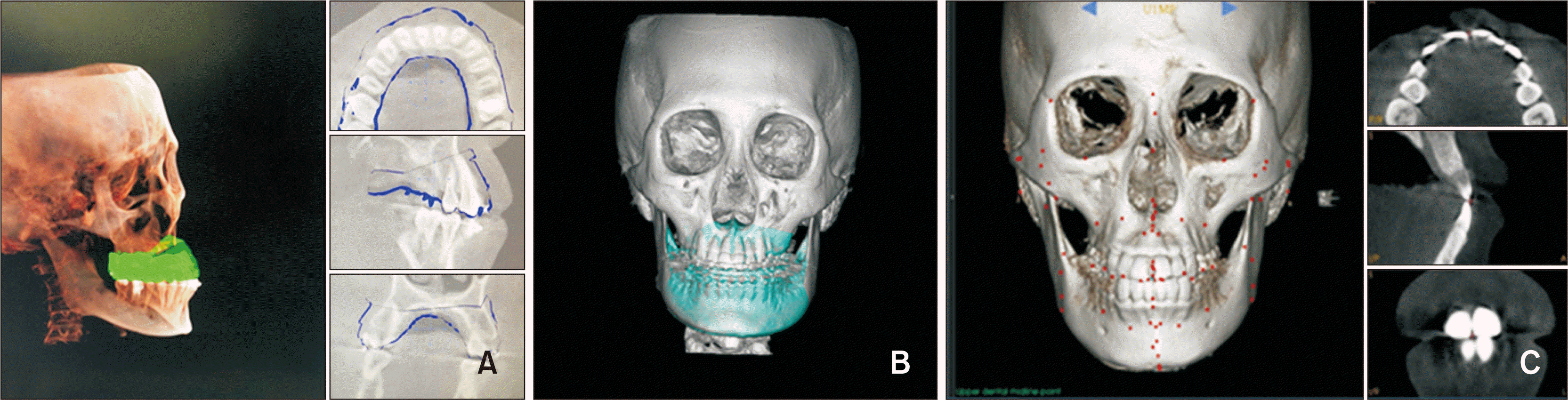 | Figure 1The process of merging between the scanned dentition and the three-dimensional cone-beam computed tomography (3D-CBCT) image and landmark digitization. A, Merging of the scanned dentition with 3D-CBCT image using three sectional views of the sagittal, frontal, and axial planes. B, Result of fine-tuned merging between dentition and 3D-CBCT images. C, Landmark digitization to identify the midpoint between the maxillary central incisors at the occlusal level (U1MP) and the mesiobuccal cusp tips (MBCTs) of the maxillary right and left first molar crowns using the three sectional views of the sagittal, frontal, and axial planes. The mean coordinate value from the two digitized MBCTs was automatically calculated by the ON3D software (3D ONS Inc., Seoul, Korea).
|
Merging the T0 scanned dentition data onto the T0 three-dimensional volume image (Figure 1)
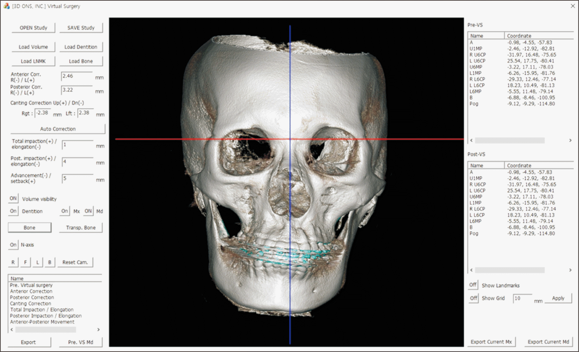
Virtual surgical simulation
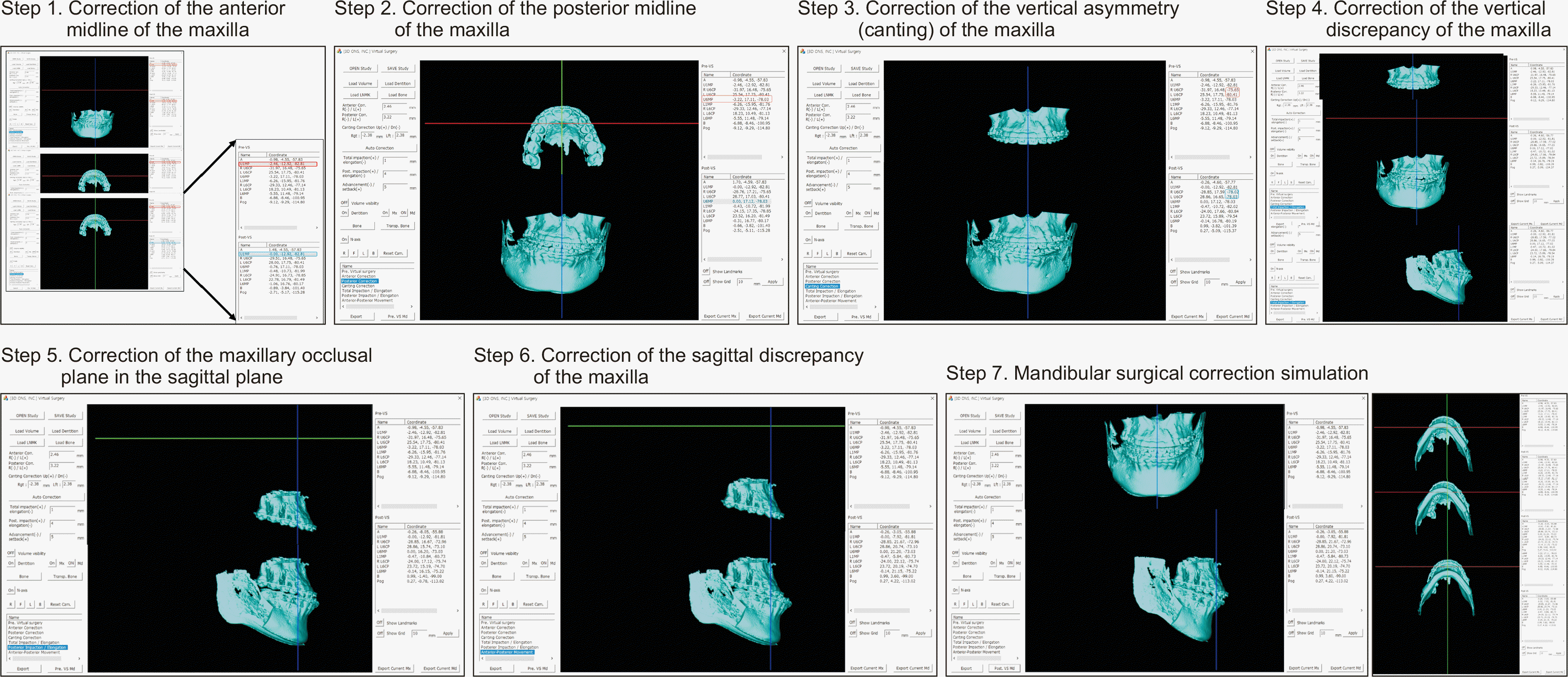 | Figure 3The step-by-step procedure of virtual surgical simulation. Step 1, Correction of the anterior midline of the maxilla. Step 2, Correction of the posterior midline of the maxilla. Step 3, Correction of the vertical asymmetry (canting) of the maxilla. Step 4, Correction of the vertical discrepancy of the maxilla. Step 5, Correction of the maxillary occlusal plane in the sagittal plane. Step 6, Correction of the sagittal discrepancy of the maxilla. Step 7, Correction of the mandible.
|
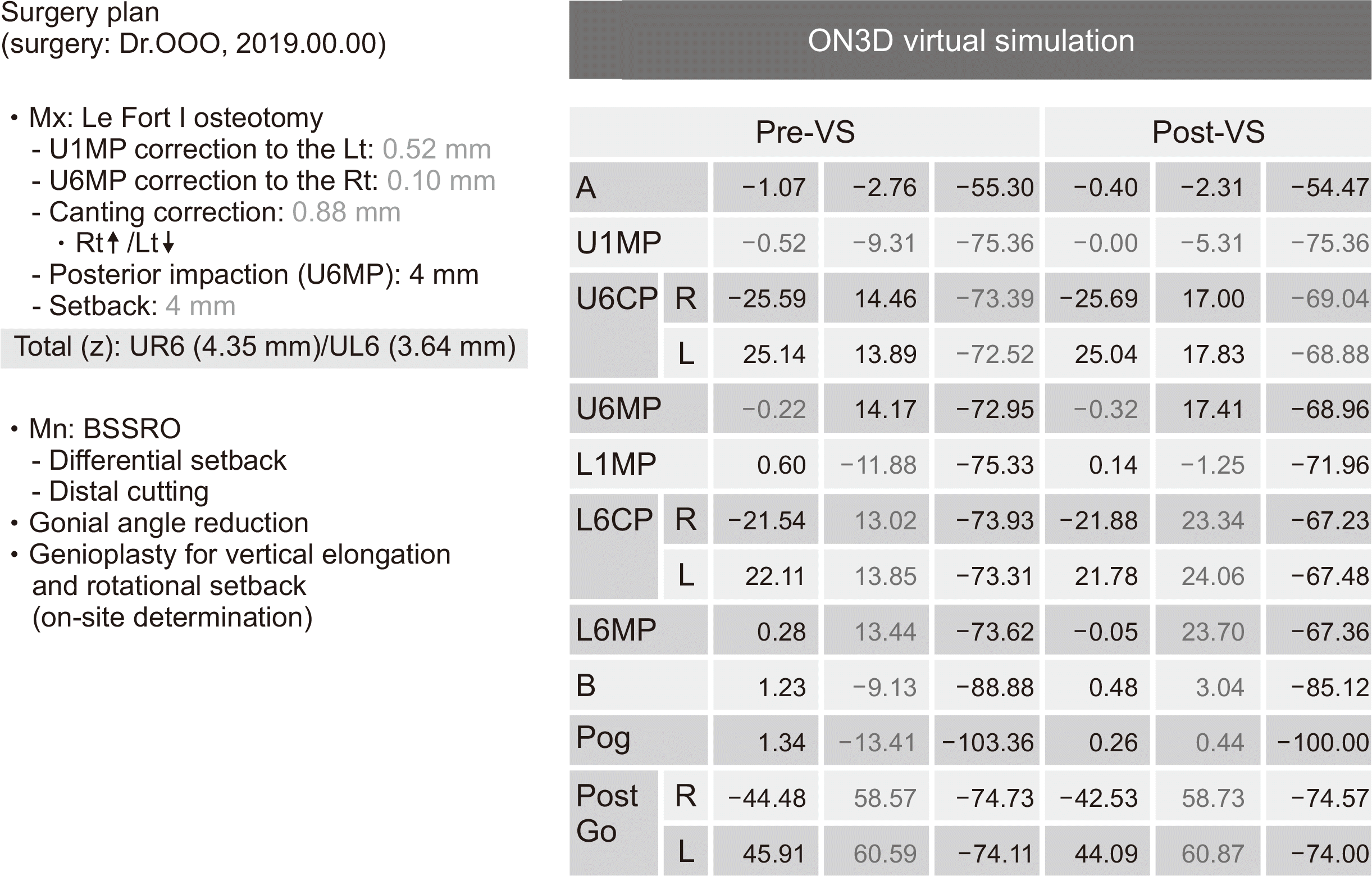 | Figure 4An example of surgical plan and coordinate values of the selected landmarks at the pre- and post-virtual surgery (VS).
Mx, maxilla; Mn, mandible; Lt, left; Rt, right; BSSRO, bilateral sagittal split ramus osteotomy; U1MP, the midpoint between the maxillary central incisors at the occlusal level; U6MP, the midpoint between the two mesiobuccal cusp tips of the maxillary right and left first molar crowns; UR6, the mesiobuccal cusp tip of the maxillary right first molar crown; UL6, the mesiobuccal cusp tip of the maxillary left first molar crown; U6CP, the mesiobuccal cusp tip of the maxillary first molar crown; L1MP, the midpoint between the mandibular central incisors at the occlusal level; L6CP, the mesiobuccal cusp tip of the mandibular first molar crown; L6MP, the midpoint between the two mesiobuccal cusp tips of the mandibular right and left first molar crowns; A, A point; B, B point; Pog, pogonion; Go, gonion.
|
Three-dimensional printing of surgical splints
Landmarks
Validation of landmark identification
Types of surgical movements of the maxilla
Measurement variables representing surgical accuracy
Statistical analysis
RESULTS
Cephalometric characteristics of the samples (Table 1)
Table 1
| Cephalometric variable | T0 stage | T1 stage | p-value | |||
|---|---|---|---|---|---|---|
|
|
|
|||||
| Mean | SD | Mean | SD | |||
| SNA (°) | 82.3 | 4.4 | 83.2 | 3.5 | 0.246 | |
| SNB (°) | 84.3 | 4.2 | 79.8 | 3.8 | 0.000*** | |
| ANB (°) | −2.0 | 2.5 | 3.4 | 1.0 | 0.000*** | |
| A-N perp (mm) | −0.3 | 4.7 | 1.3 | 2.9 | 0.073 | |
| Pog-N perp (mm) | 5.4 | 8.7 | –0.9 | 6.0 | 0.001*** | |
| FMA (°) | 25.8 | 7.8 | 29.6 | 5.3 | 0.005** | |
| U1 to FH (°) | 112.9 | 6.6 | 106.4 | 4.2 | 0.001** | |
| IMPA (°) | 85.4 | 6.7 | 83.1 | 7.3 | 0.110 | |
The samples consisted of 15 Korean young adult patients with skeletal Class III malocclusion (9 males and 6 females; mean age, 22.3 years).
SNA, Sella-nasion-point A angle; SNB, sella-nasion-point B angle; ANB, point A-nasion-poing B angle; A-N perp, point A-nasion perpendicular line; Pog-N perp, pogonion-nasion perpendicular line; FMA, Frankfort horizontal plane to mandibular plane angle; U1 to FH, maxillary incisor inclination to Frankfort horizontal plane; IMPA, incisor mandibular plane angle; SD, standard deviation.
![]()
Surgical movement types of the maxilla (Table 2)
Table 2
| Surgical movementtypes of the maxilla | Number of patients who underwent the specific type of surgical movement (%) | Virtually planned movement(mm) | Actual movement(mm) | Discrepancy between virtually plannedand actual movement† (mm) | |||||||||
|---|---|---|---|---|---|---|---|---|---|---|---|---|---|
|
|
|
|
|||||||||||
| Mean | SD | Range | Mean | SD | Range | Mean | SD | Range | p-value‡ | ||||
| Transverse | |||||||||||||
| Anterior midline correction | 14/15 (93.3) | 1.38 | 0.99 | 0.13–3.00 | 1.45 | 1.37 | −0.78–4.13 | −0.07 | 0.86 | −1.24–1.15 | |||
| Posterior midline correction | 13/15 (86.7) | 1.49 | 1.00 | 0.50–3.22 | 1.53 | 1.51 | −1.08–4.16 | −0.04 | 0.91 | −1.23–1.76 | |||
| Sagittal | |||||||||||||
| Maxillary advancement | 3/15 (20.0) | 4.33 | 1.53 | 3.00–6.00 | 3.10 | 2.20 | 0.68–4.98 | 1.24 | 0.99 | 0.37–2.32 | |||
| Maxillary setback | 10/15 (66.7) | 2.80 | 1.55 | 1.00–5.00 | 2.66 | 1.69 | −0.61–5.50 | 0.14 | 0.78 | −0.75–1.61 | 0.023* | ||
| Vertical | |||||||||||||
| Maxillary total impaction | 5/15 (33.3) | 1.20 | 0.45 | 0.99–2.00 | 0.75 | 0.76 | −0.37–1.55 | 0.44 | 0.73 | −0.55–1.37 | |||
| Maxillary anterior elongation | 6/15 (40.0) | 2.08 | 1.11 | 1.00–4.00 | 2.94 | 1.42 | 1.08–4.34 | −0.85 | 0.77 | −2.15–−0.08 | |||
| Maxillary posterior impaction | 12/15 (80.0) | 2.38 | 1.23 | 1.00–5.00 | 1.73 | 1.54 | −1.39–4.45 | 0.65 | 0.79 | −0.65–2.39 | |||
| Total number of the specific type of surgical movement | 63 | 0.13 | 0.92 | −2.15–2.39 | |||||||||
Anterior midline correction, maxillary advancement, maxillary setback, maxillary total impaction, and maxillary anterior elongation were evaluated using U1MP, while posterior midline correction and maxillary posterior impaction were assessed using U6MP.
SD, Standard deviation; U1MP, the midpoint between maxillary central incisors at the occlusal level; U6MP, the midpoint between two mesiobuccal cusp tips of maxillary right and left first molar crowns.
Post-hoc analysis after Kruskal–Wallis test showed a significant difference between the mean discrepancy values of maxillary anterior elongation and maxillary posterior impaction (–0.85 mm [overcorrection] vs. 0.65 mm [undercorrection], *p < 0.05).
![]()
Comparison of surgical accuracy among the surgical movement types of the maxilla surgical accuracy in the overall dimensions (Tables 2 and 3)
Table 3
| Surgical movementtypes of the maxilla | Surgical achievement percentage (%) | Absolute difference value | p-value† | ||||||||
|---|---|---|---|---|---|---|---|---|---|---|---|
|
|
|
||||||||||
| Mean | SD | Range | p-value* | ≤ 2.00 mm | ≤ 1.00 mm(precision percentage‡) | ||||||
|
|
|
||||||||||
| Number of patient | Percentage | Number of patient | Percentage | ||||||||
| Transverse | |||||||||||
| Anterior midline correction | 103.4 | 149.3 | −260.0–400.0 | 14/14 | 100.0 | 9/14 | 64.3 | ||||
| Posterior midline correction | 95.8 | 112.5 | −158.8–305.0 | 13/13 | 100.0 | 9/13 | 69.2 | ||||
| Sagittal | |||||||||||
| Maxillary advancement | 65.5 | 37.3 | 22.7–90.8 | 2/3 | 66.7 | 1/3 | 33.3 | ||||
| Maxillary setback | 89.8 | 56.4 | −61.0–132.0 | 0.066 | 10/10 | 100.0 | 8/10 | 80.0 | 0.717 | ||
| Vertical | |||||||||||
| Maxillary total impaction | 63.1 | 70.7 | −37.0–155.0 | 5/5 | 100.0 | 4/5 | 80.0 | ||||
| Maxillary anterior elongation | 142.6 | 36.8 | 108.0–207.5 | 5/6 | 83.3 | 3/6 | 50.0 | ||||
| Maxillary posterior impaction | 58.9 | 68.3 | −139.0–132.5 | 11/12 | 91.7 | 9/12 | 75.0 | ||||
| Total (n=63) | 89.9 | 97.4 | −260.0–400.0 | 60/63 | 95.2 | 43/63 | 68.3 | ||||
Surgical achievement percentage, (amount of movement in actual surgery/amount of movement in virtual surgery) × 100 (%); Precision percentage, [number of precise samples (≤ 1.00 mm)/number of samples] × 100 (%).
Anterior midline correction, maxillary advancement, maxillary setback, maxillary total impaction, and maxillary anterior elongation were evaluated using U1MP, while posterior midline correction and maxillary posterior impaction were assessed using U6MP.
SD, Standard deviation; U1MP, the midpoint between maxillary central incisors at the occlusal level; U6MP, the midpoint between two mesiobuccal cusp tips of maxillary right and left first molar crowns; T1, post-surgery.
‡A discrepancy of less than 1 mm between virtual surgical simulation and T1 landmarks was considered as a precise outcome. Adapted from the article of Choi et al. (Angle Orthod 2009;79:306-311).4
![]()
 XML Download