Journal List > Korean J Orthod > v.50(4) > 1145701

Coban, Yavuz, Karadas, and Demirbas: Three-dimensional assessment of nasal changes after maxillary advancement with impaction using stereophotogrammetry

Abstract

Objective

To evaluate the changes in the nose in three dimensions after Le Fort I osteotomy in patients with skeletal Class III malocclusion.

Methods

The subjects were 40 adult patients (20 females and 20 males; mean age, 20.3 ± 3.0 years; range, 17.0 to 31.1 years) who underwent one-piece Le Fort I osteotomy with maxillary advancement and impaction treatment for maxillary hypoplasia. The mean maxillary advancement was 4.56 ± 1.34 mm, and the mean maxillary impaction was 2.03 ± 1.04 mm. Stereophotogrammetry was used to acquire three-dimensional images before and at least 6 months after surgery.

Results

Alare (Al) and alare curvature (Ac) points had moved vertically and antero-laterally postoperatively. A significant increase was observed in the nasal ala width and alar base width, and no changes were noted in the columellar length, nasolabial angle, and nasal area. There was a significant relationship between maxillary impaction and nasal ala width and horizontal and sagittal positions of the bilateral Al and Ac. The only relationship found was between maxillary advancement and postoperative sagittal location of the subnasale and pronasale.

Conclusions

Nasal soft tissues were highly affected by the vertical movement of the maxilla; however, the soft tissue responses were individual-dependent.

INTRODUCTION

The major goals of orthognathic surgery treatment are as follows: formation of facial esthetics and functional occlusion, improvement and/or maintenance of airway volume and periodontal health, healthy temporomandibular joints, and elimination of the patient's major complaint.1 For determination of facial aesthetics, the nose is a very important structure as the center of the face,2 and all surgical interventions on the midface affect the nose. Undesirable changes in the appearance of the nose were the most important cause of patient dissatisfaction in maxillary orthognathic surgery.3 Therefore, careful preoperative examinations of the nose and implementation of a treatment plan based on these findings will yield effective results.
Previous articles have used two-dimensional (2D) methods, such as radiographs and photographs, to show the aesthetic proportion of the profile and front of the face.4 However, since the human body is three-dimensional (3D), face depth and shape cannot be accurately evaluated using 2D measures. Recent developments in technology have produced a variety of 3D techniques, such as 3D computed tomography,5 laser scanning,6 3D facial morphometry,7 and stereophotogrammetry,5,8 to capture facial topography and overcome the disadvantages of traditional photographic and radiographic methods. Stereophotogrammetry is a fast, accurate, highly reproducible, and non-invasive 3D digital camera system that can easily yield not only linear but also topographic, angular, and areal measurements in 3D.5,8
The traditional belief is that maxillary advancement often leads to shortening of the nasal ridge and an increase in the nasolabial angle due to nasal tip elevation,9 and maxillary impaction with10 or without maxillary advancement11 often leads to widening of the nose. This may cause an unpleasant appearance in patients with a wide nose preoperatively.
Many studies have been conducted with 3D digital imaging systems to examine changes in the nose after orthognathic surgery in relation to maxillary surgery and the nasal tip, nasolabial angle, alar base width, and coordinate pronasale changes.
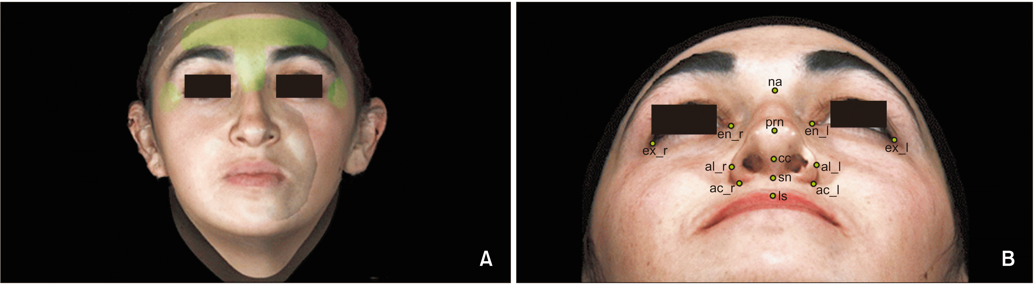
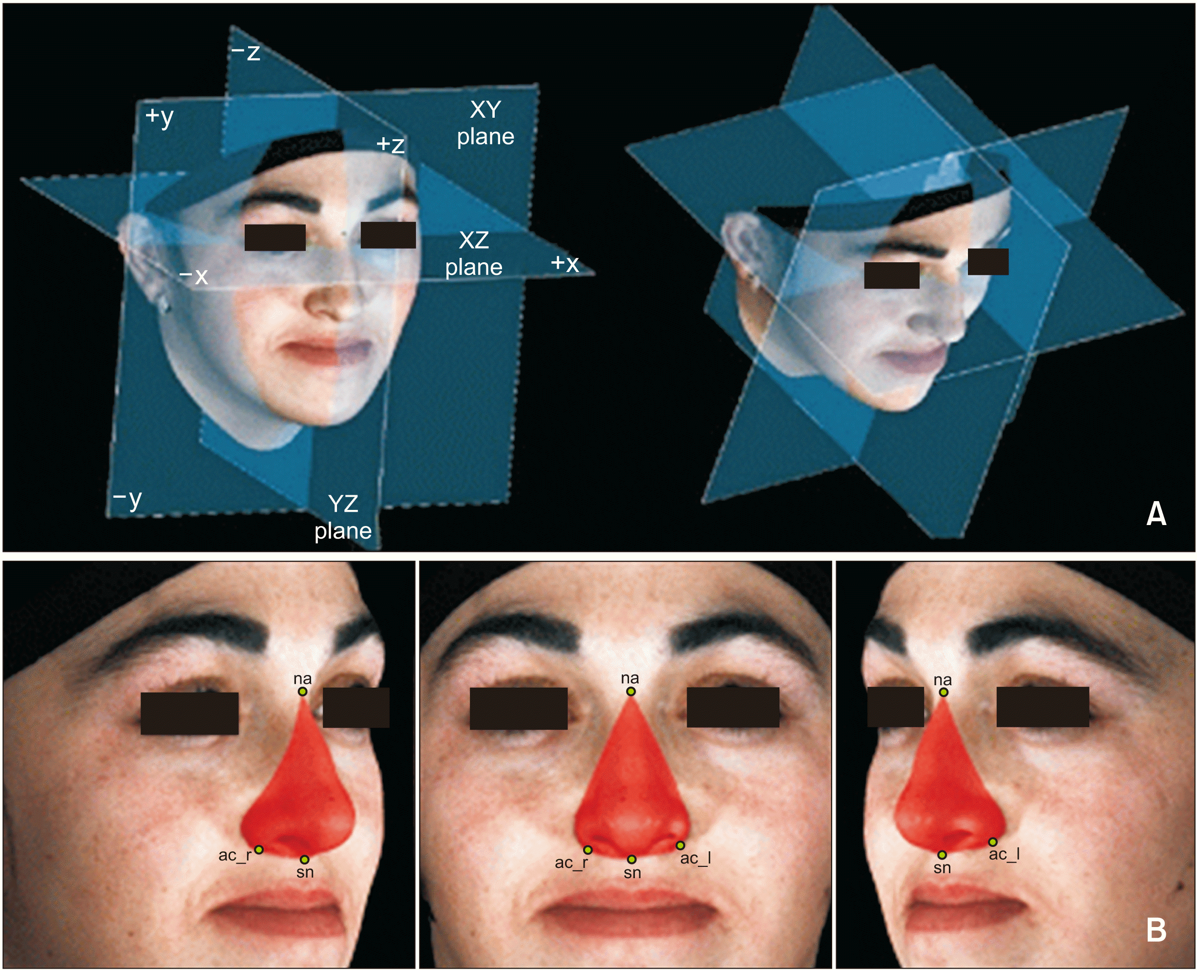
5,8,12,13 However, these studies failed to investigate 3D nose symmetry in detail after orthognathic surgery treatment. Therefore, the aims of this study were as follows: (1) perform a comprehensive evaluation of the 3D changes of the nose using stereophotogrammetry after maxillary advancement with impaction in patients with skeletal Class III malocclusion; (2) assess the magnitude of 3D symmetry and post-operative changes of the alare and alare curvature points according to the reference planes; and (3) explore the correlation between the hard and the soft tissue changes after orthognathic surgery treatment.

MATERIALS AND METHODS

Subjects

A retrospective study was designed and implemented to address the research objectives. The study protocol was approved by the Erciyes University Clinical Research Ethics Committee (Approval No: 2018/83). The alar base width for the first ten patients was evaluated in a pilot study, and a power analysis (G*Power Software ver. 3.1.9.2; Universität Düsseldorf, Düsseldorf, Germany) was performed. A sample size of 39 patients had 90% power to detect significant differences at a 0.05 significance level. The study population consisted of all patients presenting for evaluation and management of skeletal Class III malocclusion and aged between 17 years 0 months and 31 years 1 month.
The subjects were 40 adult Turkish patients (20 females and 20 males; mean age, 20.3 ± 3.0 years) who underwent orthodontic treatment and maxillary surgery for skeletal Class III malocclusion at Department of Orthodontics and Oral Maxillofacial Surgery, Faculty of Dentistry, Erciyes University, Kayseri, Turkey. All patients underwent maxillary orthognathic surgery, including one-piece Le Fort type I maxillary advancement and maxillary total impaction, to achieve a Class I dentoskeletal relationship. The mean maxillary advancement was 4.56 ± 1.34 mm and the mean maxillary impaction was 2.03 ± 1.04 mm. To measure the amount of surgical movement on the cephalometric film, a horizontal reference plane (HRP) was formed 7° from the sella-nasion plane, and the vertical reference plane (VRP) passing through the sella was drawn perpendicular to this horizontal plane (Figure 1).6 The perpendicular distances of the hard tissue A point to VRP and the posterior nasal spina (PNS) to HRP between before and after the orthognathic surgery revealed the amount of movement. The hard tissue A point was used for evaluation of advancement of the maxilla and the PNS point was used to evaluate the impaction. The occlusal plane angle was measured to check impaction and was 15.36° ± 7.31º pre-surgery and 14.97° ± 5.31º post-surgery.
None of the patients had severe facial asymmetry, history of trauma, congenital anomalies, and/or other craniofacial disorders. Patients who underwent an anterior nasal spina (ANS) reduction, an alar base cinch suture, or a rhinoplasty surgery were excluded. All patients had a body mass index between 18 and 26 kg/m2. The 3D images of the patients were acquired with stereophotogrammetry (3dMD Face; 3dMD, Atlanta, GA, USA) 1 day before and at least 6 months (8.91 ± 2.81 months) after surgery on the same day that the cephalometric films were taken, in the natural head position (NHP), centric occlusion, and with the lips at rest. For acquisition of NHP,14 the patients were firstly recommended to walk around and relax. Next, the patients performed gradually decreasing forward and backward head oscillations until they settled their heads into a self-balanced position. They were then asked to look at the reflection of their eyes in a mirror. Soft tissue nasal changes were evaluated using the 3dMDvultus (3dMD) software, and the file format was “.tsb.”
For the initial registration, the 3D images of the patients before and after surgery were aligned manually, before using the automatic registration feature of the software for fine-tuning the alignment. After the 3D images were adequately positioned manually, the most optimal surface areas of the face that would not be influenced by surgery were selected: the widest surface on the forehead, the surface from the nasal root to the nasal hump, and the lateral regions of the exocanthion (Figure 2A).15 The 3D image superimpositions were shown to be reliable with an average root mean square (RMS) error of 0.24 and a range of 0.12 to 0.45 (smaller than 0.5). These data were clinically acceptable and consistent with previously used methods.16
The landmarks for the soft tissues (Figure 2B) and reference planes (Figure 3A) are defined15 in Table 1. The 3D coordinates of the facial soft tissue landmarks were manually placed using 3dMDvultus (3dMD) software. The following facial soft tissue landmarks were considered:
  • Median points: nasion, na; pronasale, prn; subnasale, sn; columella constructed, cc; labiale superior, ls.

  • Lateral points: (right and left sides indicated by subscripts _r and _l, respectively) endocanthion, en_r and en_l; exocanthion, ex_r and ex_l; alare, al_r and al_l; alar curvature, ac_r and ac_l.

In determining the 3D coordinate system settings,17 left and right pupil points are connected by a segment that defines the x (left-right)-axis. The YZ plane perpendicularly bisects the x-axis. The z-axis is the anteroposterior axis of the face on the YZ plane. The y (vertical)-axis segment passes perpendicularly through the center of the z-axis on the YZ plane. The imaginary center point of 3D facial geometry is the zero point (y = 0, z = 0, and yz = 0). The xz plane is perpendicular to the yz plane and passes through the zero point. The xy plane passes through the zero point and is perpendicular to the yz and xz planes (Figure 3A). The 3D coordinate values (x, y, and z) of the landmarks were acquired using the coordinate system of the imaginary center of facial geometry (the zero point).
The changes in the 3D coordinates and directions of landmarks are shown in Table 2. The preoperative differences in each right and left al and ac points according to the reference planes were compared to the postoperative differences (Table 3). Table 3 shows the changes in bilateral Al and Ac between reference planes and patients after orthognathic surgery treatment.
The external nasal surface area was the computed sum of the surfaces between na, the following soft tissue border of the right alar-facial sulcus, the groove and right ac, sn, and left ac, the following soft tissue border of the left alar-facial groove, the sulcus and na (Figure 3B). The changes in the 6 coordinate values, 6 difference values, and the 5 linear, 1 angular, and 1 areal measurements (Tables 2,3 and 4) were calculated, and the correlations between hard and soft tissues were evaluated, (Table 5) in this study.

Statistical analysis

All the data in this study were normally distributed according to the Shapiro–Wilk test. The samples were evaluated as a single group and analyzed by the paired-samples t-test, since there was no statistically significant difference in the soft tissue changes between male and female patients in the independent-samples t-test. A p-value < 0.05 was considered statistically significant. The correlations between the soft-tissue and hard-tissue movements in the horizontal, vertical, and sagittal planes were analyzed by Pearson correlation analysis. The measurements of ten patient’s digital images were performed twice by the same examiner (GC) one month apart. Intraclass correlation coefficients for nasal measurements were between 0.85 and 0.98, indicating a strong positive correlation and sufficient reliability.18

RESULTS

The 3D coordinate (x, y, and z) values of the landmarks were obtained by using a coordinate system. In the horizontal (x) plane, bilateral al and ac showed significant lateral movement while there were no significant changes in prn and sn. In the vertical (y) plane, all soft tissue landmarks moved upward, and in the sagittal (z) plane, all soft tissue landmarks moved forward significantly (Table 2). The average increase of 0.4° in the occlusal plane angle was not statistically significant. Table 3 shows the difference between pre- and postoperative measurements to the reference planes of the right and left al and ac in 3 dimensions. Only the vertical (y) preoperative difference between the right and left ac increased significantly after the operation (Table 3). There was a significant increase in the 3D linear measurements of the nose alar width and alar base width, and there were no significant changes in the nasal bridge length, columellar length, nasolabial angle, nasal tip protrusion, and external nasal surface area (Table 4). Table 5 shows the correlation between hard tissue and soft tissue changes. There was a significant relationship between maxillary impaction and nasal alar width, and lateral and anterior movements of the bilateral al and ac. The only significant positive correlation was noted between maxillary advancement and anterior movement of sn and prn.

DISCUSSION

The aim of this study was to evaluate the effect of maxillary advancement and impaction on 3D nasal symmetry and changes in the soft tissues of the nose. We used 3D stereophotogrammetry, a fast and reliable technique5 that is accurate enough for clinical use with a clinical error of 0.2 to 1.0 mm8 compared to 0.5–1.0 mm for laser scanning.6
Imaging is a commonly used method in orthodontics and maxillofacial surgery to record, evaluate, and display the size and shape of craniofacial structures. The 3D imaging systems include 3D computed tomography,5 laser scanning,6 3D facial morphometry,7 and stereophotogrammetry.5,8 The advantages of stereophotogrammetry include fast capture times, which decrease the likelihood that 3D images will be affected by body movements; minimal cooperation required from patients; no exposure to radiation; accurate and precise measurements obtained by rotating the images in the desired direction; high reproducibility; the provision for linear, topographic, areal, and volumetric measurements; and the facility to perform registration on pre- and post-surgery images.19 Disadvantages are the need for additional imaging methods (such as cone-beam computed tomography and cephalometric roentgens) for evaluation of hard tissues,20 high cost, the requirement of a large specialized area to set up the machinery, the difficulties of displaying complex areas and anatomical gaps, the need to calibrate the system frequently, and the impossibility of acquiring dynamic images.8 The 3D image collection and evaluations were performed at least 6 months after surgery to allow complete resolution of edema and establishment of soft-tissue adaptation to maxillary osteotomy.21 In assessments of 3D coordinates, Baik and Kim6 observed a mean 1.3-mm movement in al_r to the right side and in al_l to the left side, and both of these moved anteriorly by a mean distance of 2.3 mm, which was similar to our results for al and ac.
Consistent with previous reports, a tendency toward an upward and forward movement of the prn was observed in this study.8,9,12,20 Schendel and Williamson9 reported a mean nasal tip elevation of 2.4 mm and an impaction of 6.4 mm in patients who underwent mean maxillary advancement. In this study, the mean 1-mm upward movement of prn may have been due to the differences in surgical quantities.
The differences in the distances of the right and left al and ac points relative to the reference planes before and after treatment and horizontal changes of prn and sn were examined to evaluate 3D nasal symmetry changes. These values were considered smaller post-operatively with no deviations observed at prn and sn. Only the difference between the vertical (y) positions of the right and left ac points increased by 0.3 mm after the operation; this may be due to ac points that were closer to the osteotomy line and the soft tissues underlying these points being thin. The increases were statistically significant; however, we observed that it was clinically insignificant.
ANS is an important component of nasal tip protrusion,21 and when ANS is left untouched, as in our study, larger quantities of nasal tip movement could be seen.22 However, Gassmann et al.23 claimed that there was no significant relationship between the presence or absence of ANS and nasal tip protrusion.
An increase was reported in the nasal alar width and alar base width after maxillary advancement surgery by both McCance et al.,24 who used a 3D laser scanner, and Ferrario et al.,7 who used 3D facial morphometry. Baik and Kim6 reported a mean alar widening of 2 mm, and Chung et al.12 reported a significant increase in alar width and alar base width even in cases where a cinch suture was properly used. Interalar width was always greater post-surgery, probably because the nasal base, like sn, was pushed forward by maxillary advancement surgery.7 Honrado et al.8 reported equivocal results, both decreases and increases, with nasal tip protrusion and columellar length, and the nasolabial angle was the only measurement showing a significant increase in patients who underwent maxillary advancement and impaction. Chung et al.12 reported a mean decrease of 1 mm in nasal tip protrusion, which was significant. This study showed an insignificant decrease in nasal tip protrusion, which could have occurred because sn was closer to the underlying hard tissue than to prn.
Misir et al.22 reported that there was no significant change in the nasolabial angle in 27 patients who were treated with bimaxillary surgery. Radney and Jacobs25 indicated that the change in the nasolabial angle was associated with the amount and direction of the maxillary surgery. No clockwise or counterclockwise rotation was made in our patients, and no significant change was found in the nasolabial angle.
The nasal bridge length that was measured from the na to the prn showed a tendency to decrease post-surgery, which was possibly due to changes in the direction of the prn.10,20 There was no significant change in the nose area after orthognathic surgery, which was similar to the report by Sforza et al.26
The ratios of the soft-tissue to hard-tissue changes in the prn and sn were 31% and 33% sagittally in our study. The respective measurements in the study by Baik and Kim6 were 30% and 57% and 81% and 29% in the study by Soncul and Bamber.27 In this study, the mean amounts of maxillary advancement and impaction were 4.6 and 2.0 mm, respectively, while they were 1.0 to 3.0 mm in the study by Baik and Kim6 and 3 mm in the study by Soncul and Bamber.27 The differences in the ratios were most probably caused by the degrees of surgical movement. The reason for comparing the facial midline soft tissues following maxillary movements to the lateral soft tissues was that the semicircular form of the maxilla and the muscular attachments were more tightly connected to the bone in the midline.27 Freihofer28 observed that the sn and prn advanced by 50% and 25% with the maxilla, respectively.
Sarver and Weissman21 reported that the soft tissue changes associated with maxillary impaction were minimal. However, we found a significant vertical decrease in all nasal landmarks (prn, sn, al, and ac) measured. The ratios of the transversal al and ac changes relative to the maxillary impaction were 35% and 33%; the sagittal change ratios were 43% and 40%, respectively. There was a significant correlation between nasal alar width and maxillary impaction. Previous studies suggested that the probability of erroneous measurements in the vertical direction was greater than those in other directions, and the correlations with the vertical changes were difficult to predict.29,30 Soft tissue response differences might be due to the amounts of hard tissue movements, muscle structures, racial, and biological differences in the subjects, or unique variations in the soft tissue responses.
The limitations of the present study were the small sample size in each group and the lack of a control group. To make predictions of nasal changes according to maxillary skeletal changes, a larger sample size and control group of untreated patients would be needed in future studies.
The results of this study suggest that clinicians can predict the effect of orthognathic surgical interventions for the maxilla on the nasal structures and 3D nasal symmetry via a preliminary assessment and use this to guide surgical planning to improve patient outcomes. However, despite the increased use and advancement of 3D imaging systems, soft tissue predictions, especially in the nose, are still quite limited,8 and it is not always possible to anticipate the nasal changes precisely after maxillary surgery.2

CONCLUSION

Patients should be thoroughly informed about nasal changes after orthognathic surgery and the possibility of additional nasal surgical procedures before surgery. 3D stereophotogrammetry may be a valuable and useful tool in evaluating the nose in 3 dimensions.
The following conclusions can be reached from this study:
  • Maxillary impaction was found to be correlated with changes in the horizontal and anteroposterior location of bilateral alare and alare curvature and nasal ala width measurements, while maxillary advancement was correlated only with changes in the anteroposterior location of the pronasale and subnasale.

  • Alare and alare curvature points moved laterally, and an increase was observed in nose width.

  • Nasal symmetry did not change after surgery, with the exception of the left and right vertical position of the alare curvature.

  • Both pronasale and subnasale moved upwards; however, pronasale moved to a greater extent.

  • The external nasal surface area did not change.

Notes

How to cite this article: Coban G, Yavuz I, Karadas B, Demirbas AE. Three-dimensional assessment of nasal changes after maxillary advancement with impaction using stereophotogrammetry. Korean J Orthod 2020;50:249-257.

CONFLICTS OF INTEREST

No potential conflict of interest relevant to this article was reported.

REFERENCES

1. Arnett GW, McLaughlin RP. 2004. Facial and dental planning for orthodontists and oral surgeons. Mosby;London: p. 4.
2. Schendel SA, Carlotti AE Jr. 1991; Nasal considerations in orthognathic surgery. Am J Orthod Dentofacial Orthop. 100:197–208. DOI: 10.1016/0889-5406(91)70056-3. PMID: 1877544.
crossref
3. Naini FB, Gill DS. 2017. Orthognathic surgery: principles, planning and practice. John Wiley & Sons;Chichester: p. 417. DOI: 10.1002/9781119004370.
4. Larrabee WF Jr. 1987; Facial analysis for rhinoplasty. Otolaryngol Clin North Am. 20:653–74. PMID: 3320863.
crossref
5. van Loon B Jr, van Heerbeek N, Bierenbroodspot F, Verhamme L, Xi T, de Koning MJ, et al. 2015; Three-dimensional changes in nose and upper lip volume after orthognathic surgery. Int J Oral Maxillofac Surg. 44:83–9. DOI: 10.1016/j.ijom.2014.08.001. PMID: 25218802.
crossref
6. Baik HS, Kim SY. 2010; Facial soft-tissue changes in skeletal Class III orthognathic surgery patients analyzed with 3-dimensional laser scanning. Am J Orthod Dentofacial Orthop. 138:167–78. DOI: 10.1016/j.ajodo.2010.02.022. PMID: 20691358.
crossref
7. Ferrario VF, Sforza C, Schmitz JH, Santoro F. 1999; Three-dimensional facial morphometric assessment of soft tissue changes after orthognathic surgery. Oral Surg Oral Med Oral Pathol Oral Radiol Endod. 88:549–56. DOI: 10.1016/S1079-2104(99)70084-3. PMID: 10556748.
crossref
8. Honrado CP, Lee S, Bloomquist DS, Larrabee WF Jr. 2006; Quantitative assessment of nasal changes after maxillomandibular surgery using a 3-dimensional digital imaging system. Arch Facial Plast Surg. 8:26–35. DOI: 10.1001/archfaci.8.1.26. PMID: 16415444.
crossref
9. Schendel SA, Williamson LW. 1983; Muscle reorientation following superior repositioning of the maxilla. J Oral Maxillofac Surg. 41:235–40. DOI: 10.1016/0278-2391(83)90265-3.
crossref
10. Mitchell C, Oeltjen J, Panthaki Z, Thaller SR. 2007; Nasolabial aesthetics. J Craniofac Surg. 18:756–65. DOI: 10.1097/scs.0b013e3180684360. PMID: 17667661.
crossref
11. Guenthner TA, Sather AH, Kern EB. 1984; The effect of Le Fort I maxillary impaction on nasal airway resistance. Am J Orthod. 85:308–15. DOI: 10.1016/0002-9416(84)90188-X.
crossref
12. Chung C, Lee Y, Park KH, Park SH, Park YC, Kim KH. 2008; Nasal changes after surgical correction of skeletal Class III malocclusion in Koreans. Angle Orthod. 78:427–32. DOI: 10.2319/041207-186.1. PMID: 18416623.
crossref
13. Choi JW, Lee JY, Oh TS, Kwon SM, Yang SJ, Koh KS. 2014; Frontal soft tissue analysis using a 3 dimensional camera following two-jaw rotational orthognathic surgery in skeletal class III patients. J Craniomaxillofac Surg. 42:220–6. DOI: 10.1016/j.jcms.2013.05.004. PMID: 23870714.
crossref
14. Solow B, Tallgren A. 1971; Natural head position in standing subjects. Acta Odontol Scand. 29:591–607. DOI: 10.3109/00016357109026337. PMID: 5290983.
crossref
15. Lim YK, Chu EH, Lee DY, Yang IH, Baek SH. 2010; Three-dimensional evaluation of soft tissue change gradients after mandibular setback surgery in skeletal Class III malocclusion. Angle Orthod. 80:896–903. DOI: 10.2319/021210-90.1. PMID: 20578861.
crossref
16. Lin HH, Chiang WC, Lo LJ, Sheng-Pin Hsu S, Wang CH, Wan SY. 2013; Artifact-resistant superimposition of digital dental models and cone-beam computed tomography images. J Oral Maxillofac Surg. 71:1933–47. DOI: 10.1016/j.joms.2013.06.199. PMID: 23911142.
crossref
17. Kim YK, Lee NK, Moon SW, Jang MJ, Kim HS, Yun PY. 2015; Evaluation of soft tissue changes around the lips after bracket debonding using three-dimensional stereophotogrammetry. Angle Orthod. 85:833–40. DOI: 10.2319/090414.622.1. PMID: 26308106.
crossref
18. Liu ZY, Yu J, Dai FF, Jiang RP, Xu TM. 2019; Three-dimensional changes in lip vermilion morphology of adult female patients after extraction and non-extraction orthodontic treatment. Korean J Orthod. 49:222–34. DOI: 10.4041/kjod.2019.49.4.222. PMID: 31367577. PMCID: PMC6658897.
crossref
19. Rasse M, Forkert G, Waldhäusl P. 1991; Stereophotogrammetry of facial soft tissue. Int J Oral Maxillofac Surg. 20:163–6. DOI: 10.1016/S0901-5027(05)80008-6. PMID: 1890324.
crossref
20. Betts NJ, Vig KW, Vig P, Spalding P, Fonseca RJ. 1993; Changes in the nasal and labial soft tissues after surgical repositioning of the maxilla. Int J Adult Orthodon Orthognath Surg. 8:7–23.
21. Sarver DM, Weissman SM. 1991; Long-term soft tissue response to LeFort I maxillary superior repositioning. Angle Orthod. 61:267–76.
22. Misir AF, Manisali M, Egrioglu E, Naini FB. 2011; Retrospective analysis of nasal soft tissue profile changes with maxillary surgery. J Oral Maxillofac Surg. 69:e190–4. DOI: 10.1016/j.joms.2010.10.032. PMID: 21367507.
23. Gassmann CJ, Nishioka GJ, Van Sickels JE, Thrash WJ. 1989; A lateral cephalometric analysis of nasal morphology following Le Fort I osteotomy applying photometric analysis techniques. J Oral Maxillofac Surg. 47:926–30. DOI: 10.1016/0278-2391(89)90375-3.
crossref
24. McCance AM, Moss JP, Fright WR, James DR, Linney AD. 1992; A three dimensional analysis of soft and hard tissue changes following bimaxillary orthognathic surgery in skeletal III patients. Br J Oral Maxillofac Surg. 30:305–12. DOI: 10.1016/0266-4356(92)90180-Q. PMID: 1390562.
crossref
25. Radney LJ, Jacobs JD. 1981; Soft-tissue changes associated with surgical total maxillary intrusion. Am J Orthod. 80:191–212. DOI: 10.1016/0002-9416(81)90218-9. PMID: 6943938.
crossref
26. Sforza C, Peretta R, Grandi G, Ferronato G, Ferrario VF. 2007; Soft tissue facial volumes and shape in skeletal Class III patients before and after orthognathic surgery treatment. J Plast Reconstr Aesthet Surg. 60:130–8. DOI: 10.1016/j.bjps.2006.06.003. PMID: 17223510.
crossref
27. Soncul M, Bamber MA. 2004; Evaluation of facial soft tissue changes with optical surface scan after surgical correction of Class III deformities. J Oral Maxillofac Surg. 62:1331–40. DOI: 10.1016/j.joms.2004.04.019. PMID: 15510353.
crossref
28. Freihofer HP Jr. 1977; Changes in nasal profile after maxillary advancement in cleft and non-cleft patients. J Maxillofac Surg. 5:20–7. DOI: 10.1016/S0301-0503(77)80071-4. PMID: 265348.
crossref
29. Mansour S Jr, Burstone C, Legan H. 1983; An evaluation of soft-tissue changes resulting from Le Fort I maxillary surgery. Am J Orthod. 84:37–47. DOI: 10.1016/0002-9416(83)90146-X.
crossref
30. Koh CH, Chew MT. 2004; Predictability of soft tissue profile changes following bimaxillary surgery in skeletal class III Chinese patients. J Oral Maxillofac Surg. 62:1505–9. DOI: 10.1016/j.joms.2004.04.022. PMID: 15573350.
crossref

FIGURES AND TABLES

Figure 1
Evaluation of surgical quantities of hard tissues.
N, Nasion; S, sella; A, A point; ANS, anterior nasal spina; PNS, posterior nasal spina; HRP, horizontal reference plane; VRP, vertical reference plane.
KJOD-50-249-f1.tif
Figure 2
A, Reference markers used for superimposition: forehead area (the wide surface over the eyebrows), nasion area (the corresponding soft tissue point from the nasal root to the nasal hump), and exocanthion (the 3/4 lateral regions of the exocanthion). B, Soft tissue landmarks. na, Soft tissue nasion; en, endocanthion; ex, exocanthion; prn, pronasale; cc, columella constructed point; al, alare; ac, alare curvature; sn, subnasale; ls, labiale superior; r, right; l, left.
KJOD-50-249-f2.tif
Figure 3
A, Axial reference plane passing through both endocanthions when the patients had their natural head positions. Sagittal reference plane passing through na, perpendicular to the axial and coronal reference planes; coronal reference plane passing through both exocanthions, perpendicular to the axial and sagittal planes. B, External nasal surface area measurement was computed following soft tissue nasal borders.
na, Soft tissue nasion; ac, alare curvature; sn, subnasale; r, right; l, left.
KJOD-50-249-f3.tif
Table 1
Definitions of soft tissue landmarks and reference planes
Soft tissue landmark Abbreviation Definition
Nasion na The point in the midline of both the nasal root and the frontonasal suture
Endocanthion en The soft tissue point located at the inner commissure of the each eye fissure
Exocanthion ex The soft tissue point located at the outer commissure of the each eye fissure
Pronasale prn The most prominent point on the nasal tip
Columella constructed point cc The midpoint of the columella crest at the level of the nostril top points
Alare al The most lateral point on each alar contour
Alare curvature ac The point located at the facial insertion of each alar base
Subnasale sn The midpoint on the nasolabial soft tissue contour between the columella crest and the upper lip
Labiale superior ls The midpoint of the vermilion line of the upper lip
Reference plane
Axial plane A plane constructed with the right endocanthion and the left endocanthion on the natural head position, parallel to the floor
Coronal plane A plane constructed with the right exocanthion and the left exocanthion, perpendicular to the axial plane
Sagittal plane A plane constructed with nasion, perpendicular to the axial and coronal planes
Table 2
Changes in three-dimensional coordinates of landmarks
Variable X Y Z


∆T (T1–T0) p-value ∆T (T1–T0) p-value ∆T (T1–T0) p-value
Prn −0.08 ± 0.95 0.928 1.14 ± 1.63 0.000*** 1.35 ± 0.87 0.000***
Sn −0.14 ± 0.75 0.459 0.60 ± 0.99 0.000*** 2.24 ± 1.59 0.000***
Al_r −1.22 ± 0.90 0.000*** 1.21 ± 1.20 0.000*** 2.64 ± 1.55 0.000***
Al_l 1.25 ± 1.28 0.000*** 1.07 ± 1.72 0.002** 2.82 ± 1.29 0.000***
Ac_r −1.42 ± 0.90 0.000*** 0.78 ± 1.10 0.000*** 3.67 ± 1.43 0.000***
Ac_l 1.54 ± 1.27 0.000*** 1.12 ± 1.33 0.000*** 3.32 ± 1.73 0.000***

Values are presented as mean ± standard deviation.

Paired-samples t-test were performed.

T0, Presurgery; T1, postsurgery; ΔT, amount of change after surgery (T1–T0); x-axis, left (no sign), right (minus sign); y-axis, superior (no sign), inferior (minus sign); z-axis, anterior (no sign), posterior (minus sign).

** p < 0.01, ***p < 0.001.

See Table 1 for definitions of the other landmarks.

Table 3
Three-dimensional nasal symmetry measurements according to the reference planes
Variable T0 T1 p-value
Sagittal (x) (mm)
Al_l/Al_r −0.78 ± 1.77 −0.80 ± 2.18 0.892
Ac_l/Ac_r −1.06 ± 2.06 −1.18 ± 2.21 0.574
Axial (y) (mm)
Al_l/Al_r 0.64 ± 1.40 0.50 ± 0.86 0.487
Ac_l/Ac_r 0.39 ± 0.93 0.74 ± 0.93 0.004**
Coronal (z) (mm)
Al_l/Al_r 0.56 ± 1.48 0.38 ± 1.73 0.339
Ac_l/Ac_r 0.53 ± 1.32 0.88 ± 1.30 0.339

Values are presented as mean ± standard deviation.

Paired t-test was performed.

T0, Pre-treatment; T1, post-treatment; Al_l/r, alare left/right; Ac_l/r, alare curvature left/right; Point_l/Point_r, difference between the distances from point_l and point_r to the reference plane.

** p < 0.01.

Table 4
Changes in nasal measurements
Variable T0 T1 p-value
Nose/alar width (Al_r-Al_l) (mm) 34.70 ± 3.77 37.04 ± 3.90 0.000***
Alar base width (Ac_r-Ac_l) (mm) 29.05 ± 3.11 32.09 ± 3.56 0.000***
Nasal bridge length (Na-Prn) (mm) 46.94 ± 5.46 46.78 ± 5.46 0.500
Nasal tip protrusion (Sn-Prn) (mm) 19.97 ± 4.87 19.64 ± 4.64 0.130
Columellar length (Sn-Cc) (mm) 9.96 ± 2.58 9.90 ± 2.57 0.872
Nasolabial angle (Ls-Sn-Cc) (°) 103.41 ± 15.32 103.02 ± 11.95 0.766
Nose area (cm2) 24.81 ± 4.01 24.65 ± 4.32 0.712

Values are presented as mean ± standard deviation.

Paired t-test was performed.

T0, Pre-treatment; T1, post-treatment; Al_l/r, alare left/right; Ac_l/r, alare curvature left/right; Na, soft tissue nasion; Prn, pronasale; Sn, subnasale; Cc, columella constructed; Ls, labiale superior.

*** p < 0.001.

Table 5
Correlations of soft-tissue and hard-tissue changes
Variable Maxillary advancement
(mean 4.56 ± 1.34 mm)
Maxillary impaction
(mean 2.03 ± 1.04 mm)


Movement (%) p-value Movement (%) p-value
Prn_y 24 0.133 13 0.410
Prn_z 31 0.029* 14 0.768
Sn_y 21 0.189 14 0.525
Sn_z 33 0.034* 1 0.318
Al_x 10 0.896 35 0.026*
Al_y 12 0.588 12 0.486
Al_z 25 0.145 43 0.003**
Ac_x 25 0.434 33 0.048*
Ac_y 22 0.692 5 0.803
Ac_z 25 0.131 40 0.003**
Nasal alar width 10 0.542 36 0.038*
Alar base width 20 0.075 32 0.063

Mean values for bilateral points (Al and Ac) are presented.

Pearson correlation analysis was performed.

Prn, Pronasale; Sn, subnasale; Al, alare; Ac, alare curvature; _x, transversal direction; _y, vertical direction; _z, anteroposterior direction.

* p < 0.05, **p < 0.01.

TOOLS
Similar articles