Journal List > Diabetes Metab J > v.44(5) > 1145642

Choi, Lee, Kim, and Lee: The Association between Pulmonary Functions and Incident Diabetes: Longitudinal Analysis from the Ansung Cohort in Korea

Abstract

Background

We sought to explore whether reduced pulmonary function is an independent risk factor for incident diabetes in Koreans.

Methods

We conducted a prospective cohort study of pulmonary function as a risk factor for incident diabetes using 10-year follow-up data from 3,864 middle-aged adults from the Ansung cohort study in Korea. The incidence of diabetes was assessed using both oral glucose tolerance tests and glycosylated hemoglobin levels.

Results

During 37,118 person-years of follow-up, 583 participants developed diabetes (incidence rate: 15.7 per 1,000 person-years). The mean follow-up period was 8.0±3.7 years. Forced vital capacity (FVC; % predicted) and forced expiratory volume in 1 second (FEV1; % predicted) were significantly correlated with incident diabetes in a graded manner after adjustment for sex, age, smoking, exercise, and metabolic parameters. The adjusted hazard ratio (HR) and confidence interval (CI) for diabetes were 1.408 (1.106 to 1.792) and 1.469 (1.137 to 1.897) in the first quartiles of FVC and FEV1, respectively, when compared with the highest quartile. Furthermore, the FVC of the lowest first and second quartiles showed a significantly higher 10-year panel homeostasis model assessment of insulin resistance index, with differences of 0.095 (95% CI, 0.010 to 0.018; P=0.028) and 0.127 (95% CI, 0.044 to 0.210; P=0.003), respectively, when compared to the highest quartiles.

Conclusion

FVC and FEV1 are independent risk factors for developing diabetes in Koreans. Pulmonary factors are possible risk factors for insulin resistance and diabetes.

INTRODUCTION

Pulmonary dysfunction, particularly reduced forced vital capacity (FVC), is commonly found in patients with diabetes [12]. That is, diabetes is often accompanied by a restrictive pattern of pulmonary dysfunction [1]. Several prospective studies in Western countries have reported that decline in lung function precedes and predicts the onset of diabetes. One cohort study conducted in Malmo, Sweden reported that low vital capacity was an independent predictor of diabetes over 6 years of follow-up [3]. The Atherosclerosis Risk in Communities study also showed that lower baseline FVC predicted higher diabetes incidence after 9 years [4]. Another study found that low FVC and low forced expiratory volume in 1 second (FEV1) predicted diabetes mellitus (DM) 13 years later, independent of other risk factors such as obesity [5]. In the same cohort, the incidence of cardiovascular disease was higher among subjects with low FVC who later developed insulin resistance [6]. Finally, the National Health and Nutrition Examination Survey study showed that FVC and FEV1 were inversely correlated with the incidence of diabetes over 20 years of follow- up [7]. Cross-sectional studies have also found that decreased FVC and FEV1 were associated with an increased prevalence of diabetes and that they were closely correlated with components of metabolic syndrome, including blood pressure, glucose, triglyceride, high-density lipoprotein cholesterol (HDL-C), and waist circumference [8910]. One prospective study showed that a reduction in FVC over 6 years of follow-up was linked to an increase in the incidence of metabolic syndrome, and that the association was mainly due to an increase in central obesity [11]. In addition, several prospective studies have reported that a decline in FVC and FEV1 is associated with increased morbidity and mortality due to cardiovascular disease and diabetes [12].
Several Asian studies have also revealed significant associations between lower pulmonary functions and the prevalence of type 2 DM. However, all such studies were of cross-sectional design, so they could not ascertain the causal relationship [13141516]. One hospital-based cohort study demonstrated an association between reduced FVC and FEV1 and increased diabetes prevalence in young Korean men who regularly undertook workplace health checkups [17]. However, the results cannot be extrapolated to the general population, as they addressed only young men with specific occupations [17]. No prospective studies have addressed decreased lung function as a determinant of incident diabetes in Asian populations. Considering that DM shows ethnic differences, it is essential that such studies be carried out on Asians. Furthermore, most previous studies have used fasting blood glucose or self-report to diagnose diabetes, so they may have been limited by confirmation bias and underdiagnosis of diabetes.
In the present background, we used data from the Ansung community-based cohort study to determine whether reductions in lung functions in Koreans were independently associated with increased diabetes and consistently correlated with various metabolic indicators. Secondly, we investigated whether the changes of FVC and FEV1 differed between patients who later developed diabetes and controls. Thirdly, we analyzed the interaction between FVC and FEV1, as well as sequential changes in insulin resistance over the follow-up period, to identify the mechanisms that may link diabetes occurrence to lower FVC and FEV1.

METHODS

Subjects

The data from Ansung cohort were used in this analysis. The Korean government conducted a large, community-based epidemiological study (Korean Genome and Epidemiology study [KoGES]) to investigate chronic, non-communicable diseases such as diabetes among Koreans. The Ansung cohort was part of the KoGES and was selected to represent Korean rural areas [18]. In particular, the Ansung cohort study focused on diabetes and its related metabolic problems. All subjects in the cohort underwent biannual 75-g oral glucose tolerance tests during the study follow-up if they were not already diagnosed as having diabetes. The Korean Centers for Disease Control and Prevention (KCDC) obtained written informed consent from all participants, and the Institutional Review Board of Eulji University Hospital approved the study protocol (IRB No. EMCS 2019-05-008). After IRB approval was granted, the KCDC provided us the data for analysis after removing the participants' names and registry numbers (approval No. 2019- EPI-41). The study protocol adhered to the tenets of the Declaration of Helsinki. The baseline examination of the Ansung cohort was conducted between 2001 and 2003. The present study included 5,018 eligible adults who were 40 to 69 years of age. Follow-up surveys were conducted at 2-year intervals for 10 years. Subjects who did not undergo pulmonary function testing at baseline were excluded (n=142), as were additional subjects with a past or current medical history of asthma (n=129), chronic obstructive pulmonary disease (n=42), or pulmonary tuberculosis (n=225). Of the remaining subjects (n=4,520), a further 656 with baseline DM were excluded, because the Kaplan-Meier survival and Cox proportional hazard analysis sought only to assess diabetes that developed during follow-up.

Assessment of clinical characteristics and parameters

Anthropometric parameters and blood pressure were measured using standard methods. A series of surveys were conducted to collect information regarding demographics, past and present medical history, medication use, and a variety of health-related variables. The collected blood samples were sent to the central laboratory for analysis. Fasting plasma glucose, creatinine, aspartame aminotransferase (AST), alanine aminotransferase (ALT), total cholesterol, triglycerides, and HDL-C, were measured using a Hitachi 747 chemistry analyzer (Hitachi Ltd., Tokyo, Japan). Glycosylated hemoglobin (HbA1c) was measured using high-performance liquid chromatography (Variant II; BioRad Laboratories, Hercules, CA, USA), C-reactive protein (CRP) using immunoradiometric assay (ADVIA 1650; Bayer Diagnostics, Tarrytown, NY, USA), and the plasma insulin concentration using radioimmunoassay (Linco kit; Linco Research, St Charles, MO, USA). The homeostasis model assessment of insulin resistance index (HOMA-IR) was calculated as follows: HOMA-IR=(fasting glucose [mmol/L])×(fasting insulin [µU/mL])/22.5 [19]. Metabolic syndrome was identified when three or more of the following criteria were present, according to modified Adult Treatment Panel III guidelines: (1) central obesity (waist circumference cutoff points of >90 cm for men and >80 cm for women), (2) elevated triglyceride level (≥1.7 mmol/L or drug treatment for elevated triglyceride levels), (3) reduced HDL-C levels (<1.0 mmol/L in men, <1.3 mmol/L in women, or drug treatment for low HDL-C), (4) elevated blood pressure (≥130 mm Hg systolic, ≥85 mm Hg diastolic, or antihypertensive drug treatment), and (5) elevated fasting plasma glucose (≥5.6 mmol/L or drug treatment for diabetes) [20]. With regards to cigarette smoking, the subjects were categorized as never-smokers, former smokers, light smokers (less than 1 pack per day), or heavy smokers (more than 1 pack per day) using a standard questionnaire. Physical activity was defined as the number of episodes of exercise per week, categorized into three groups (none, 1–3 times, 4–7 times a week).
Pulmonary function tests were performed by a trained technician using a spirometer (Vmax-2130; Sensor Medics, Yorba Linda, CA, USA) according to the American Thoracic Society standard protocol [21]. The spirometry tests were calibrated quality controlled regularly according to the American Thoracic Society guidelines. All participants underwent pulmonary function testing three times at baseline (year 0), and at years 2 and 4.

Definition of diabetes

Individuals were categorized as having diabetes if one or more of the following criteria were met, adapted from 2019 American Diabetes Association: (1) fasting glucose level ≥7.0 mmol/L, (2) post-challenge 2-hour glucose level ≥1.1 mmol/L, (3) HbA1c ≥6.5%, or (4) current use of anti-diabetic medications [22]. Persons classified as having diabetes at baseline were excluded from the Kaplan-Meier survival and Cox proportional hazard analysis of incident DM.

Statistics

All values are expressed as means±standard deviations. The FEV1 (% predicted) and FVC (% predicted) of the baseline study population were normally distributed. We categorized the FEV1 and FVC into quartiles for further analysis. Continuous and categorical variables were analyzed using analysis of variance (ANOVA) and the chi-square test, respectively. The time to incident diabetes was assessed in a survival analysis using quartiles of FEV1 and FVC. A Kaplan-Meier plot of the cumulative proportions of diabetes was constructed according to the quartiles of FEV1 and FVC among subjects without diabetes at baseline. Cox proportional hazards models were used in a multivariate analysis. Relative hazard ratios (HRs) were used to compare the incidence of diabetes in the lower three versus the highest quartiles of FEV1 and FVC, after adjustment for confounding factors. Model 1 was adjusted for age, sex, smoking, exercise, and body mass index (BMI), Model 2 for the same factors as Model 1, plus HbA1c, white blood cell (WBC) count, and CRP, and Model 3 for the same factors as Model 2, plus total cholesterol, AST, ALT, and creatinine. We calculated HRs with 95% confidence intervals (CIs). Longitudinal data were analyzed using the panel data analysis model provided by the STATA statistical package. We compared changes of FEV1 and FVC between subjects who progressed to DM and those who did not. Panel data were used to analyze FEV1 and FVC in the first 4-year period, during which pulmonary function had been measured at three time points (baseline [year 0], year 2, and year 4). The panel data of HOMA-IR over 10 years were used to compare the differences among the quartiles of FEV1 and FVC. HOMA-IR was also measured at 2-year intervals. A P values <0.05 were considered to be statistically significant. Statistical analyses were performed using STATA version 13.1 (StataCorp, College Station, TX, USA).

RESULTS

Baseline characteristics

The subjects' baseline characteristics are shown in Table 1 by FVC quartiles (% predicted). At baseline, 656 (14.5%) of 4,520 subjects were diabetes patients, while 1,802 (39.7%) were current (n=1,232) or ex-smokers (n=570). Subjects with lower FVC (% predicted) were significantly more likely to be older, to be male, to be current or ex-smokers, to exercise less, and to have higher WBC count, blood glucose, total cholesterol, triglyceride, AST, ALT, creatinine, waist and hip circumferences, BMI, systolic blood pressure (SBP), and diastolic blood pressure (DBP). Subjects with lower FEV1 showed similar characteristics as those with lower FVC, although their exercise, total cholesterol, SBP, and waist circumference differed. Importantly, individuals with lower FVC or FEV1 had a higher prevalence of metabolic syndrome and diabetes (Table 1).
To determine whether lower FVC or FEV1 at baseline was associated with components of insulin resistance syndrome, linear regression analyses were conducted. Lower FVC (% predicted) was independently associated with age, sex, HbA1c, total cholesterol, AST, creatinine, SBP, and BMI in a multivariate analysis (Table 2). Lower FEV1 was independently associated with age, sex, smoking status, HbA1c, AST, creatinine, and SBP (Table 2).

Incident diabetes

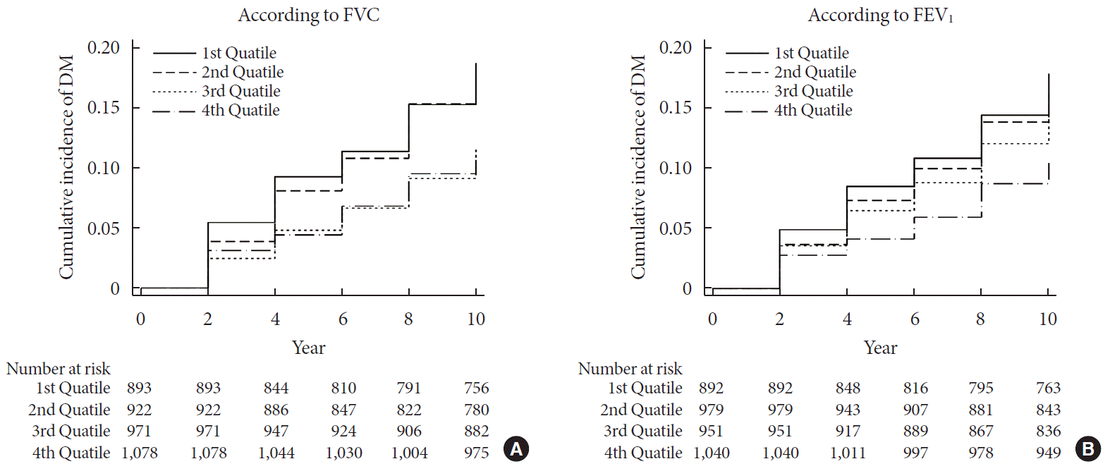
During 37,118 person-years of follow-up, 583 subjects developed diabetes (incidence rate: 15.7 per 1,000 person-years). The mean follow-up period was 8.0±3.7 years. There were inverse relationships between FVC and FEV1 and incidence of diabetes (Fig. 1).
In the Cox regression analysis, the first and second quartiles of FVC and the first to third quartiles of FEV1 showed a significantly increased incidence of diabetes, even after adjustment for sex, age, smoking, BMI, HbA1c, WBC count, CRP, total cholesterol, AST, ALT, and creatinine (Table 3).

Comparison of FVC and FEV1 reductions according to diabetes development
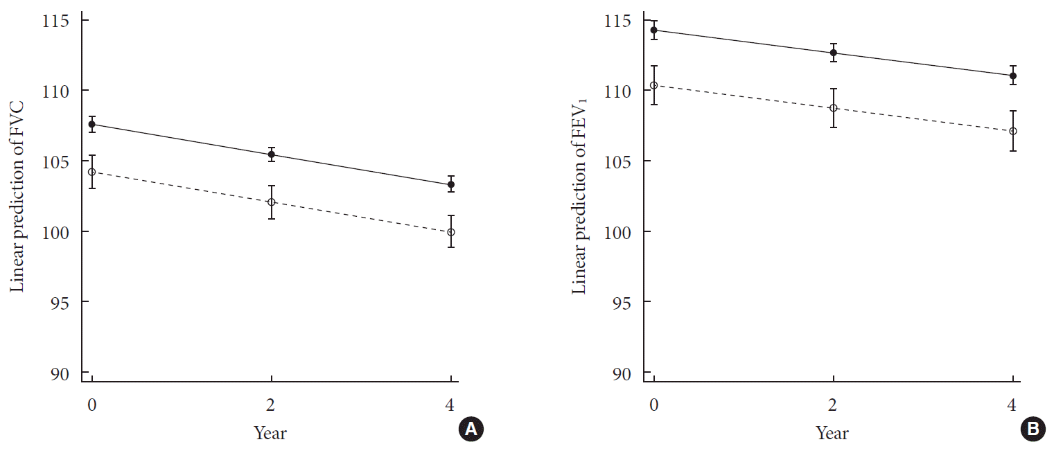
Among subjects who did not have diabetes at baseline, those who developed diabetes during the 10-year follow-up period were further classified as DM progressors, whereas those who did not were defined as non-DM progressors. The differences in FVC and FEV1 changes between DM progressors and non-DM progressors were analyzed using a random effects panel data regression model. Over 4 years, the FVC tended to decrease by −1.06% per year in the overall population (95% CI, −1.16% to −0.95%; P<0.001). The FVC was −3.38% lower in the DM progressors (95% CI, −4.51% to −2.14%; P<0.001) than in the non-DM progressors. FEV1 showed similar results as FVC, decreasing by −0.81% per year (95% CI, −0.92% to −0.69%; P<0.001) in the overall group. In DM progressors, the FEV1 was 3.91% lower than in non-DM progressors (95% CI, −5.45% to −2.37%; P<0.001). The interaction between DM development and time was not significant to either FVC or FEV1. Fig. 2 shows a similar decreasing trend in both the DM progressor and non-progressor groups.

Panel HOMA-IR results according to quartiles of FVC (% predicted)

The differences in the HOMA-IR panel results were analyzed among the FVC and FEV1 quartiles over 10 years using a random effects panel data regression model. HOMA-IRs were measured at 2-year intervals during the 10-year follow-up period. The FVC (% predicted) of the lowest first and second quartiles showed significantly higher panel HOMA-IR results, with differences of 0.095 (95% CI, 0.010 to 0.018; P=0.028) and 0.127 (95% CI, 0.044 to 0.210; P=0.003), respectively, when compared to the highest quartile as a reference group (Table 4). The results remained significant after adjustment for age, sex, smoking and exercise status (Table 4). The results of FEV1 were not robust (Table 4).

DISCUSSION

In this prospective cohort study of otherwise healthy, community-dwelling Korean subjects, a lower FVC and FEV1 predicted subsequent diabetes. The association was graded and was independent of many potential covariates. Moreover, subjects with lower FVC or FEV1 had many aspects of insulin resistance at baseline; they showed higher blood levels of glucose and triglyceride, as well as higher waist circumference and blood pressure. Our results are generally in accord with previous prospective studies that have shown associations with lower pulmonary function and diabetes development [45679]. To our knowledge, this was the first study of the relationship between pulmonary function and diabetes incidence in Asian populations.
The exact mechanisms of these associations are not well known. However, several observations have suggested that insulin resistance and increased inflammation are mediators of this association [623]. For example, previous studies have demonstrated that raised plasma CRP levels are associated with future diabetes, and that interleukin 6 (IL-6) may be important for glucose and insulin metabolism [242526]. Inflammation is also known to be associated with atherosclerosis development [27]. It follows that systemic, low-grade inflammation could be the link between reduced FVC, development of insulin resistance, and subsequent cardiovascular disease.
Another explanation is mitochondrial dysfunction, which is an important mechanism in the pathogenesis of insulin resistance [282930]. Basically, the pulmonary function test measures both lung function and the metabolic rate of the whole body. If the metabolic rate of the whole body is reduced, pulmonary function will also decrease [31]. Persons with a low metabolic rate become obese, which can lead to diabetes [32]. In support of this hypothesis, one previous study showed that low FVC is inversely related with fat mass in the trunk or central area, even after adjustment for multiple covariates [33]. Metabolic rate depends on body mass and the body's mitochondrial density and function [34], so we speculated that mitochondrial dysfunction is also an important factor in the association between lung function and diabetes. The accumulation of environmental pollutants in the body is known to cause both inflammation and mitochondrial dysfunction [34].
Ventilatory dysfunction in patients with diabetes is more likely to have a restrictive pattern than an obstructive one. Indeed, the FVC is lower in patients with diabetes than in controls, but the FEV1/FVC is conserved [293536]. Autopsy studies and transbronchial lung biopsies have long shown that, as in diabetic kidney disease, microangiopathic changes occur in the pulmonary structure of patients with diabetes. Specifically, histopathology has shown changes in microvascular structures, such as fibrosis and basal lamina thickening, which are thought to cause restrictive pattern dysfunction [37383940]. Several studies have reported that poor blood glucose control and duration of diabetes are associated with pulmonary function [83536]. These observations suggest that pulmonary dysfunction is an important diabetic complication. In the baseline data of the present study, the proportion of restrictive pattern lung dysfunction (both [FEV1/FVC] ≥0.7 and FVC [% predicted] <80%) was significantly higher in subjects with diabetes (6.9%) than in those without diabetes (4.6%; P<0.008), whereas there was no difference in obstructive pattern lung dysfunction (FEV1/FVC <0.7; diabetes: 1.2% vs. non-diabetes: 1.1%; P=0.778).
The present study showed that pulmonary function decline in early diabetes did not differ between DM progressors and non-DM progressors, although FVC and FEV1 remained lower in the DM progressors than in the non-DM progressors throughout the observations. Several previous studies have analyzed changes in pulmonary function in patients with diabetes, but have not reached a consistent conclusion [28936]. Some studies have reported that the pulmonary function reduction in patients with diabetes is steeper than in the control group, and that it varies according to diabetes severity, diabetes duration, and anti-diabetic medications [28]. Conversely, some studies have reported that lung function changes in diabetic patients do not differ from the control group [936]. These discrepancies may have arisen because the patients differed across the studies in terms of diabetes severity and presence of microangiopathy, and because the studies used only fasting blood glucose or self-report to diagnose diabetes, so underdiagnosis may have occurred.
The lungs depend on strictly controlled immune and inflammatory processes. They are constantly exposed to various airborne infection sources, harmful gases, and particulates in the process of gas exchange. As a response, inflammatory and immune cells in the lungs produce various inflammatory mediators and cytokines. In this sense, the lung itself may serve as a source of low-grade inflammation, even when overt lung diseases are absent. Furthermore, fine dust and air pollution, which have emerged as major environmental problems in recent years, may increase the inflammatory reaction. Consistent with this hypothesis, the FVC and FEV1 were significantly correlated with baseline WBC count in the present study.
The FVC quartiles showed interactions with changes in insulin resistance during the course of diabetes development in our panel analysis. The mechanism by which pulmonary function is associated with DM is elusive for the present. However, the present study was the first to identify the interaction between FVC and insulin resistance over time.
The present study had several limitations. Firstly, selection bias may have occurred because all the participants were residents of Ansung: a rural area in Korea. Therefore, the results of the present study may not be generalizable to all populations.
The strengths of the present study were that we included both men and women, as well as both smokers and non-smokers. Secondly, the incidence of diabetes was evaluated thoroughly using both the oral glucose tolerance test and HbA1c level. Moreover, our data consisted of both baseline data and 10-year follow-up time points, and we assessed serial HOMA-IR changes according to FVC and FEV1 quartiles, providing valuable insight into the complex interplay between vital capacity and diabetes development. Finally, we adjusted for heavy smoking, which is independently associated with diabetes incidence [41].
In conclusion, lower pulmonary function is an independent risk factor for incident diabetes in Koreans. Pulmonary factors are possible risk factors for insulin resistance and diabetes.

ACKNOWLEDGMENTS

This study was supported by a 2019 Research Grant from Kangwon National University.

Notes

CONFLICTS OF INTEREST

No potential conflict of interest relevant to this article was reported.

AUTHOR CONTRIBUTIONS

Conception or design: H.S.C., S.W.L., J.T.K., H.K.L.

Acquisition, analysis, or interpretation of data: H.S.C., S.W.L., J.T.K., H.K.L.

Drafting the work or revising: H.S.C., J.T.K., H.K.L.

Final approval of the manuscript: H.S.C., S.W.L., J.T.K., H.K.L.

References

1. van den Borst B, Gosker HR, Zeegers MP, Schols AM. Pulmonary function in diabetes: a metaanalysis. Chest. 2010; 138:393–406.
2. Yeh HC, Punjabi NM, Wang NY, Pankow JS, Duncan BB, Cox CE, Selvin E, Brancati FL. Cross-sectional and prospective study of lung function in adults with type 2 diabetes: the Atherosclerosis Risk in Communities (ARIC) study. Diabetes Care. 2008; 31:741–746.
crossref
3. Eriksson KF, Lindgarde F. Poor physical fitness, and impaired early insulin response but late hyperinsulinaemia, as predictors of NIDDM in middle-aged Swedish men. Diabetologia. 1996; 39:573–579.
crossref
4. Yeh HC, Punjabi NM, Wang NY, Pankow JS, Duncan BB, Brancati FL. Vital capacity as a predictor of incident type 2 diabetes: the Atherosclerosis Risk in Communities study. Diabetes Care. 2005; 28:1472–1479.
crossref
5. Engstrom G, Janzon L. Risk of developing diabetes is inversely related to lung function: a population-based cohort study. Diabet Med. 2002; 19:167–170.
6. Engstrom G, Hedblad B, Nilsson P, Wollmer P, Berglund G, Janzon L. Lung function, insulin resistance and incidence of cardiovascular disease: a longitudinal cohort study. J Intern Med. 2003; 253:574–581.
crossref
7. Ford ES, Mannino DM. National Health and Nutrition Examination Survey Epidemiologic Follow-up Study. Prospective association between lung function and the incidence of diabetes: findings from the National Health and Nutrition Examination Survey Epidemiologic Follow-up Study. Diabetes Care. 2004; 27:2966–2970.
crossref
8. Davis TM, Knuiman M, Kendall P, Vu H, Davis WA. Reduced pulmonary function and its associations in type 2 diabetes: the Fremantle Diabetes Study. Diabetes Res Clin Pract. 2000; 50:153–159.
crossref
9. Litonjua AA, Lazarus R, Sparrow D, Demolles D, Weiss ST. Lung function in type 2 diabetes: the Normative Aging Study. Respir Med. 2005; 99:1583–1590.
crossref
10. Lawlor DA, Ebrahim S, Smith GD. Associations of measures of lung function with insulin resistance and type 2 diabetes: findings from the British Women’s Heart and Health Study. Diabetologia. 2004; 47:195–203.
crossref
11. Kim SK, Bae JC, Baek JH, Jee JH, Hur KY, Lee MK, Kim JH. Decline in lung function rather than baseline lung function is associated with the development of metabolic syndrome: a sixyear longitudinal study. PLoS One. 2017; 12:e0174228.
crossref
12. Wannamethee SG, Shaper AG, Rumley A, Sattar N, Whincup PH, Thomas MC, Lowe GD. Lung function and risk of type 2 diabetes and fatal and nonfatal major coronary heart disease events: possible associations with inflammation. Diabetes Care. 2010; 33:1990–1996.
crossref
13. Oda E, Kawai R. A cross-sectional relationship between vital capacity and diabetes in Japanese men. Diabetes Res Clin Pract. 2009; 85:111–116.
crossref
14. Oda E, Kawai R. A cross-sectional relationship between vital capacity and metabolic syndrome and between vital capacity and diabetes in a sample Japanese population. Environ Health Prev Med. 2009; 14:284–291.
crossref
15. Kim SK, Hur KY, Choi YH, Kim SW, Chung JH, Kim HK, Lee MK, Min YK, Kim KW, Kim JH. The relationship between lung function and metabolic syndrome in obese and non-obese Korean adult males. Korean Diabetes J. 2010; 34:253–260.
crossref
16. Lim SY, Rhee EJ, Sung KC. Metabolic syndrome, insulin resistance and systemic inflammation as risk factors for reduced lung function in Korean nonsmoking males. J Korean Med Sci. 2010; 25:1480–1486.
crossref
17. Kwon CH, Rhee EJ, Song JU, Kim JT, Kwag HJ, Sung KC. Reduced lung function is independently associated with increased risk of type 2 diabetes in Korean men. Cardiovasc Diabetol. 2012; 11:38.
crossref
18. Kim Y, Han BG. KoGES group. Cohort profile: the Korean Genome and Epidemiology Study (KoGES) Consortium. Int J Epidemiol. 2017; 46:e20.
crossref
19. Quon MJ. Limitations of the fasting glucose to insulin ratio as an index of insulin sensitivity. J Clin Endocrinol Metab. 2001; 86:4615–4617.
crossref
20. Expert Panel on Detection, Evaluation, and Treatment of High Blood Cholesterol in Adults. Executive summary of the third report of the National Cholesterol Education Program (NCEP) Expert Panel on Detection, Evaluation, and Treatment of High Blood Cholesterol in Adults (Adult Treatment Panel III). JAMA. 2001; 285:2486–2497.
21. American Thoracic Society. Standardization of spirometry, 1994 update. Am J Respir Crit Care Med. 1995; 152:1107–1136.
22. American Diabetes Association. 2. Classification and diagnosis of diabetes: standards of medical care in diabetes. 2019. Diabetes Care. 2019; 42:Suppl 1. S13–S28.
23. Duncan BB, Schmidt MI, Pankow JS, Ballantyne CM, Couper D, Vigo A, Hoogeveen R, Folsom AR, Heiss G. Atherosclerosis Risk in Communities Study. Low-grade systemic inflammation and the development of type 2 diabetes: the atherosclerosis risk in communities study. Diabetes. 2003; 52:1799–1805.
crossref
24. Chambers JC, Eda S, Bassett P, Karim Y, Thompson SG, Gallimore JR, Pepys MB, Kooner JS. C-reactive protein, insulin resistance, central obesity, and coronary heart disease risk in Indian Asians from the United Kingdom compared with European whites. Circulation. 2001; 104:145–150.
crossref
25. Kuller LH, Tracy RP, Shaten J, Meilahn EN. Relation of C-reactive protein and coronary heart disease in the MRFIT nested case-control study. Multiple Risk Factor Intervention Trial. Am J Epidemiol. 1996; 144:537–547.
26. Pradhan AD, Manson JE, Rifai N, Buring JE, Ridker PM. C-reactive protein, interleukin 6, and risk of developing type 2 diabetes mellitus. JAMA. 2001; 286:327–334.
crossref
27. Libby P, Ridker PM. Inflammation and atherosclerosis: role of C-reactive protein in risk assessment. Am J Med. 2004; 116 Suppl 6A:9S–16S.
crossref
28. Petersen KF, Dufour S, Befroy D, Garcia R, Shulman GI. Impaired mitochondrial activity in the insulin-resistant offspring of patients with type 2 diabetes. N Engl J Med. 2004; 350:664–671.
crossref
29. Petersen KF, Befroy D, Dufour S, Dziura J, Ariyan C, Rothman DL, DiPietro L, Cline GW, Shulman GI. Mitochondrial dysfunction in the elderly: possible role in insulin resistance. Science. 2003; 300:1140–1142.
crossref
30. Song J, Oh JY, Sung YA, Pak YK, Park KS, Lee HK. Peripheral blood mitochondrial DNA content is related to insulin sensitivity in offspring of type 2 diabetic patients. Diabetes Care. 2001; 24:865–869.
crossref
31. Choi JW, Pai SH. Brief communication: respiratory function is closely associated with basal metabolic rate in elderly persons. Ann Clin Lab Sci. 2004; 34:99–102.
32. Weyer C, Pratley RE, Salbe AD, Bogardus C, Ravussin E, Tataranni PA. Energy expenditure, fat oxidation, and body weight regulation: a study of metabolic adaptation to long-term weight change. J Clin Endocrinol Metab. 2000; 85:1087–1094.
33. Lim S, Kwon SY, Yoon JW, Kim SY, Choi SH, Park YJ, Yoon HI, Chang YS, Lee JH, Lee CT, Kim KW, Park KS, Jang HC. Association between body composition and pulmonary function in elderly people: the Korean Longitudinal Study on Health and Aging. Obesity (Silver Spring). 2011; 19:631–638.
crossref
34. Kim JT, Lee HK. Metabolic syndrome and the environmental pollutants from mitochondrial perspectives. Rev Endocr Metab Disord. 2014; 15:253–262.
crossref
35. Walter RE, Beiser A, Givelber RJ, O’Connor GT, Gottlieb DJ. Association between glycemic state and lung function: the Framingham Heart Study. Am J Respir Crit Care Med. 2003; 167:911–916.
36. Lange P, Groth S, Kastrup J, Mortensen J, Appleyard M, Nyboe J, Jensen G, Schnohr P. Diabetes mellitus, plasma glucose and lung function in a cross-sectional population study. Eur Respir J. 1989; 2:14–19.
37. Watanabe K, Senju S, Toyoshima H, Yoshida M. Thickness of the basement membrane of bronchial epithelial cells in lung diseases as determined by transbronchial biopsy. Respir Med. 1997; 91:406–410.
crossref
38. Vracko R, Thorning D, Huang TW. Basal lamina of alveolar epithelium and capillaries: quantitative changes with aging and in diabetes mellitus. Am Rev Respir Dis. 1979; 120:973–983.
39. Weynand B, Jonckheere A, Frans A, Rahier J. Diabetes mellitus induces a thickening of the pulmonary basal lamina. Respiration. 1999; 66:14–19.
crossref
40. Kodolova IM, Lysenko LV, Saltykov BB. Changes in the lungs in diabetes mellitus. Arkh Patol. 1982; 44:35–40.
41. Cho NH, Chan JC, Jang HC, Lim S, Kim HL, Choi SH. Cigarette smoking is an independent risk factor for type 2 diabetes: a four-year community-based prospective study. Clin Endocrinol (Oxf). 2009; 71:679–685.
crossref
Fig. 1

Kaplan-Meier plot for the cumulative proportions of diabetes mellitus (DM) according to quartiles of (A) forced vital capacity (FVC) (% predicted) and (B) forced expiratory volume in 1 second (FEV1) (% predicted).

dmj-2019-0109f1.tif
Fig. 2

(A) Forced vital capacity (FVC) (% predicted) and (B) forced expiratory volume in 1 second (FEV1) (% predicted) according to diabetes mellitus (DM) development and time. Markers represents mean values at each time, and vertical lines represent 95% confidential intervals. Solid line with circle marker represents non-DM progressors, and dashed line with hollow circle marker represents DM progressors.

dmj-2019-0109f2.tif
Table 1.
Baseline characteristics according to the quartiles of FVC (% predicted) and FEV1 (% predicted)
Characteristic Quartiles of FVC (% predicted)
Quartiles of FEV1 (% predicted)
1st 2nd 3rd 4th P value/P for trend 1st 2nd 3rd 4th P valuea/P for trendb
No. of cases 1,105 1,088 1,125 1,202 1,074 1,152 1,112 1,180
Values, % predicted 86.33±9.15 101.68±2.82 111.24±2.85 126.61±9.39 88.63±9.99 107.37±3.65 119.36±3.39 137.61±10.46
Age, yr 55.3±8.8 54.5±9.0 55.1±8.7 56.3±8.6 <0.001/0.002 55.3±8.8 54.3±8.8 54.6±8.7 56.9±8.5 <0.001/<0.001
Male sex, % 53.3 44.5 45.2 33.7 <0.001/<0.001 60.6 51.8 37.9 26.8 <0.001/<0.001
BMI, kg/m2 24.68±3.50 24.70±3.36 24.48±3.10 24.28±3.03 0.005/<0.001 24.21±3.35 24.70±3.34 24.65±3.13 24.53±3.17 0.002/0.035
WC, cm 85.2±9.3 84.8±8.8 84.5±8.1 84.0±8.4 0.006/<0.001 84.4±8.9 85.1±8.6 84.8±8.2 84.3±8.8 0.114/0.564
Hip circumference, cm 91.7±6.3 91.8±6.2 91.5±5.8 91.1±6.0 0.030/0.004 91.3±6.1 92.0±6.1 91.7±5.9 91.1±6.2 0.002/0.189
WH ratio, % 0.93±0.06 0.92±0.07 0.92±0.06 0.92±0.06 0.044/0.005 0.92±0.06 0.92±0.07 0.92±0.06 0.92±0.06 0.758/0.815
Met SD, % 46.0 44.9 41.0 40.5 0.015/0.002 39.8 43.5 44.5 44.2 0.100/0.038
DM at baseline, % 19.2 15.3 13.7 10.3 <0.001/<0.001 17.0 15.0 14.5 11.9 0.007/0.001
SBP, mm Hg 130.3±18.7 130.0±18.9 128.4±18.9 128.3±19.3 0.018/0.001 129.7±18.9 129.4±18.9 129.3±18.8 128.4±19.3 0.394/0.091
DBP, mm Hg 84.7±10.9 83.8±11.3 83.4±11.4 82.7±11.5 0.001/<0.001 84.5±10.9 83.7±11.3 83.6±11.6 82.8±11.4 0.006/<0.001
Smoking, % <0.001/<0.001 <0.001/<0.001
 None 53.2 58.7 60.4 67.2 44.4 52.9 65.5 76.1
 Ex 15.6 11.6 12.9 10.8 15.0 15.0 12.0 9.2
 Light 11.2 11.2 11.3 10.0 14.3 12.2 10.6 6.7
 Heavy 20.0 18.5 15.4 12.0 26.3 19.9 11.9 8.0
Exercise, % 0.028/0.005 0.053/0.028
 None 77.0 79.2 79.0 81.7 79.2 77.2 78.6 82.2
 1–3/wk 12.4 11.4 13.3 11.1 11.7 12.6 12.9 11.0
 4–7/wk 10.6 9.4 7.7 7.2 9.1 10.2 8.5 6.8
FBS, mg/dL 88.8±23.9 85.9±14.4 85.4±19.2 83.3±13.2 <0.001/<0.001 87.6±22.4 86.5±17.1 85.4±18.2 84.0±14.3 <0.001/<0.001
Fasting insulin, µU/mL 7.71±4.36 8.52±5.84 8.29±6.53 8.06±5.44 0.008/0.569 7.48±4.43 8.26±5.74 8.44±5.55 8.35±6.42 <0.001/<0.001
HbA1c, % 5.98±1.07 5.85±0.90 5.82±0.93 5.75±0.88 <0.001/<0.001 5.94±1.06 5.86±0.91 5.81±0.93 5.78±0.88 <0.001/<0.001
HOMA-IR 1.76±1.99 1.82±1.31 1.77±1.51 1.67±1.22 0.132/0.122 1.65±1.18 1.81±2.00 1.80±1.31 1.75±1.45 0.074/0.039
Total cholesterol, mg/dL 188.6±36.6 186.2±35.1 185.9±33.1 183.6±34.0 0.008/<0.001 185.0±35.2 186.4±34.2 186.6±35.1 186.1±34.6 0.723/0.765
Triglyceride, mg/dL 173.8±111.6 165.4±107.4 165.9±112.5 157.9±100.1 0.006/0.001 168.7±107.3 171.4±116.3 163.8±101.7 159.2±105.7 0.033/0.008
HDL-C, mg/dL 44.4±10.3 44.3±10.1 44.7±10.3 44.5±9.9 0.861/0.495 44.9±10.5 43.8±10.1 44.5±9.8 44.7±10.1 0.051/0.761
AST, IU/L 32.5±30.8 30.5±16.2 30.0±13.1 28.2±12.9 <0.001/<0.001 32.4±28.2 30.8±20.5 29.2±15.3 27.7±10.5 <0.001/<0.001
ALT, IU/L 30.0±27.8 28.9±27.8 26.8±18.5 24.4±13.4 <0.001/<0.001 29.1±22.6 29.5±25.8 27.3±26.3 24.1±13.6 <0.001/<0.001
Creatinine, mg/dL 0.84±0.20 0.82±0.21 0.81±0.15 0.78±0.13 <0.001/<0.001 0.84±0.20 0.83±0.21 0.80±0.15 0.77±0.13 <0.001/<0.001
WBC, ×103/mL 6.64±1.82 6.52±1.83 6.36±1.79 6.26±1.82 <0.001/<0.001 6.74±1.85 6.53±1.80 6.37±1.84 6.14±1.74 <0.001/<0.001
CRP, mg/dL 0.29±0.74 0.23±0.39 0.26±0.83 0.22±0.50 0.448a/0.216 0.25±0.47 0.28±0.77 0.26±0.84 0.22±0.36 0.417a/0.318

Values are presented as mean±standard deviation.

FVC, forced vital capacity; FEV1, forced expiratory volume in 1 second; BMI, body mass index; WC, waist circumference; WH, waist hip; Met SD, metabolic syndrome standard deviation; DM, diabetes mellitus; SBP, systolic blood pressure; DBP, diastolic blood pressure; FBS, fasting blood sugar; HbA1c, glycosylated hemoglobin; HOMA-IR, homeostasis model assessment of insulin resistance; HDL-C, high-density lipoprotein cholesterol; AST, aspartame aminotransferase; ALT, alanine aminotransferase; WBC, white blood cell; CRP, C-reactive protein.

a P for nonparametric analysis using Kruskal-Wallis H test,

b P for trends were calculated by Wilcoxon rank-sum test.

Table 2.
Linear regression analysis between FVC and FEV1 and clinical correlates
Factor FVC
FEV1
Univariate
Multivariatea
Univariate
Multivariatea
Beta-coeff SE P value Beta-coeff SE P value Beta-coeff SE P value Beta-coeff SE P value
Age, yr 0.108 0.028 <0.001 0.149 0.03 <0.001 0.192 0.033 <0.001 0.236 0.035 <0.001
Male sex –4.461 0.484 <0.001 –3.951 0.842 <0.001 –10.843 0.558 <0.001 –6.863 0.972 <0.001
BMI, kg/m2 –0.243 0.075 0.001 –0.218 0.082 0.008 0.205 0.089 0.021 0.013 0.095 0.890
WC, cm –0.094 0.028 0.001 –0.008 0.033 0.806
Hip circumference, cm –0.109 0.040 0.006 –0.058 0.047 0.221
WH ratio, % –7.801 3.870 0.044 5.205 4.603 0.258
SBP, mm Hg –0.036 0.013 0.005 –0.034 0.014 0.013 –0.010 0.015 0.490 –0.038 0.016 0.018
DBP, mm Hg –0.087 0.021 <0.001 –0.071 0.025 0.005
Smoking
 Ex –3.419 0.751 <0.001 0.793 0.986 0.421 –8.399 0.867 <0.001 –2.041 1.138 0.073
 Light –1.985 0.801 0.013 1.404 0.976 0.150 –9.469 0.924 <0.001 –3.587 1.127 0.001
 Heavy –4.016 0.679 <0.001 0.349 0.961 0.716 –12.095 0.783 <0.001 –4.600 1.109 <0.001
Exercise, /wk
 1–3 –0.521 0.750 0.487 –0.719 0.891 0.419
 4–7 –2.614 0.867 0.003 –2.348 1.032 0.023
FBS, mg/dL –0.091 0.014 <0.001 –0.084 0.016 <0.001
Fasting insulin, μU/mL 0.046 0.044 0.301 0.170 0.052 0.001
HbA1c, % –1.359 0.255 <0.001 –1.244 0.264 <0.001 –1.261 0.303 <0.001 –1.354 0.305 <0.001
HOMA-IR –0.221 0.163 0.175 0.233 0.193 0.229
Total cholesterol, mg/dL –0.019 0.007 0.007 –0.017 0.007 0.020 0.011 0.008 0.182 –0.004 0.009 0.610
Triglyceride, mg/dL –0.008 0.002 0.001 0.002 0.002 0.377 –0.007 0.003 0.005 0.003 0.003 0.232
HDL-C, mg/dL 0.005 0.024 0.824 0.013 0.028 0.643
AST, IU/L –0.067 0.012 <0.001 –0.065 0.020 0.001 –0.086 0.015 <0.001 –0.068 0.023 0.003
ALT, IU/L –0.061 0.011 <0.001 0.012 0.018 0.492 –0.069 0.013 <0.001 0.036 0.020 0.076
Creatinine, mg/dL –11.016 1.355 <0.001 –6.004 1.521 <0.001 –17.714 1.600 <0.001 –5.396 1.756 0.002
WBC, ×103/mL –0.618 0.133 <0.001 –0.260 0.141 0.065 –1.220 0.157 <0.001 –0.588 0.163 <0.001
CRP, mg/dL –0.884 0.379 0.020 –0.674 0.377 0.074 –0.750 0.451 0.096 –0.338 0.435 0.437

FVC, forced vital capacity; FEV1, forced expiratory volume in 1 second; Beta-coeff, beta-coefficient; SE, standard error; BMI, body mass index; WC, waist circumference; WH, waist hip; SBP, systolic blood pressure; DBP, diastolic blood pressure; FBS, fasting blood sugar; HbA1c, glycosylated hemoglobin; HOMA-IR, homeostasis model assessment of insulin resistance; HDL-C, high-density lipoprotein cholesterol; AST, aspartame aminotransferase; ALT, alanine aminotransferase; WBC, white blood cell; CRP, C-reactive protein.

a Multivariate analysis was performed with adjustment for age, sex, smoking status, HbA1c, total cholesterol, triglyceride, AST, ALT, creatinine, WBC, CRP, SBP, and BMI.

Table 3.
Proportional hazards models of diabetes as a function of quartiles of FVC and FEV1
Variable FVC
FEV1
1st 2nd 3rd 1st 2nd 3rd
Univariate 1.751 (1.385–2.213) 1.665 (1.316–2.106) 1.043 (0.807–1.349) 1.772 (1.389–2.261) 1.592 (1.248–2.032) 1.421 (1.106–1.826)
Model 1a 1.623 (1.277–2.064) 1.568 (1.234–1.993) 1.034 (0.797–1.341) 1.675 (1.299–2.161) 1.470 (1.142–1.893) 1.426 (1.105–1.841)
Model 2b 1.389 (1.092–1.768) 1.390 (1.092–1.768) 1.016 (0.783–1.318) 1.452 (1.124–1.877) 1.358 (1.053–1.751) 1.430 (1.108–1.846)
Model 3c 1.408 (1.106–1.793) 1.381 (1.084–1.759) 1.025 (0.790–1.330) 1.469 (1.137–1.898) 1.334 (1.034–1.722) 1.449 (1.122–1.870)

Hazard ratios and 95% confidence intervals in parentheses were presented, using the 4th quartile group as a reference group.

FVC, forced vital capacity; FEV1, forced expiratory volume in 1 second.

a Model 1 included age, sex, smoking status (never-smoker, ex-smoker, light smoker and heavy smoker), exercise and body mass index,

b Model 2 included Model 1 and glycosylated hemoglobin, white blood cell, and C-reactive protein,

c Model 3 included Model 2 and total cholesterol, aspartame aminotransferase, alanine aminotransferase, and creatinine.

Table 4.
Panel homeostasis model assessment of insulin resistance results with quartiles of FVC and FEV1 as independent variables
Variable FVC
FEV1
Univariate
Multivariate
Univariate
Multivariate
Coefficients (95% CI) P valuea Coefficients (95% CI) P valueb Coefficients (95% CI) P valuea Coefficients (95% CI) P valueb
1st 0.095 (0.010 to 0.180) 0.028 0.129 (0.044 to 0.215) 0.003 0.003 (–0.083 to 0.089) 0.944 0.064 (–0.024 to 0.153) 0.156
2nd 0.127 (0.044 to 0.210) 0.003 0.139 (0.055 to 0.223) 0.001 0.790 (–0.004 to 0.162) 0.061 0.114 (0.029 to 0.199) 0.009
3rd 0.018 (–0.064 to 0.100) 0.661 0.044 (–0.039 to 0.126) 0.298 0.036 (–0.047 to 0.119) 0.396 0.045 (–0.038 to 0.129) 0.290

FVC, forced vital capacity; FEV1, forced expiratory volume in 1 second; CI, confidence interval.

a Coefficients and P values were calculated by random effects panel data regression model, using the 4th quartile group as a reference group,

b Multivariate analyses were performed with adjustment for age, sex, smoking and exercise status.

TOOLS
Similar articles