Journal List > Restor Dent Endod > v.45(3) > 1145634

Ugurlu: Bonding of a resin-modified glass ionomer cement to dentin using universal adhesives

Abstract

Objectives

This study aims to assess the effect of universal adhesives pretreatment on the bond strength of resin-modified glass ionomer cement to dentin.

Materials and Methods

Fifty caries-free human third molars were employed. The teeth were randomly assigned into five groups (n = 10) based on dentin surface pretreatments: Single Bond Universal (3M Oral Care), Gluma Bond Universal (Heraeus Kulzer), Prime&Bond Elect (Dentsply), Cavity Conditioner (GC) and control (no surface treatment). After Fuji II LC (GC) was bonded to the dentin surfaces, the specimens were stored for 7 days at 37°C. The specimens were segmented into microspecimens, and the microspecimens were subjugated to microtensile bond strength testing (1.0 mm/min). The modes of failure analyzed using a stereomicroscope and scanning electron microscopy. Data were statistically analyzed with one-way analysis of variance and Duncan tests (p = 0.05).

Results

The surface pretreatments with the universal adhesives and conditioner increased the bond strength of Fuji II LC to dentin (p < 0.05). Single Bond Universal and Gluma Bond Universal provided higher bond strength to Fuji II LC than Cavity Conditioner (p < 0.05). The bond strengths obtained from Prime&Bond Elect and Cavity Conditioner were not statistically different (p > 0.05).

Conclusions

The universal adhesives and polyacrylic acid conditioner could increase the bond strength of resin-modified glass ionomer cement (RMGIC) to dentin. The use of universal adhesives before the application of RMGIC may be more beneficial in improving bond strength.

INTRODUCTION

The glass-ionomer cements (GICs) have been introduced in dental practice by Wilson and Kent in the early 1970s [1]. The GICs have been widely used in dentistry since their favorable properties, such as biocompatibility, chemical adhesion, and prevention of secondary caries by fluoride-releasing [2]. However, the high moisture sensitivity and low mechanical properties of GICs limit their indications for clinical use [23]. Resin-modified glass ionomer cement (RMGIC) was developed to increase the mechanical and esthetic features of the conventional GICs via the adjunct of hydrophilic monomer, 2-hydroxyethyl methacrylate (HEMA), and photo-initiators to the conventional GIC [3]. Its lower sensitivity to moisture and increased mechanical properties made it a successful alternative to composite resins at especially the restoration of cervical lesions [45].
The RMGIC is a material that does not require extra procedures for adhesion, as it bonds directly to dental hard tissues [6]. Adhesion of the RMGIC to dental hard tissues takes place through 2 different mechanisms: 1) a chemical bonding which occurred polyalkenoic acid chains and calcium ions in hydroxyapatite, 2) a micro-mechanical retention procured by the infiltration of the organic components into a partially demineralized dentin surface created by the self-etching characteristic of RMGIC [457]. Pretreatment with a polyacrylic-acid conditioner is recommended to maximize the bonding of GICs [6]. Previous studies concluded that the use of a polyacrylic-acid conditioner before the application of RMGIC provided stronger dentin bond strength [478]. It has also been reported that the use of etch-and-rinse [59] and self-etch adhesives [101112131415] improved the bond strength of RMGIC to dentin.
The developments in adhesive dentistry aim to ease bonding procedures through decreasing application steps, abridging clinical application time, and reducing technique sensitivity [16]. The latest generation of adhesives is termed universal adhesives or multi-mode adhesives [17]. The adhesives can be used either in an etch-and-rinse or self-etch mode and can also be used with a selective enamel etching approach to achieve better bond durability to enamel [1819]. The use of universal adhesives in each mode provides choosing a procedure according to the clinical case to increase clinical success [20]. The universal adhesives are single-component and one-step adhesives [19]. Although the universal adhesives resemble one-step self-etch adhesives, the composition of universal adhesive differs from the current one-step self-etch adhesives by the addition of functional monomers that are up to promote chemical and micromechanical adhesion to the dental hard tissues [2021]. The universal adhesives could provide substantial bonding to dentin in both etch-and-rinse and self-etch mode [18]. However, it has been stated that the chemical bonding of universal adhesives to dentin occurred more in self-etch mode via their functional monomers [22]. The manufacturers state that universal adhesives can be used for the placement of both direct and indirect restorations, including metals, zirconia, porcelain, and composite. But it is still unclear what the effect of the application of universal adhesives on the bond strength of RMGIC to dentin.
Therefore, the purpose of this study was to assess the impact of universal adhesives pretreatment on the bond strength of RMGIC to dentin. The null hypothesis to be tested was that the use of universal adhesives would not influence the bond strength of RMGIC to dentin.

MATERIALS and METHODS

Tooth selection and preparation

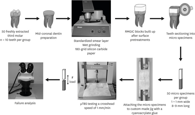
Fifty human third molars without caries were collected after ethical approval (Ref. No. 2019/328). The teeth were kept in 0.5% chloramine-T at 4°C and employed within 1 month following extraction. The mid-coronal dentin surface of all teeth was prepared using a diamond saw (Minitom, Struers, Ballerup, Denmark) under water cooling. The dentin surfaces were examined under a stereomicroscope (S4E, Leica Microsystems, Wetzlar, Germany) to control if there was enamel and/or pulp tissue. The dentin surfaces were polished with wet #180 grit silicon carbide paper for 60 seconds to produce a standardized smear layer in all teeth (Figure 1).
Figure 1

Schematic illustrating the experimental study design.

µTBS, microtensile bond strength; RMGIC, resin-modified glass ionomer cement.
rde-45-e36-g001

Experimental design and specimen preparation

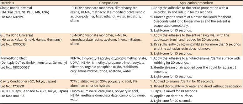
The teeth were randomly divided into five groups (n = 10) based on the surface pretreatment before the application of RMGIC. The different surface pretreatments were employed: Single Bond Universal Adhesive (3M Oral Care, St. Paul, MN, USA, also known as Scotchbond Universal in some countries), Gluma Bond Universal (Heraeus Kulzer GmbH, Hanau, Germany), Prime&Bond Elect (Dentsply DeTrey GmbH, Konstanz, Germany) and Cavity Conditioner (GC, Tokyo, Japan). In the control group, the dentin surfaces did not receive any surface pretreatment. The three universal adhesive systems were tested in self-etch mode and all of the materials were used based on the manufacturer's instructions (Table 1).
Table 1

Chemical composition and application procedure of the tested materials

rde-45-e36-i001
Materials Composition Application procedure
Single Bond Universal (3M Oral Care, St. Paul, MN, USA) 10-MDP phosphate monomer, dimethacrylate resins, HEMA, methacrylate-modified polyalkenoic acid co-polymer, filler, ethanol, water, initiators, silane 1. Apply the adhesive to the entire preparation with a microbrush and rub it in for 20 seconds.
Lot No.: 602724 2. Direct a gentle stream of air over the liquid for about 5 seconds until it no longer moves and the solvent is evaporated completely.
3. Light-cure for 10 seconds.
Gluma Bond Universal (Heraeus Kulzer GmbH, Hanau, Germany) 10-MDP phosphate monomer, 4-META, dimethacrylate resins, acetone, fillers, initiators, silane 1. Apply the adhesive to the entire cavity wall with the applicator brush and rubbed for 20 seconds.
Lot No.: K010033 2. Dry sufficiently by blowing mild air for more than 5 seconds until the adhesive resin does not move.
3. Light-cure for 10 seconds.
Prime&Bond Elect (Dentsply DeTrey GmbH, Konstanz, Germany) PENTA, 2-hydroxy-3 acryloyloxypropyl methacrylate, UDMA, HEMA, trimethylolpropane trimethacrylate, diketone, organic phosphine oxide, stabilizers, cetylamine hydrofluoride, acetone, water 1. Apply the adhesive to air-dried enamel/dentin surface with rubbing for 20 seconds.
Lot No.: 1802000551 2. Gentle stream of air applied over the liquid for at least 5 seconds.
3. Light-cure for 10 seconds.
Cavity Conditioner (GC, Tokyo, Japan) 77% distilled water, 20% polyacrylic acid, 3% aluminum chloride hydrate 1. Applied to enamel/dentin for 10 seconds.
Lot No.: 1708231 2. Rinsed thoroughly with water and dried without desiccation.
Fuji II LC Capsule shade A2 (GC, Tokyo, Japan) Fluoro-alumino-silicate glass, polyacrylic acid, HEMA, urethane dimethacrylate, camphorquinone, water 1. Capsule mixed for 10 seconds.
Lot No.: 180110A 2. Applied on dentin surface.
3. Light-cure for 20 seconds.
10-MDP, 10-methacryloyloxydecyl dihydrogen phosphate; HEMA, hydroxyethylmethacrylate; 4-META, 4-methacryloxyethyl trimellitate anhydride; UDMA, urethane dimethacrylate; PENTA, dipentaerythritol penta acrylate monophosphate; Bis-GMA, bisphenol-glycidyl methacrylate.
After the surface pretreatments, the RMGIC, Fuji II LC Capsule (GC), was mixed using a capsule mixer (Silver Mix, Stomamed, Bratislava, Slovakia) for 10 seconds and incrementally applied on each testing surface in 3–4 layers and up to a height of 5–6 mm using a cylindrical transparent plastic mold. Each increment was light-cured for 20 seconds using a halogen light-curing unit (Optilux 500, Kerr, Orange, CA, USA) with a light output not lower than 600 mW/cm2. After 1-week storage in distilled water at 37°C, the specimens were segmented longitudinally in mesiodistal and buccal-lingual across the bonded interface using the slow-speed diamond saw (Minitom, Struers) to obtain micro tensile test specimens of approximately 1 × 1 mm wide and 8–9 mm long, measured with a digital caliper (Digimatic Caliper, Mitutoyo, Tokyo, Japan). For reducing the effect of regional dentin variability, the five beams were chosen from the center of each tooth and tested for microtensile bond strength (µTBS) test. A total of 50 specimens were tested per each group (n = 50).

µTBS test

After the checking of the cross-sectional area of each beam, the beams were mounted to a custom made microtensile test apparatus using cyanoacrylate glue (Loctite Super Glue, Henkel, Dusseldorf, Germany) and stressed in tension on a universal testing machine (Autograph AGS-X, Shimadzu, Kyoto, Japan) at a crosshead speed of 1 mm/minute until failure. The µTBS values were calculated in MPa by dividing the imposed force (in N) at the time of fracture by the bonding area (in mm2).

Statistical analyses

The mean of µTBS of the beams producing from the same tooth was calculated in each experimental group, and the mean bond strength was taken as one unit for statistical analysis. Statistical analyses were done with the SPSS Program, version 20.0 (Statistical Package for the Social Sciences; SPSS, Chicago, IL, USA). The normal distribution of data was confirmed by Kolmogorov-Smirnov test. The data were analyzed using one-way analysis of variance (ANOVA). For post-hoc multiple comparisons, Duncan test was performed. In all statistical analyses, the significance level was p < 0.05.

Failure analyses

The failure modes were analyzed with a stereomicroscope (S4E, Leica Microsystems) at ×80 magnification. The modes of failure were categorized as adhesive failure, cohesive failure in material, cohesive failure in dentin, or mixed failure (combinations of failure modes). Some representative samples were selected for scanning electron microscope (SEM) analysis. The samples were placed in an aluminum sample holder and fixed with carbon tape and viewed with SEM (Quanta Feg 250, FEI, Eindhoven, The Netherlands).

RESULTS

The one-way ANOVA revealed statistically significant differences between the experimental groups (Table 2). The mean µTBS values of all groups and the standard deviations are presented in Table 3. The statistical analysis results of multiple comparisons are also shown in Table 3. Any pre-test failures were not detected.
Table 2

One-way analysis of variance results for microtensile bond strength test

rde-45-e36-i002
Source Sum of squares df Mean square F p
Between groups 1,656.849 4 414.212 28.332 0.000*
Within groups 657.900 45 14.620
Total 2,314.749 49
*Statistically significant differences (p < 0.05).
Table 3

The mean microtensile bond strengths of the different experimental groups

rde-45-e36-i003
Experimental groups MPa ± SD
Single Bond Universal 31.45 ± 4.02ab
Gluma Bond Universal 34.13 ± 4.63b
Prime&Bond Elect 28.15 ± 3.81ac
Cavity Conditioner 25.85 ± 3.40c
Control 17.35 ± 3.07d
Total number of specimens for each experimental group (n = 50).
SD, standard deviation.
Same superscript small letter indicates no statistically significant difference in the column.
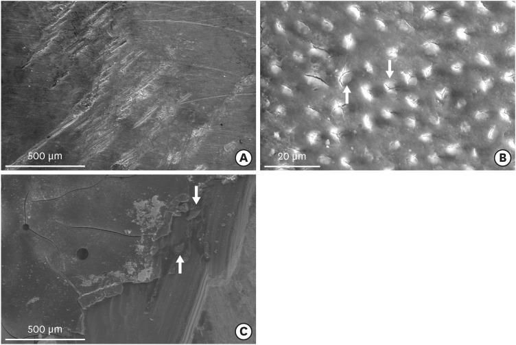
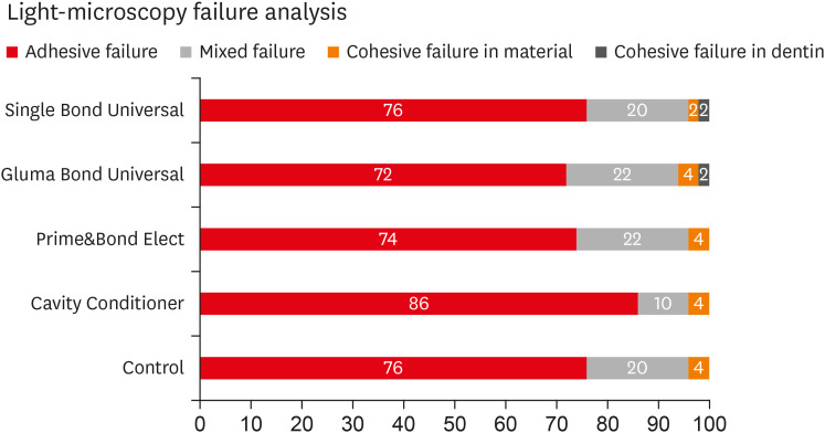
The surface pretreatments with the universal adhesives and conditioner increased the bond strength of the RMGIC to dentin compared to the control group (p < 0.05). Single Bond Universal and Gluma Bond Universal provided higher bond strength to RMGIC than Cavity Conditioner (p < 0.05). The bond strengths obtained from Prime&Bond Elect and Cavity Conditioner were not statistically different (p > 0.05). The failure mode results are shown in Figure 2. The adhesive type failures were mostly seen in each group. The representative SE photomicrographs are illustrated in Figure 3.
Figure 2

Failure mode frequencies (%) observed using light microscopy.

rde-45-e36-g002
Figure 3

Scanning electron photomicrographs of the debonded specimens along the dentin side. (A) An adhesive failure in the control group, showing most of the dentinal tubules occluded by the smear layer, and the remaining scratches from the surface grinding are visible. (B) An adhesive failure of Cavity Conditioner. The partially open dentinal tubules are visible with almost no smear layer. (C) An adhesive failure of Prime&Bond Elect. The arrows point to a resin area covering the dentin surface.

rde-45-e36-g003

DISCUSSION

The results of this study showed that the pretreatment with universal adhesives before the application of RMGIC enhanced the dentin bond strength of the RMGIC. Therefore, the null hypothesis that the use of universal adhesives would not influence the bond strength of RMGIC to dentin was rejected. In the present study, the microtensile test method was used for bond strength tests since its various benefits, such as providing in better stress distribution at the adhesive interface by a smaller surface area. When the surface areas are larger, the flaws and voids could form in the adhesive layer, which causes high-stress concentrations in these areas that result in lower bond strengths [520].
There are two bonding mechanisms in the adhesion of the CICs to dentin, including chemical interaction and micro-mechanical interlocking [67]. The quality of the bond strength is crucial for the clinical longevity of RMGIC restorations [23]. The RMGICs had superior bonding performance than the conventional GICs because the RMGICs contain HEMA, which ensures a high wetting ability of dentin and increases mechanical interlocking with dentinal tubules [1124]. A smear layer forms on the surface during cavity preparation using rotary instruments [25]. If the smear is not removed from the dentin surface before the application of RMGIC, the layer may block the chemical bonding of RMGIC to intertubular dentin and limit any micromechanical interlocking to collagen [45]. The smear layer does not tightly adhere to the tooth surface, and it may be removed or dissolved by conditioners and adhesives to achieve adequate bonding to the tooth surface [16]. However, the RMGICs may bond to the smear layer in different ways. The polyacrylic acid in RMGIC may slightly dissolve the smear layer by act as a mild self-conditioner [5]. The smear layer involves calcium ions, which might ensure chemical bonding with the polyalkenoic acid chains in RMGIC [5]. The inherent dentin irregularities additionally created during specimen preparation could provide micromechanical interlocking [5].
The polyacrylic-acid conditioner, Cavity Conditioner, partially removes the smear layer and demineralize the underlying dentin without entirely unplugging dentin tubules, thus enhancing the bond strength of RMGIC through increasing surface area and micro porosities [4511]. Besides, the exposed calcium ions within hydroxyapatite bonds chemically to carboxyl groups of the polyalkenoic acid, and the exposed collagen provides adjunct micromechanical retention [5]. The aluminum chloride in Cavity Conditioner might procure better penetration of the RMGIC by stabilizing the collagen matrix during demineralization [5]. It has been reported that when dentin was treated with Cavity Conditioner, the formation of resin tags and a submicron hybrid layer which similar to the one produced by mild self-etch adhesives [826]. It has been concluded that a gel phase occurred as an intermediate formation settled over the hybrid layer of polyacrylic acid-conditioned dentin surface [727]. The formation of the gel phase is thought to symbolize the accumulation of a calcium-polycarboxylate salt, welding from a chemical reaction between the carboxyl groups of polyacrylic acid in the conditioner and calcium in hydroxyapatite [27]. Therefore, the chemical interaction could play an essential role in the bonding mechanism of RMGIC than the micro-mechanical interlocking originated from dentin hybridization [7]. It has also been stated that the use of Cavity Conditioner was unimportant to improving the bond strength of RMGIC to dentin in the absence of smear layer [7]. In this study, the standardized smear layer was created on the flat dentin surfaces in all experimental groups, and the Cavity Conditioner increased the dentin bond strength of RMGIC compared to the control group, as concluded in previous studies [4711]. Furthermore, the removal of the smear layer might provide an adjunct resource of water for the acid-base setting reaction of GIC by increasing dentin permeability [28]. Thus, the bonding of RMGIC to dentin could become more resistant to degradation in the time due to inducing the maturation of GIC at the interface [29].
This study has just demonstrated that universal adhesives enhanced the bonding performance of RMGIC to dentin. The dentin bond strength could be enhanced by improving the chemical adhesion of RMGIC via different functional monomers in the universal adhesives, as attributed in previous studies [511121314]. The adhesives may form a chemical interaction with RMGIC through the existence of HEMA and other resins in RMGIC [510]. It has been reported that a conditioner (Self Conditioner, GC) containing 4-methacryloxyethyl trimellitate anhydride (4-META) and HEMA enhanced the dentin bond strength of RMGIC [458]. This increased bond strength has been attributed to enhancing dentin wettability and allowing better monomer penetration, thereby increasing the quality of the hybrid layer, which is the main source of bonding [458]. 4-META is a functional monomer that chemically interacts with the hydroxyapatite in dentin, and HEMA provides an improvement in dentin wettability and penetration of RMGIC into the dentin [830]. It has been reported that the presence of a polyalkenoic acid co-polymer or an acidic monomer in an adhesive could positively affect the bond strength of RMGIC [10]. Single Bond Universal and Gluma Bond Universal include 10-methacryloyloxydecyl dihydrogen phosphate (10-MDP) as the acidic functional monomer, which creates surface micro-retention and interacts chemically with calcium in hydroxyapatite [1630]. It has been stated that the acidic functional monomer, 10-MDP, provided superior bond strength for RMGIC [12]. Gluma Bond Universal contains acidic monomer 4-META involving 2 carboxylic groups in addition to 10-MDP. Single Bond Universal also involves a polyalkenoic acid co-polymer, which can also bond chemically to hydroxyapatite. However, it has been reported that the polyalkenoic-acid co-polymer potentially contests with the 10-MDP functional monomer for calcium-binding sites in hydroxyapatite, and may also inhibit monomer polymerization due to its high molecular weight [17]. The main functional monomer of Prime&Bond Elect is dipentaerythritol penta acrylate monophosphate (PENTA). In this study, Single Bond Universal and Gluma Bond Universal significantly more increased the bond strength than Cavity Conditioner though the bond strengths obtained from Prime&Bond Elect and Cavity Conditioner were not different. The obtained high bond strengths could be due to the monomers in their content are different.
Moreover, the pH of the pretreatment agent may be effective on the bond strength of RMGIC [5]. If the pH is adequately low, it might partly remove the smear layer, thereby it allows the penetration of the matrix of RMGIC to the superficial dentin layer and creates a cement-matrix dentin interdiffusion zone [5]. The pH of Cavity Conditioner is 1.2 [8]. The pH of tested universal adhesives Single Bond Universal, Gluma Bond Universal and Prime&Bond Elect is 2.7, 1.8 and 2.5, respectively. The mild (pH ≈ 2) and ultra-mild (pH > 2.5) self-etch adhesives are not able to entirely remove the smear layer [16]. These adhesives dissolve the smear layer without too deeply demineralizing the tooth surface, thereby preserving hydroxyapatite at the interface [16]. The presence of the smear layer and the hydroxyapatite at the interface could create an additional resource for the chemical bonding of RMGIC, doing so improving bonding efficacy [5]. When the use of the Cavity Conditioner before the application of RMGIC, any layer has not formed, but an adhesive layer has occurred following the use of adhesive, as shown in Figure 3. The dentin surface on which has an adhesive layer may be more suitable than the conditioned dentin surface by Cavity Conditioner for the effective adhesion of RMGIC.
Previous studies reported variability in the failure modes for RMGIC [41213]. It has been stated that adhesive failure might mostly be determined early, but the number of cohesive failures might increase with aging [6]. The high incidence of the mixed and cohesive failure types could increase due to the presence of voids that forms with air entrapment within the material during hand mixing [10]. Since the capsule form was used in the present study, the failure mode was the predominantly adhesive failure. Nonetheless, more relevant studies should be conducted to support the findings of this study. Also, different aging conditions may be considered in further studies.

CONCLUSIONS

Under the limitations of this study, it can be concluded that the pretreatment with universal adhesives and polyacrylic acid conditioner increased the bond strength of RMGIC to dentin. The use of universal adhesives before the application of RMGIC could be more beneficial in improving bond strength. The adhesive failure mode was mostly detected.

ACKNOWLEDGEMENTS

The author thanks to 3M Oral Care, Heraeus Kulzer, Dentsply and GC Corporation, for the material support of this study.

Notes

Conflict of Interest: No potential conflict of interest relevant to this article was reported.

References

1. Wilson AD, Kent BE. A new translucent cement for dentistry. The glass ionomer cement. Br Dent J. 1972; 132:133–135. PMID: 4501690.
crossref
2. Garoushi S, Vallittu PK, Lassila L. Characterization of fluoride releasing restorative dental materials. Dent Mater J. 2018; 37:293–300. PMID: 29279547.
crossref
3. Burgess J, Norling B, Summitt J. Resin ionomer restorative materials: the new generation. J Esthet Dent. 1994; 6:207–215. PMID: 8593217.
crossref
4. Saad A, Inoue G, Nikaido T, Ikeda M, Burrow MF, Tagami J. Microtensile bond strength of resin-modified glass ionomer cement to sound and artificial caries-affected root dentin with different conditioning. Oper Dent. 2017; 42:626–635. PMID: 28857710.
crossref
5. Imbery TA, Namboodiri A, Duncan A, Amos R, Best AM, Moon PC. Evaluating dentin surface treatments for resin-modified glass ionomer restorative materials. Oper Dent. 2013; 38:429–438. PMID: 23088188.
crossref
6. Hoshika S, De Munck J, Sano H, Sidhu SK, Van Meerbeek B. Effect of conditioning and aging on the bond strength and interfacial morphology of glass-ionomer cement bonded to dentin. J Adhes Dent. 2015; 17:141–146. PMID: 25859569.
7. Cardoso MV, Delmé KI, Mine A, Neves Ade A, Coutinho E, De Moor RJ, Van Meerbeek B. Towards a better understanding of the adhesion mechanism of resin-modified glass-ionomers by bonding to differently prepared dentin. J Dent. 2010; 38:921–929. PMID: 20728505.
crossref
8. Coutinho E, Van Landuyt K, De Munck J, Poitevin A, Yoshida Y, Inoue S, Peumans M, Suzuki K, Lambrechts P, Van Meerbeek B. Development of a self-etch adhesive for resin-modified glass ionomers. J Dent Res. 2006; 85:349–353. PMID: 16567557.
crossref
9. Vargas MA, Fortin D, Swift EJ Jr. Bond strengths of glass ionomers using a dentin adhesive. Am J Dent. 1995; 8:197–200. PMID: 7576387.
10. El-Askary F, Nassif M. Bonding nano-filled resin-modified glass ionomer to dentin using different self-etch adhesives. Oper Dent. 2011; 36:413–421. PMID: 21819202.
crossref
11. Poggio C, Beltrami R, Scribante A, Colombo M, Lombardini M. Effects of dentin surface treatments on shear bond strength of glass-ionomer cements. Ann Stomatol (Roma). 2014; 5:15–22.
crossref
12. Saad A, Inoue G, Nikaido T, Abdou AMA, Sayed M, Burrow MF, Tagami J. Effect of dentin contamination with two hemostatic agents on bond strength of resin-modified glass ionomer cement with different conditioning. Dent Mater J. 2019; 38:257–263. PMID: 30504695.
crossref
13. Dursun E, Attal JP. Combination of a self-etching adhesive and a resin-modified glass ionomer: effect of water and saliva contamination on bond strength to dentin. J Adhes Dent. 2011; 13:439–443. PMID: 20978640.
14. Besnault C, Attal JP, Ruse D, Degrange M. Self-etching adhesives improve the shear bond strength of a resin-modified glass-ionomer cement to dentin. J Adhes Dent. 2004; 6:55–59. PMID: 15119588.
15. Pereira PN, Yamada T, Inokoshi S, Burrow MF, Sano H, Tagami J. Adhesion of resin-modified glass ionomer cements using resin bonding systems. J Dent. 1998; 26:479–485. PMID: 9699441.
crossref
16. Van Meerbeek B, Yoshihara K, Yoshida Y, Mine A, De Munck J, Van Landuyt KL. State of the art of self-etch adhesives. Dent Mater. 2011; 27:17–28. PMID: 21109301.
crossref
17. Wagner A, Wendler M, Petschelt A, Belli R, Lohbauer U. Bonding performance of universal adhesives in different etching modes. J Dent. 2014; 42:800–807. PMID: 24814138.
crossref
18. Cuevas-Suárez CE, da Rosa WLO, Lund RG, da Silva AF, Piva E. Bonding performance of universal adhesives: an updated systematic review and meta-analysis. J Adhes Dent. 2019; 21:7–26. PMID: 30799468.
19. Rosa WL, Piva E, Silva AF. Bond strength of universal adhesives: a systematic review and meta-analysis. J Dent. 2015; 43:765–776. PMID: 25882585.
crossref
20. Sofan E, Sofan A, Palaia G, Tenore G, Romeo U, Migliau G. Classification review of dental adhesive systems: from the IV generation to the universal type. Ann Stomatol (Roma). 2017; 8:1–17. PMID: 28736601.
21. Hanabusa M, Mine A, Kuboki T, Momoi Y, Van Ende A, Van Meerbeek B, De Munck J. Bonding effectiveness of a new ‘multi-mode’ adhesive to enamel and dentine. J Dent. 2012; 40:475–484. PMID: 22381614.
crossref
22. Takamizawa T, Imai A, Hirokane E, Tsujimoto A, Barkmeier WW, Erickson RL, Latta MA, Miyazaki M. SEM observation of novel characteristic of the dentin bond interfaces of universal adhesives. Dent Mater. 2019; 35:1791–1804. PMID: 31727447.
crossref
23. Cook NB, Feitosa SA, Patel A, Alfawaz Y, Eckert GJ, Bottino MC. Bonding ability of paste-paste glass ionomer systems to tooth structure: in vitro studies. Oper Dent. 2015; 40:304–312. PMID: 25535780.
24. Nakanuma K, Hayakawa T, Tomita T, Yamazaki M. Effect of the application of dentin primers and a dentin bonding agent on the adhesion between the resin-modified glass-ionomer cement and dentin. Dent Mater. 1998; 14:281–286. PMID: 10379257.
crossref
25. Pashley DH. Smear layer: overview of structure and function. Proc Finn Dent Soc. 1992; 88(Supplement 1):215–224. PMID: 1508877.
26. Abdalla AI. Morphological interface between hybrid ionomers and dentin with and without smear-layer removal. J Oral Rehabil. 2000; 27:808–814. PMID: 11012857.
crossref
27. Coutinho E, Yoshida Y, Inoue S, Fukuda R, Snauwaert J, Nakayama Y, De Munck J, Lambrechts P, Suzuki K, Van Meerbeek B. Gel phase formation at resin-modified glass-ionomer/tooth interfaces. J Dent Res. 2007; 86:656–661. PMID: 17586714.
crossref
28. Yiu CK, Tay FR, King NM, Pashley DH, Carvalho RM, Carrilho MR. Interaction of resin-modified glass-ionomer cements with moist dentine. J Dent. 2004; 32:521–530. PMID: 15304297.
crossref
29. Marquezan M, Fagundes TC, Toledano M, Navarro MF, Osorio R. Differential bonds degradation of two resin-modified glass-ionomer cements in primary and permanent teeth. J Dent. 2009; 37:857–864. PMID: 19631441.
crossref
30. Yoshida Y, Nagakane K, Fukuda R, Nakayama Y, Okazaki M, Shintani H, Inoue S, Tagawa Y, Suzuki K, De Munck J, Van Meerbeek B. Comparative study on adhesive performance of functional monomers. J Dent Res. 2004; 83:454–458. PMID: 15153451.
crossref
TOOLS
Similar articles