

 PDF
PDF Citation
 Print
Print


INTRODUCTION
The main goal of the root canal obturation is to provide an appropriate filling of anatomical irregularities with minimal voids [1]. In order to achieve this objective, gutta-percha cones are associated with endodontic sealers [2]. More recently, calcium silicate-based sealers have been introduced in endodontic practice [3]. Novel calcium silicate sealers are presented in a premixed-syringe with an intracanal tip designed to be used with a single-cone obturation technique [1]. Among these new materials, Bio-C Sealer (Angelus, PR, Brazil) showed short setting time, alkalinization capacity, proper flow and radiopacity, besides cytocompatibility [45]. However, there is no study evaluating the filling ability of this sealer.
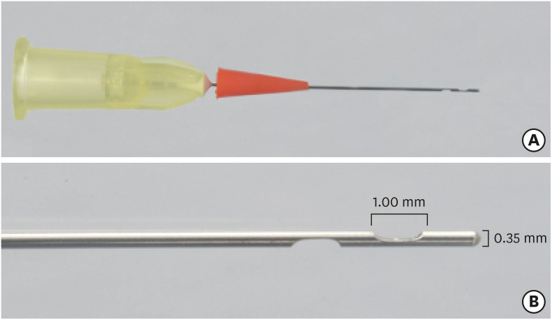
During the root canal obturation, an important issue is the sealer extrusion to the periradicular tissues or into the surrounding anatomical structures, which could cause postoperative pain [6]. Calcium silicate-based sealers have been associated with a significantly higher incidence of extrusion [78]. This fact could be associated with their application with an endodontic tip provided by the manufacturer [7]. Recently, a new Sealer Injection System (SIS) (Angelus) was developed to be used with ready to use premixed bioceramic sealers. SIS has needles with 2 lateral openings, located between 1 and 4 mm from the tip, without an apical opening. These side openings are oval 1 mm long and the needle is 0.35 mm wide. According to the manufacturer, this system provides better tridimensional filling of the root canal system. Nevertheless, up to now, no study was performed evaluating this new system.
High-resolution micro-computed tomography (micro-CT) has been used in endodontic research due to its accuracy and nondestructive characteristic, allowing detailed imaging of the root canal system [39]. Micro-CT can be applied as a reliable tool for assessments of artificial canals in Endodontic research [10]. Regarding the main advantages of using artificial canals, the anatomical variation is minimized, ensuring standardization of the samples [11].
Since the filling ability of all the root canal system and the apical extrusion of the sealers are important issues in endodontics, the aim of this study was to investigate by micro-CT the presence of voids besides the apical extrusion in simulated root canals filled by Bio-C using its conventional system to deliver the sealer or a new SIS with side openings. The null hypothesis was that there would be no differences between the systems regarding filling ability and apical sealer extrusion.
Go to : 
MATERIALS AND METHODS
Specimen selection and root canal instrumentation
Acrylic resin models with a main artificial canal and 3 lateral simulated canals (cervical, middle, and apical thirds) were used (n = 16). The main artificial canal had a standard size (19 mm), 60° angle of curvature and 5 mm radius and the center of the curvature were 5 mm from the end of the canal.
The working length (WL) was determined using a size 10 K-file (Dentsply, Maillefer, Ballaigues, Switzerland) until the apical foramen and was recorded 1 mm short of this length (WL = 18 mm). A single operator who was trained and calibrated prepared all the specimens using the ProDesign Logic system (Easy Equipamentos Odontológicos, Belo Horizonte, MG, Brazil) operated with an endodontic motor (VDW Silver, VDW GmbH, Munich, Germany). The files were used in continuous rotation using in-and-out movement up to the WL following the sequence: 25/0.01, 25/0.04, 25/0.06, 35/0.01, and 35/0.05. The 25/0.01 and 35/0.01 files were used at 350 rpm speed and 1 Ncm torque, while the 25/0.04, 25/0.06, and 35/0.05 were used at 600 rpm speed and 4 Ncm torque. The canals were irrigated with 2.5 mL of distilled water after each instrument by using a disposable syringe and a 27-G NaviTip needle (Ultradent, South Jordan, UT, USA). The root canals were dried only with absorbent paper cones without causing excessive dryness. After, in order to create some resistance to avoid a great amount of apical extrusion of the sealer, the root apexes were enveloped by acrylic wool.
Root canal filling
The prepared canals were divided into 2 groups (n = 8). The canals were filled by a single cone technique with the premixed ready to use calcium silicate-based Bio-C Sealer. In the first group, Bio-C was injected into the root canal by the conventional method, according to the manufacturer's instruction. The syringe was properly positioned, with the tip inserted until 4 mm shorter from the WL and Bio-C Sealer was applied in the apical third of the root canal. The plunger was lightly pressed until a complete filling of the root canals. In the second group, the SIS with 0.35 mm of tip size and side openings was used (Figure 1) and threaded to the Bio-C Sealer syringe. A silicone plug at the base of the SIS was fitted at the beginning of the root canal. The SIS tip was inserted into the root canal until the WL. When the pressure was observed in the syringe plunger, the SIS tip was indented from 3 to 5 mm until the filling of all thirds of the root canal. Subsequently, for both groups, 35/0.05 gutta-percha cones (Tanari industry Ltda., São Paulo, SP, Brazil), which were previously selected in a profilometer (Profile Projector Nikon Model 6C-2), were inserted into each canal up to the WL. Then, the gutta-percha cones were cut at the cervical level and the remaining material was slightly compacted with a heated plugger. Digital periapical radiographs were taken (Kodak RVG 6100 Digital Radiography System, Marne-la-Vallée, France) to confirm the complete filling of root canals. The coronal portion was sealed with a provisional restorative material (Coltosol, Vigodent, Rio de Janeiro, RJ, Brazil). All specimens were stored in an oven at 37°C in relative humidity for 7 days to allow the sealers to set completely. As Bio-C Sealer requires moisture for setting, 2 pieces of wet cloth were placed over the samples, according to Tanomaru-Filho et al. [12].
Micro-CT scanning and analysis
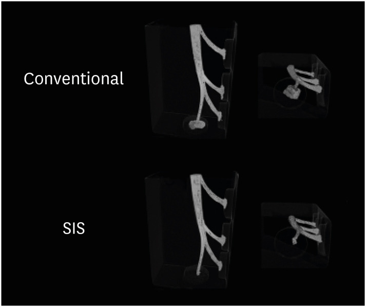
The samples were scanned using micro-CT (SkyScan 1272, Bruker, Kontich, Belgium). The micro-CT parameters were 10 µm voxel size, 80 kV, 125 µA, rotation step 0.2, frame 4, 1.0 mm aluminum filter, and 180° scanning. The images obtained were reconstructed using the NRecon software (NRecon v.1.6.10.4, Bruker), and analyzed by the CTAn software (CTAn v1.15.4.0, Bruker). Root canal volume, filling material volume (gutta-percha and sealer), and voids percentage were quantified at all extension for the main root canal. The volume of apical extrusion of the sealer was recorded. The volume percentage of voids in the lateral canals was also quantified in the cervical, middle, and apical thirds. The grayscale range required to recognize each object under study was determined in a density histogram by using an adaptive threshold method. Qualitative analyses were performed by means of models obtained by using CTVox software (CTVox v. 3.2, Bruker) (Figure 2).
Statistical analysis
The normality of the data was tested using the Kolmogorov-Smirnov test. Statistical analysis was performed with t-test with a significance level of 5%.
Go to :
RESULTS
The results are presented in Tables 1 and 2. Both obturation systems presented similar filling capacity in the main root canal (p > 0.05). However, the conventional system showed the highest apical extrusion of the sealer (p < 0.05). Regarding the filling of the lateral canals, there was a similar percentage of voids in the canals obturated with both systems in the middle and apical thirds (p > 0.05). Nevertheless, SIS had the best filling ability of the cervical third of the lateral canal (p < 0.05). Three-dimensional models illustrating the filling of the root canals and the apical extrusion of the sealer can be observed in Figure 2.

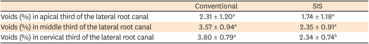
Table 1
Volume percentage of voids in the main root canal and volume of apical extrusion after root canal filling using the conventional or Sealer Injection System (SIS) systems
| Conventional | SIS | |
|---|---|---|
| Total voids (%) in the main root canal | 0.52 ± 0.13a | 0.42 ± 0.14a |
| Apical sealer extrusion (mm3) | 3.31 ± 0.65a | 0.14 ± 0.10b |
The values are mean ± standard deviation. Different letters on the same line indicate a statistically significant difference between the groups (p < 0.05).
![]()
Table 2
Volume percentage of voids in all thirds of the lateral root canals after root canal filling using the conventional or Sealer Injection System (SIS) systems
The values are mean ± standard deviation. Different letters on the same line indicate a statistically significant difference between the groups (p < 0.05).
![]()
Go to :
DISCUSSION
An appropriate root canal filling provided by the association between gutta-percha cones and endodontic sealers is clinically valuable [9]. Therefore, different techniques are used in order to obtain adequate obturation with minimal voids formation [1]. This is the first study evaluating a new SIS with side openings regarding its filling ability and apical extrusion. Our results showed the satisfactory filling ability of the main and lateral root canals, besides a low sealer apical extrusion for this system. Since differences between the conventional obturation system and SIS were observed in some analyses, our null hypothesis was partially rejected.
Simulated artificial canals have already been validated as a satisfactory model to studies in the endodontic field, due to the standardization of the canal anatomy [13]. In the current study, the main artificial canal was prepared by the ProDesign Logic system up to 35.05 file, since the apical enlargement with this instrument decreases the percentage of untouched root canal surface, without causing deviations or procedural errors [14]. This standardized instrumentation with a proper apical enlargement probably favored the adequate filling of the apical third for both systems evaluated, which showed a volume percentage of voids in the main root canal below 1%.
The appropriate filling of the main root canal for both systems could also be related to the high flow of Bio-C Sealer [4], since this is an important characteristic to seal spaces between gutta-percha cone and dentinal wall [15]. Moreover, the flow property of root canal sealers determines how effectively accessory canals will be obturated, influencing in the presence of the voids [16]. Although a high flow should facilitate the sealer penetrability into accessory anatomy, there is an increase in the risk of periapical extrusion [16]. Therefore, a balance should be struck in selecting a sealer with the appropriate flow without causing apical extrusion [16]. Our study showed that SIS generated the least apical extrusion of the sealer. This finding is in agreement with previous studies that compared the apically extruded debris proportionated with open-ended or side-vented needle tips and observed that open-ended needles were associated with significantly more extrusion [1718]. Although the higher apical extrusion of ready-to-use calcium silicate-based sealers would not be related with pain [7], and could not have any significant effect on the treatment outcome [8], the sealer extrusion can lead to a neurogenic inflammation, which may underlie the flare-up occurrences [19]. Therefore, the use of SIS could be safer than the conventional system. Additionally, SIS had the greatest filling ability of the lateral canal in the cervical third, probably due to the lateral pressure exerted during its use. Based on these results, SIS proves to be a system capable of filling irregularities in the root canal system, with easy clinical application.
Many studies have been proposed the use of micro-CT for the evaluation of endodontic materials [13412202122]. Micro-CT allows a nondestructive, quantitative and qualitative high-resolution imaging-based assessment [3]. Some limitation related to the micro-CT method is that different radiopacity and rheological properties of the root canal sealers could affect its detection of voids [9]. Therefore, our study used only Bio-C as an endodontic sealer in order to have only the obturation systems as a variable. This is the first study to evaluate the filling ability of Bio-C. However, a previous study showed a low percentage of voids for this sealer, even after immersion in distilled water or phosphate buffered saline [22]. Moreover, the scanning resolution of the micro-CT images is fundamental for small voids detection. Therefore, our study used 10 µm as the voxel size since 11.2 µm is a reliable cutoff value for the evaluation of root canal filling voids in micro-CT imaging [23].
Go to :
 XML Download